Background
Viral infections in pregnancy are major causes of maternal and fetal morbidity and mortality. Infections can develop in the neonate transplacentally, perinatally (from vaginal secretions or blood), or postnatally (from breast milk or other sources). The clinical manifestations of neonatal infections vary depending on the viral agent and the gestational age at exposure. The risk for infection is usually inversely related to gestational age at acquisition, some resulting in a congenital malformation syndrome.
Infections known to produce congenital defects have been described with the acronym TORCH (Toxoplasma, others, rubella, cytomegalovirus [CMV], herpes). The "others" category has rapidly expanded to include several viruses known to cause neonatal disease.
Traditionally, the only viral infections of concern during pregnancy were those caused by rubella virus, CMV, and herpes simplex virus (HSV). Other viruses now known to cause congenital infections include parvovirus B19 (B19V), varicella-zoster virus (VZV), West Nile virus, measles virus, enteroviruses, adenovirus, human immunodeficiency virus (HIV), and Zika virus.
Also of importance is hepatitis E virus because of the high mortality rate associated with infection in pregnant women. Recently, lymphocytic choriomeningitis virus (LCMV) has been implicated as a teratogenic rodent-borne arenavirus.
Worldwide, congenital HIV infection is now a major cause of infant and childhood morbidity and mortality, responsible for an estimated 4 million deaths since the start of the HIV pandemic. The breadth and depth of this problem is beyond the scope of this article.
With emerging concerns for an influenza pandemic, attention has also now been directed to the effects of influenza on pregnant women. Pregnant women are more likely to develop severe disease, perhaps related to physiological changes in pregnancy, such as decreased lung capacity, increased oxygen needs, and increased heart rate. Currently, inactivated influenza vaccine is recommended in all trimesters of pregnancy. [1] One study found that influenza vaccination of high-risk pregnant patients also provides some protective immunity for newborns and reduces subsequent hospitalizations in the infants. [2] Influenza has historically been shown to produce significant morbidity and mortality in this population (see Influenza and H1N1 Influenza [Swine Flu]).
With the recent Ebola-related deaths in the United States, there is some suggestion that pregnant people may be more susceptible to severe disease and death from Ebola.
Zika virus
Zika virus, first introduced in South America in 2015, has since spread throughout the Americas. The virus has been shown to cause severe congenital abnormalities, including microcephaly. There is an ongoing Zika virus outbreak in the Americas, the Caribbean, and the Pacific. The World Health Organization (WHO) declared Zika virus a Public Health Emergency of International Concern between February and November 2016.
Cytomegalovirus
Cytomegalovirus (CMV) infection is the most common perinatal viral infection in the United States. CMV is a double-stranded DNA herpes virus and represents the most common congenital viral infection. The CMV seropositivity rate increases with age. Geographic location, socioeconomic class, and work exposure are other factors that influence the risk of infection. CMV infection requires intimate contact through saliva, urine, and/or other body fluids. Possible routes of transmission include sexual contact, organ transplantation, transplacental transmission, transmission via breast milk, and blood transfusion (rare).
Primary, reactivation, or recurrent CMV infection can occur in pregnancy and can lead to congenital CMV infection. Transplacental infection can result in intrauterine growth restriction, sensorineural hearing loss, intracranial calcifications, microcephaly, hydrocephalus, hepatosplenomegaly, delayed psychomotor development, and/or optic atrophy.
Vertical transmission of CMV can occur at any stage of pregnancy; however, severe sequelae are more common with infection in the first trimester, while the overall risk of infection is greatest in the third trimester. The risk for transmission to the fetus in primary infection is 30% to 40%. Most (90%) CMV infections cause no symptoms, but 10% result in signs and symptoms such as microcephaly, thrombocytopenia, hepatosplenomegaly, intrauterine growth restriction, or a combination thereof.
Thirty percent of infants with severe CMV infection die; among survivors, more than half eventually develop neurological sequelae, including microcephaly, intellectual disability, and/or sensorineural hearing loss. Seven percent of asymptomatic neonates develop sensorineural hearing loss or developmental delays during the first 2 years of life. [3, 4, 5, 6, 7] Five percent eventually develop microcephaly and neuromuscular defects, and 2% develop chorioretinitis. Congenital hearing loss is the most common sequela of recurrent CMV infection.
Herpes simplex virus
Thirty to 60% of individuals receiving obstetric care have serologic evidence of past HSV infection. Although both HSV-1 and HSV-2 may cause neonatal herpes, HSV-2 is responsible for 70% of cases. Neonatal herpetic infection is defined as infection within 28 days of birth. Ninety percent of infections are perinatally transmitted in the birth canal. HSV infection acquired in this manner carries a 70% risk for dissemination and is associated with three distinct syndromes, each with its own typical outcome. The first and most common (45%) is localized skin, eye, or mouth disease. Approximately 30% of cases manifest as central nervous system (CNS) disease, including meningitis or encephalitis, with evidence of HSV DNA in the cerebrospinal fluid (CSF). Finally, 25% of neonatal herpetic infections manifest as disseminated disease that involves multiple organs. Initial symptoms of this disease usually present during the first 4 weeks of life.
Approximately 10% of infections are congenital, usually a consequence of the mother acquiring primary HSV infection during pregnancy and the fetus acquiring the infection transplacentally or via an ascending infection from the cervix. Intrauterine infection is associated with intrauterine growth restriction, preterm labor, and miscarriage. [8, 9] The risk for neonatal herpes and death is highest in infants born to mothers who have not seroconverted by the time of delivery.
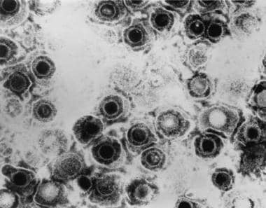 Viral infections and pregnancy. Transmission electron micrograph of herpes simplex virus. Some nucleocapsids are empty, as shown by penetration of electron-dense stain. Image and caption from US Centers for Disease Control and Prevention Public Health Image Library, available at: http://phil.cdc.gov/Phil/search.asp. Use Image ID 281.
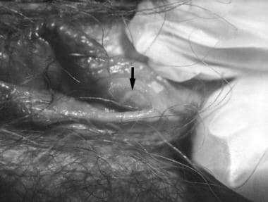 Viral infections and pregnancy. Blisters on the vulva due to a recurring herpes II (HSV-2) virus infection. Image and caption from US Centers for Disease Control and Prevention Public Health Image Library, available at: http://phil.cdc.gov/Phil/search.asp. Use Image ID 2319.
Rubella
Rubella is one of the more teratogenic viruses. Congenital rubella syndrome (CRS) is characterized by intrauterine growth restriction, intracranial calcifications, microcephaly, cataracts, cardiac defects (most commonly patent ductus arteriosus or pulmonary arterial hypoplasia), neurologic disease (with a broad range of presentations, from behavior disorders to meningoencephalitis), osteitis, and hepatosplenomegaly.
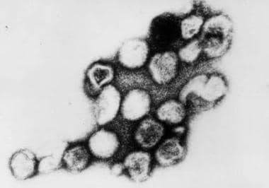 Viral infections and pregnancy. Transmission electron micrograph of rubella virus. Image and caption from US Centers for Disease Control and Prevention Public Health Image Library, available at: http://phil.cdc.gov/Phil/search.asp. Use Image ID 269.
Neonates with rubella may have a "blueberry muffin" appearance caused by purpuric skin lesions that result from extramedullary hematopoiesis. Heart defects in these infants include ventricular septal defects, patent ductus arteriosus, pulmonary stenosis, and coarctation of the aorta. The presentation of rubella at birth varies greatly. Most of these complications develop in infants born to mothers who acquire rubella infection during the first 16 weeks of pregnancy. Ninety percent of infants present with some finding of congenital rubella if infection occurs within the first 12 weeks, and 20% present with congenital disease if the infection occurs between weeks 12 and 16. [10] Cataracts results when infection occurs between the third and eighth week of gestation, deafness between the 3rd and 18th week, and heart abnormalities between the 3rd and 10th week. [11]
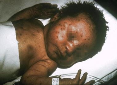 Viral infections and pregnancy. Infant with congenital rubella and blueberry muffin skin lesions. Lesions are sites of extramedullary hematopoiesis and can be associated with several different congenital viral infections and hematologic diseases. Image and caption from US Centers for Disease Control and Prevention Public Health Image Library, available at: http://phil.cdc.gov/Phil/search.asp. Use Image ID 713.
Parvovirus B19
B19V causes erythema infectiosum (fifth disease). Although most adults with B19V infection are asymptomatic, the effects of this virus on the fetus are much greater and include miscarriage, fetal anemia, hydrops fetalis, myocarditis, and/or intrauterine fetal death. Infection occurs most frequently in the winter and spring. B19V infection accounts for 15% to 20% of cases of nonimmune hydrops fetalis. Thirty to 40% of pregnant women are seronegative for B19V and are thus susceptible to infection.
Various studies have estimated that 3% to 14% of intrauterine fetal deaths occur in the setting of B19V infection. Second-trimester infections have been studied most frequently because infection in this trimester carries a 1% to 3% risk for hydrops; however, infection in any trimester may result in intrauterine fetal loss. The critical period for the development of fetal hydrops is when maternal B19V infection is acquired between the 13th and 16th week of gestation, possibly because of the relative immaturity of the fetal immune response, as well as the shortened life span of the red blood cells at this gestational age.
Varicella-zoster virus
VZV is a common virus that carries risks for both the mother and fetus during pregnancy. Morbidity and mortality rates associated with VZV infection are much higher in adults than in children. Primary varicella infection during pregnancy is considered a medical emergency. Pneumonitis due to VZV infection is 25 times more common in adults than in children; in the third trimester, the risk for life-threatening ventilatory compromise is significant, with a mortality rate of 14%. Before the development of antiretrovirals, pneumonitis in pregnant women carried a mortality rate of 45%. Other risk factors for the development of pneumonitis include smoking and a large lesion burden (>100 lesions). [12]
Congenital varicella syndrome (CVS) results in spontaneous abortion, chorioretinitis, cataracts, limb atrophy, cerebral cortical atrophy, and/or neurological disability. Spontaneous abortion has been reported in 3% to 8% of first-trimester VZV infections, and CVS has been reported in 12%. [13] Acquisition of infection by the mother in the perinatal period, specifically 5 days prior to delivery or 2 days afterward, poses a risk for severe neonatal varicella, which carries a mortality rate of 30%. Infection at this time prevents development of maternal antibodies that avert transplacental transfer of immunoglobulin G (IgG) antibodies, which confer passive immunity to the fetus.
Enterovirus
Enterovirus infections are not believed to cross the placenta and cause fetal disease. [14] However, some studies have linked coxsackievirus and echovirus to miscarriage, neurodevelopmental delay, myocarditis, and cortical necrosis. [15, 16] One study linked the presence of coxsackievirus in the third trimester with respiratory failure and global cognitive defects. [17]
Measles virus
Measles virus infection (rubeola) during pregnancy, as with VZV infection, tends to be severe, with pneumonitis predominating. Although it is not known to be teratogenic, rubeola has been associated with spontaneous abortion, premature labor, and low birth weight. Neonates born to mothers with active measles virus infection are at risk of developing neonatal measles, but no congenital syndrome has been described. [18]
Lymphocytic choriomeningitis virus
LCMV has been associated with sporadic cases of congenital infection worldwide. Affected infants demonstrate chorioretinitis, hydrocephalus, intellectual disability, and/or visual impairment; in addition, intrauterine death is possible. Unlike congenital CMV and rubella infections, hearing deficits and hepatosplenomegaly are rarely seen in congenital LCMV.
Other viruses
Other viruses postulated to cause congenital infections include echovirus, hepatitis B virus, hepatitis C virus, and adenovirus. [19] In 2002, an article in Morbidity and Mortality Weekly Report discussed a single case of West Nile virus infection in a mother and associated chorioretinitis in her newborn. [20] A causal link has not been determined. Since then, the Centers for Disease Control and Prevention (CDC) has maintained a registry of West Nile virus infections during pregnancy. Other congenital malformations have been described in this registry, but a direct cause-effect relationship has not yet been established. Infants born to mothers who develop symptomatic West Nile virus infection within 3 weeks prior to delivery may develop symptomatic West Nile virus disease shortly after birth.
Influenza poses a significant threat to the health of the mother and infant. Historic reports of the 1918 Spanish flu pandemic and the 1957 Asian flu pandemic reported a mortality rate of approximately 50% among infected pregnant people. [21] During the 2009 H1N1 pandemic, pregnant people accounted for 5% of all deaths and were more likely to be hospitalized than the general population. [22]
Pathophysiology
Human CMV is the largest of the beta herpes viruses and can cause lytic and productive infection. Like other herpes viruses, it can be latent and reactivate. CMV infection in pregnancy can be primary (initial acquisition in pregnancy) or recurrent. Vertical transmission can occur transplacentally; in addition, the virus can be transmitted via cervicovaginal secretions at the time of delivery or by ingestion of breast milk postpartum. Transplacental transmission is associated with congenital CMV infection. Maternal shedding at time of delivery is associated with a 50% risk for infection. [23] CMV infection acquired through exposure to infected cervical secretions or human milk is usually asymptomatic and is not associated with neonatal sequelae.
Herpes may be transmitted to the fetus in the peripartum period (as the neonate passes through the birth canal [85%]), via intrauterine transmission (either from ascending infection through the cervical canal or transplacentally [5%]), or via postnatal transmission (10%). Both HSV and VZV have tropisms for neural tissue. Peripartum transmission leads to disseminated disease in 70% of infants and is characterized by skin lesions, encephalitis, and neurological disability. The risk for neonatal herpetic infection is much higher in women with primary infection (ACOG Practice Bulletin). Primary infection carries a transmission rate of 25% to 50%, whereas recurrent maternal herpes infection carries a transmission rate of less than 1%. [24] The difference in transmission rates may be due to the presence of antibodies and lower viral loads with recurrent infection.
In addition to miscarriage, B19V can cause fetal anemia due to effects on fetal red blood cell precursors, which can lead to hydrops. B19V has a tropism for the fetal bone marrow and liver, causing apoptosis of erythroid precursors and thus inhibiting erythropoiesis. Fetal liver erythroblasts exhibit viral DNA and pathognomonic changes of B19V infection. The myocardium has also been affected, causing myocarditis and resultant heart failure.
VZV is a DNA herpes virus. Following primary VZV infection, it can remain latent in the dorsal root ganglia. Primary varicella usually confers lifelong immunity. VZV is most often transmitted to the fetus transplacentally; however, ascending infection from lesions in the birth canal has been reported. [25] The mechanism of in utero VZV infection is unknown. Infection of developing nerve bundles may explain limb atrophy and chorioretinitis in CVS.
Rubella is an RNA virus found to infect only humans. It is spread by airborne respiratory secretions and is most common in late winter and early spring. The virus travels from the upper respiratory tract to the cervical lymph nodes and is then disseminated throughout the body. The incubation period is 2 to 3 weeks. Antibodies against rubella do not appear in the serum until after the rash has developed. Fetal infection results from transplacental vertical transmission.
Ebola is a zoonotic infection, and the natural reservoir is thought to be fruit bats. This virus is transmitted person-to-person via direct skin contact or mucous membranes with blood or bloody fluids of infected patients. The incubation period can range from 2 to 21 days.
Zika virus is an arthropod-borne flavivirus that is similar to other flaviviruses such as dengue fever, yellow fever, and West Nile virus. The virus spreads to humans through infected Aedes species of mosquitoes, sexual contact, and perinatal transmission to fetus in pregnancy. Both male-to-female and female-to-male sexual transmissions have been reported. It remains unclear whether pregnant women are more susceptible to infection.
Frequency
CMV is the most common virus known to be transmitted in utero, affecting approximately 0.5% to 1.5% of births. [7] Approximately 40% of maternal CMV infections during pregnancy result in congenital infection. [26]
Depending on the demographic population, neonatal herpes infection affects 1 per 1700 to 1 per 12,500 live births. [24] The rate of HSV-2 seroconversion during pregnancy is estimated to be 0.2% to 4%.
The estimated incidence of primary B19V infection in pregnancy ranges from 1% to 5%.
Varicella occurs in approximately one to seven per 10,000 pregnancies. [13]
In 1999, the incidence of rubella was 0.1 per 100,000. The incidence of congenital rubella syndrome has decreased dramatically in the United States because of rubella vaccination; currently, fewer than 50 cases occur each year. [27]
LCMV infection occurs in the Americas and Europe in areas where people are exposed to the host species of hamsters, Mus domesticus and Mus musculus. Infections tend to occur in focal geographic areas in autumn.
Morbidity and mortality
The risk for primary maternal CMV infection leading to congenital CMV infection is approximately 40%. Of neonates with congenital CMV infection, 85% to 90% are asymptomatic at birth, yet 10% to 15% eventually present with developmental, visual, hearing, or dental abnormalities in the first years of life. Of those who are symptomatic at birth, about half will present with some isolated findings, whereas the other half will present with cytomegalic inclusion disease. CMV disease in this group carries a mortality rate of around 30%; up to 80% of affected infants develop late complications, including developmental, visual, or hearing delay.
Morbidity and mortality rates are higher in patients infected with HSV-2 than in those with HSV-1. Neonatal disseminated HSV infection acquired perinatally carries a 65% mortality rate if untreated and a 25% mortality rate if treated.
Congenital varicella syndrome (CVS) carries a 30% mortality rate. [13] Acquisition of varicella infection by the mother in the immediate perinatal period, specifically from 5 days before or 2 days after delivery, poses the greatest risk for severe neonatal varicella infection, as maternal antibodies have not yet developed to confer passive immunity to the fetus. Reactivation of the virus results in zoster infection, commonly known as shingles. No evidence has shown that herpes zoster infection causes a more severe infection in pregnancy or results in congenital malformations.
Fifty to 80% of infants exposed to rubella virus within 12 weeks of conception show signs of congenital infection. [27] The rate of congenital infection drops dramatically with advancing gestational age, such that the risk of congenital infection is very small if infection occurs after 18 weeks of gestation.
LCMV infection is rarely fatal in the adult host, but fetal acquisition may lead to intrauterine death.
Up to 20% of pregnant women who acquire hepatitis E develop fulminant hepatic failure.
Historic data from previous pandemics suggest a mortality rate of up to 50% among pregnant women. During the 2009 H1N1 pandemic, pregnant individuals were more likely to be hospitalized and accounted for 5% of all influenza mortality. [28]
Congenital Zika virus infection can be associated with neurological abnormalities, positional abnormalities, hearing loss, and multiple ocular abnormalities, including, but not limited to, retinal dysplasia, glaucoma, optic nerve abnormalities, and nystagmus. These cannot be detected antenatally.
Clinical Presentation
The hallmark of diagnosis of congenital disease is maternal history and history of any recent exposures to ill individuals, workplace or daycare exposures, and/or travel; physical findings in the newborn; and appropriate laboratory testing. The maternal immunization history is also extremely important.
History
Cytomegalovirus
Maternal CMV infection is most likely due to reactivation of latent virus and thus causes no symptoms or manifests as low-grade fever, malaise, and myalgias. Primary CMV infection is usually asymptomatic but may manifest as a mononucleosislike picture, with fever, fatigue, and lymphadenopathy. Pregnant individuals who are in close contact with toddlers or preschool-aged children, daycare workers, or healthcare workers are at a higher risk for CMV infection.
Herpes simplex virus
Asking about previous HSV lesions is important; however, approximately 70% of women who have been exposed to HSV do not know they are infected. The three stages of HSV infection include primary, nonprimary first episode, and recurrent infection, categorized on the basis of clinical presentation and serologic findings. Primary infection demonstrates a more severe symptomatic picture. One third of patients with primary infection report multiple painful vesicular eruptions on the vulva and perineum. In rare cases, a systemic flulike illness develops. Rare forms of disseminated disease are associated with hepatitis, pneumonia, or encephalitis. [29] Nonprimary infections and recurrent infections due to reactivation of latent virus are associated with fewer systemic manifestations, fewer lesions, less pain, and a shorter duration of viral shedding than primary infection.
Parvovirus B19
Adults with parvovirus infection may present with fever, arthralgias, and flulike symptoms; however, 20% to 30% are asymptomatic. A faint macular rash associated with arthralgias may be a clue to B19V infection in the mother. The clinician should ask about exposure to an infected child with the classic facial rash that manifests as erythema of the cheeks (slapped-cheek appearance).
Varicella
The incubation period of chickenpox is 10 to 21 days. Primary infection is associated with a maculopapular and vesicular rash accompanied by constitutional symptoms that last 3 to 5 days. Varicella pneumonia manifests as nonproductive cough, dyspnea, fever, and pleuritic chest pain.
Rubella
A history of typical rubella rash starting on the face or neck, along with suboccipital lymphadenopathy, arthralgias, fever, and cough, suggests rubella. Obtaining an immunization history and rubella titers (usually obtained at the outset of pregnancy) are important. Immigrants from developing countries are often inadequately immunized; thus, the alert clinician inquires about rash acquired during early pregnancy in this population. Of note, 20% to 50% of infected patients are asymptomatic.
Measles
Measles virus infection is also associated with inadequate immunization and is characterized by cough, coryza, and conjunctivitis. Koplik spots are pathognomonic for measles and appear as bluish-gray spots on a red base in the buccal mucosa. [18] The rash begins several days prior to fever and spreads from the head downward to cover most of the body.
LCMV
This infection may also present as nonspecific flulike symptoms, including fever, malaise, myalgias, and headache. It may progress to aseptic meningitis in adults but is usually self-limited in nonpregnant adults, with resolution within 2 to 3 weeks.
Influenza
Pregnant individuals with influenza present with symptoms that are similar to those in the general population, including fever, cough, rhinorrhea, headache, sore throat, myalgia, and shortness of breath.
Zika virus
Zika virus causes clinical manifestations in 20% of infected patients. Patients who report residence or travel to an area where mosquito-borne Zika virus transmission has been reported, who have had sexual contact with a Zika-infected person, who reside in or have traveled to an area where mosquito-borne transmission of Zika virus infection has been reported, or who report a mosquito bite after residency or travel to such an area are at risk of infection. Patients with Zika virus infection may present with rash, arthralgia, conjunctivitis, and fever.
Physical
CMV
Most infants with congenital CMV infection are asymptomatic at birth but may develop sequelae later in life. Symptomatic infants may have splenomegaly, petechiae, or jaundice. Congenital CMV infection, occurring in 5% to 10% of infants, is characterized by jaundice, hepatosplenomegaly, petechial rash, respiratory distress, and neurologic involvement, which may include microcephaly, motor delay, cerebral calcifications, lethargy, and seizures.
HSV
Most infants exposed to HSV during gestation appear healthy at birth. Findings in those who develop clinical disease may include fever or temperature instability, respiratory distress, lethargy, and poor feeding. HSV infection can also rapidly lead to sepsis and septic shock. The classic skin findings of vesicular lesions may be absent or may appear late.
B19V
The mother may present with a photosensitive erythematous rash on the face that spares the periorbital and nasal area. She may develop painful, swollen, stiff joints, especially of the wrist, hand, knees and ankle that may persist up to 1 to 3 weeks. [30] This infection may lead to hydrops fetalis, which is characterized by two or more fluid filled-cavities in the fetus (pleural effusion, ascites, skin edema, hydropic placenta, pericardial effusion, cardiomegaly, or heart failure). These findings are usually identified with ultrasonography. In some cases, the infection resolves spontaneously, leaving the fetus or infant unaffected. Although a congenital parvovirus infection syndrome has been described in premature infants, most intrauterine parvovirus infections do not have a teratogenic effect. [31]
Varicella
Mothers with VZV infection may experience fever, malaise, and myalgia prior to the onset of rash, which is vesicular and may affect the face, trunk, oropharynx, and scalp. Adults are more prone to complications, including bacterial superinfection of the vesicles, pneumonitis, and CNS abnormalities such as Guillain-Barré syndrome. Manifestations of CVS in infants may include multiple reddish pigmented areas or dermatoma scarring, hypoplastic limbs or other limb abnormalities, chorioretinitis, optic nerve atrophy, and failure to thrive. Most of these cases occur if the mother was infected between 8 and 20 weeks’ gestation. [32] Neonatal VZV infection that occurs when the mother has been infected within 2 weeks of delivery may manifest as fever and a vesicular eruption. Some cases of herpes zoster have also been described in infants born to mothers who had varicella in pregnancy. [33]
Rubella
In adults, rubella may manifest as fever and maculopapular rash on the face that spreads cephalad to the feet. Although infected patients may present with suboccipital lymphadenopathy, arthralgias, conjunctivitis, and cough, 20% to 50% are asymptomatic. Congenital rubella syndrome (CRS) is associated with four common anomalies: deafness (60%-70% of fetuses), central nervous system abnormalities (10%-25%), eye defects such as cataracts (10%-30%), and cardiac malformations (10%-20%). At birth, many infants with congenital rubella show evidence of growth restriction and bone disease. Hepatosplenomegaly may also be present. Extramedullary hematopoiesis results in a "blueberry muffin" appearance in many infected infants. Physical examination may also reveal findings of cataracts or evidence of congenital heart disease. Other associated abnormalities include intellectual disability, microcephaly, and blood abnormalities such as anemia and thrombocytopenia.
Influenza
Like the general population, pregnant individuals with influenza may exhibit fever, runny nose, cough, sore throat, and myalgias. It remains unclear whether influenza has any effect on the fetus and whether there is a risk of congenital abnormalities, although transplacental transmission of influenza appears to be rare. Hyperthermia associated with influenza, particularly in the first trimester, may increase the risk for certain birth defects. [34]
Ebola
Ebola infection should be suspected in patients with recent travel to West Africa. Illness is characterized by sudden onset of fever, chills, myalgia, weakness, fatigue, and gastrointestinal disturbances.
Zika virus
Symptoms of Zika virus infection appear 3 to 14 days after exposure and include acute onset of low-grade fever, conjunctivitis, arthralgia, and maculopapular rash.
Workup
Laboratory studies
Careful interpretation of serologic markers for most of these infections is important. Immunoglobulin M (IgM) can persist for up to a year, leading to difficulty in determining fetal exposure during pregnancy. Furthermore, the specificity and positive predictive value of some of these tests may vary depending on the method used, requiring that positive findings be confirmed by a specialized laboratory.
Serology for CMV can be difficult to interpret. Although 50% to 80% of women may have serologic findings of a past infection, this is not completely protective against reinfection. A fourfold or greater rise in the CMV-IgG titer within 2 weeks is consistent with a recent infection. Assessing for avidity of the IgG antibody has also been useful to differentiate primary and recurrent CMV infection. Low-to-moderate avidity IgG antibody is more likely to represent an acute infection. CMV infection is also diagnosed with urine culture or polymerase chain reaction (PCR) using urine or serum. Rapid virus isolation in cell cultures (shell vial) also is highly sensitive and specific. To summarize, primary CMV infection is suspected in a pregnant person if they have IgG seroconversion or positive CMV IgM, IgG, and low IgG avidity.
Type-specific antibodies to HSV-1 and HSV-2 are used to confirm past exposure and current infection in the mother; however, because of the high prevalence of HSV infection, results may be difficult to interpret in terms of diagnosing neonatal disease. The most sensitive test for detecting HSV is cell culture, which is used to isolate the virus in tissue. PCR can be used to diagnose lesions found during pregnancy. Papanicolaou tests and Tzanck tests are poor HSV-screening tests. When a neonate has been exposed to HSV lesions, some groups advocate swabbing the skin and mucous membranes at 5- to 10-day intervals to screen for development of infection. HSV PCR of amniotic fluid is sensitive but may not correlate with neonatal HSV infection. [35] In newborns with suspected disease, cultures of the skin lesions, mouth, eyes, urine, blood, stool, rectum, and CSF should be obtained. PCR can be used to detect HSV in the spinal fluid.
Traditionally, B19V infection has been confirmed with serologic testing with IgM and IgG reactivity against virus capsid proteins. IgM may be present 10 to 12 days after exposure and can persist for up to 6 months, while IgG antibodies are formed by 3 weeks and may persist for years, potentially conferring lifelong immunity. In women with negative serology, repeat IgM and IgG 2 to 3 weeks later is recommended. Other laboratory abnormalities in pregnant patients with parvovirus infection may include anemia, leukopenia, transaminitis, and elevated lactate dehydrogenase (LDH) levels. To test the fetus, PCR has been shown to be a more sensitive diagnostic study and can be used with amniotic fluid, cord blood, maternal serum, or placental tissue. [36] Cordocentesis, which is recommended in fetuses with signs of anemia, may also reveal fetal thrombocytopenia.
Serology can be used to confirm VZV infection or previous exposure in the mother. A known lack of exposure should prompt further testing of the antibody response. [13] Primary varicella confers lifelong immunity. However, the diagnosis is usually made clinically. IgM can appear as soon as 3 days after symptom onset. Viral culture can be performed using skin lesions, or PCR for VZV DNA can be performed using specimens obtained from unroofed skin lesions. [37] Prenatal diagnosis is possible by detecting VZV antibodies via percutaneous blood sampling or DNA in fetal blood or amniotic fluid. Unfortunately, although serologic identification is possible, there is poor correlation with fetal sequelae from VZV infection.
Diagnosis of VZV infection in the infant is difficult because only 27% have an IgM response. Serology for VZV IgG can be performed after the sixth month of life. Viral culture in infants has not been found to be helpful. PCR of skin tissue may be useful. [13]
Rubella virus infection in the mother is confirmed with IgM and IgG serology. [10] Serum IgM levels peak 7 to 10 days after the onset of clinical illness and can persist for 6 weeks before declining. IgG can be detected within 2 to 3 weeks of infection. IgG antibodies persist throughout life. PCR and viral culture of amniotic fluid has been used for prenatal diagnosis in difficult cases. Chorionic villi sampling and cordocentesis can also be used to test for rubella antigen with PCR. However, cordocentesis is difficult before 20 weeks’ gestation, and fetal immunoglobulins usually go undetected before 22 weeks’ gestation. Although these tests can reveal rubella virus in the fetus, they do not indicate the degree of fetal injury. Infection in infants can be diagnosed with acute and convalescent serology, especially using rubella IgM or viral cultures of the throat, nasal secretions, urine, or CSF.
Coxsackievirus infection can be confirmed by serology in the mother. In situ hybridization or reverse-transcriptase PCR of tissue can be performed on the newborn. [17]
Measles virus infection can be confirmed by IgM serology. [18]
LCMV infection can be diagnosed based on an IgM enzyme-linked immunosorbent assay (ELISA) of CSF or serum. CSF pressure is generally increased, with protein levels of 50 to 300 mg/dL and lymphocytes. Patients may also exhibit leukopenia or thrombocytopenia.
Diagnosis of influenza in pregnancy should be based on clinical symptoms without waiting for diagnostic tests due to the high morbidity and mortality during pregnancy. Rapid influenza diagnostic tests may not be sensitive enough to rule out infection, but more sensitive tests are more time consuming. [38]
Zika virus testing is recommended in all pregnant women with possible Zika virus exposure, even if they are asymptomatic. The challenge of laboratory testing for Zika virus is that the window of time for Zika virus identification in blood or urine via PCR is within the first 2 weeks of infection. In addition, testing can be difficult, particularly for patients who have resided in dengue-endemic areas, as there is cross-reactivity between Zika virus and other flaviviruses, especially dengue viruses. ACOG recommends combined Zika virus real-time reverse-transcription PCR (rRT-PCR) and IgM and plaque reduction neutralization test (PRNT) testing. [39] Zika testing may also yield both false-positive and false-negative results, so exclusion of the infection is not always possible.
Imaging
Fetal ultrasonography can be used to diagnose growth restriction and may reveal specific findings associated with perinatal viral infections but cannot be used to diagnose fetal infection.
In a large cohort of 600 women with primary CMV infection, almost 15% of fetuses had an ultrasound abnormality, yielding a positive predictive value of 35%. [40] Ultrasonographic findings associated with CMV infection include intracranial calcifications, microcephaly, echogenic bowel, and fetal growth restriction. However, normal ultrasonographic findings do not guarantee normal neurodevelopment outcome.
If a pregnant person tests positive for B19V IgM and negative for IgG, suggesting a new infection, they should undergo serial ultrasonography to monitor for development of fetal anemia for 10 to 12 weeks after exposure. Fetal demise is most likely if infection occurs before 20 weeks' gestation, approaching 10%. [30] Doppler flow of the fetal middle cerebral artery (peak systolic velocity) is the most sensitive noninvasive test for fetal anemia. The anemic fetus tries to preserve oxygen delivery to the brain by increasing flow of low-viscosity blood. As such, Doppler assessment shows elevated middle cerebral artery peak systolic velocity (MCA-PSV) in cases of fetal anemia. [41] Elevated MCA-PSV values warrant fetal blood sampling (cordocentesis) to assess the degree of anemia and intrauterine transfusion, if necessary.
Chest radiography should be performed in any pregnant patient with a recent VZV infection and respiratory symptoms to rule out pneumonia. Pneumonia demonstrates classic viral signs, with diffuse peribronchial nodular infiltrates and interstitial pneumonitis.
Prenatal diagnosis of varicella infection is possible with examination at 5 or more weeks after the initial time of suspected VZV infection in the first trimester. Ultrasonographic findings include limb abnormalities such as hypoplasia, stippling of the epiphyseal plates, and club-foot deformities. Ventriculomegaly may also be present. Fetal echocardiography should be performed to assess for fetal cardiac abnormalities, with a follow-up postnatally in infants with CRS to evaluate for cardiac defects, including patent ductus arteriosus.
Detailed fetal ultrasonography may also be used to identify fetal injury associated with congenital rubella syndrome and include intracranial calcifications, hydrocephalus, microcephaly and cardiac defects. Fetal growth evaluations may also show intrauterine growth restriction (IUGR). Fetal echocardiogram is recommended to diagnose cardiac abnormalities, followed by postnatal echocardiogram evaluation. The most common neonatal cardiac abnormality is patent ductus arteriosus.
Ultrasonography can be used to assess fetal anatomy and growth when congenital Zika virus infection is suspected. The Centers for Disease Control and Prevention (CDC) and ACOG suggest fetal ultrasonographic examination in pregnant women with recent Zika virus infection, recent flavivirus infection, and/or presumed recent Zika virus infection. The intracranial anatomy is most important, and ultrasonographic findings may include microcephaly, ventriculomegaly, and intracranial calcifications. Other brain abnormalities may include abnormalities of the corpus callosum and cerebellum. Abnormalities of limbs, such as clubbed feet and arthrogryposis, have been reported. Negative ultrasonography findings may not reflect late-appearing abnormalities or growth restriction; as such, serial ultrasonography every 4 weeks should be considered. If imaging findings suggest congenital Zika virus infection, amniocentesis is recommended.
Diagnostic procedures
Amniocentesis or chorionic villous sampling can assist in confirming infections with rubella virus, CMV, B19V, Zika virus, varicella, and, possibly, HSV.
Amniocentesis for CMV PCR can be performed after 21 weeks’ gestation and at more than 6 weeks from maternal infection. Before this point, the fetus does not mount an immunologic response. Testing fetal serum for IgM antibodies is highly sensitive for congenital infection; however, it must be performed after 21 weeks’ gestation, it carries a significant risk to the pregnancy (fetal loss related to cordocentesis), and it is technically very difficult. [7, 42, 43] As such, identification of CMV DNA in the amniotic fluid via PCR can allow for diagnosis. When amniocentesis is performed after 21 weeks' gestation and greater than 6 weeks from primary maternal infection, a negative PCR for CMV has a specificity of 97% to 100%. [40] It is unclear whether the amount of detected viral load is associated with severity of infection; as such, severity of disease cannot be determined with amniocentesis.
Positive Zika virus rRT-PCR in amniotic fluid is diagnostic of fetal viral exposure. As with CMV, positive results are not predictive of outcome. The interval between maternal infection and transmission to the fetus is unknown.
Treatment & Management
Medical care
The treatment of these infections had been limited in the past. However, many studies of antivirals have shown that treatment may yield benefit in select cases. Among these newly described treatments, the most studied include ganciclovir in congenital CMV infection and acyclovir in maternal varicella infections.
Other treatment options that have shown to be lifesaving in small case reports include intrauterine blood transfusions to treat hydrops fetalis due to B19V infection. The procedure is performed with continuous ultrasound guidance, and the umbilical vein is preferred. Cordocentesis has been associated with a 1.2% to 4.9% fetal loss rate. Nonetheless, most studies have shown that this procedure may confer survival and outcome advantages in patients with hydrops fetalis. [44]
There is currently no specific treatment for Ebola infection. Treatment consists of supportive care, including hydration and correction of electrolytic disturbance and coagulopathy. Prompt isolation and infection control measures are critical. [45]
There is no treatment for maternal Zika virus infection. Supportive measures include acetaminophen for fever and prevention of dehydration. Feeding the infant with human milk is not contraindicated, as no cases of transmission via human milk have been reported. Zika virus rRT-PCR positivity in amniotic fluid is diagnostic of fetal viral exposure but is not predictive of outcome.
Surgical care
If primary or recurrent HSV genital infection occurs late in pregnancy, elective cesarean delivery is performed to prevent neonatal infection, although neonatal infection is still possible via transplacental passage of HSV antenatally.
Consultations
The treatment of all these infections should involve a team of well-experienced high-risk obstetricians, as well as infectious disease specialists and neonatologists.
Follow-up
Further inpatient care
Some pregnant patients with varicella may require admission for treatment if pneumonitis is suspected.
Further outpatient care
Infants with confirmed congenital CMV infection, even if asymptomatic at birth, should undergo frequent audiometric evaluations through at least age 6 years. [7]
Infants who survive intrauterine B19V infection generally have an excellent long-term prognosis. Isolated reports have described neurologic deficits in children who have received several intrauterine transfusions; as such, these children should undergo follow-up to monitor psychomotor development. [27]
Infants with congenital rubella syndrome have a guarded prognosis and require close follow-up for psychomotor development and audiometric evaluation. Half of these children eventually need to attend schools for the hearing impaired. [46]
Deterrence and prevention
Hand washing is the most important measure for reducing the spread of CMV. Among women with children in daycare, women who care for young children, and healthcare workers, the CDC recommends hand washing after contact with saliva, nasal secretions, or urine. Women should avoid placing objects in their mouths that children have had in their mouths. A study of 840 women with young children who were pregnant or who planned to become pregnant in the next 12 months found that overall, they had positive attitudes about practicing behaviors that prevent the transmission of CMV, such as hand washing, but they had less favorable attitudes about refraining from kissing their child on the lips or sharing food and utensils. [47]
Animal studies of CMV immunization have shown promising results in the prevention of congenital CMV infection and its complications. [48, 49] CMV vaccines currently in various stages of preclinical and clinical testing include protein subunit vaccines, DNA vaccines, vectored vaccines using viral vectors, peptide vaccines, and live attenuated vaccines. [50]
Congenital CMV infection is an important cause of hearing, cognitive, and motor impairments in newborns. A phase II, placebo-controlled, randomized, double blind trial by Pass et al evaluated a recombinant CMV vaccine (enveloped glycoprotein B with MF59 adjuvant). Three doses of the CMV vaccine or placebo were administered at 0, 1, and 6 months to 464 CMV-seronegative women within 1 year after they had given birth. After a minimum of 1-year follow-up, 49 confirmed infections were noted, 18 in the vaccine group and 31 in the placebo group. One congenital infection among infants of the study subjects occurred in the vaccine group, and three infections occurred in the placebo group. Ongoing research continues to evaluate the potential for a CMV vaccine to decrease maternal and congenital CMV infection. [51]
Pregnant people who are seronegative for HSV can prevent infection by abstaining from sex. An alternative would be the use of condoms and abstinence from oral-genital sex. As mentioned above, the results of several trials suggest that the use of acyclovir or famciclovir near term decreases the expression of genital herpes and, thus, the need for a cesarean delivery.
VZV immunization in unexposed women or teenage girls helps prevent CVS, but varicella vaccine (live attenuated virus) is not administered during pregnancy. Inadvertent vaccination of a pregnant woman is not an indication for pregnancy termination. The Varicella Vaccination in Pregnancy Registry, a prospective outcome monitoring system, has not shown any adverse risk related to the varicella vaccine in pregnancy.
Varicella-zoster immunoglobulin (VZIG) therapy after known exposure to varicella has been the mainstay of disease prevention in pregnant individuals. However, VZIG can be obtained only from the manufacturer. VariZIG, a similar product, is available under expanded access for use in pregnant patients at a high risk of developing varicella. If this is not an option, IVIG can also be administered. VZIG should be administered within 96 hours of exposure; however, according to the CDC, when VZIG is not given within the ideal window, it may still be administered up to 10 days after exposure. VZIG administration may decrease the severity of neonatal disease in infants born to mothers with active varicella at delivery. Again, the use of this medication may be limited by availability, and VariZIG can be used under expanded access protocol. A clinical history of varicella infection is very sensitive; however, serology for varicella antibody can be obtained, and, when positive, it reflects life-long immunity. In pregnant patients who are not immune, the first dose of varicella vaccine can be given in the immediate postpartum period and the second dose 4 to 8 weeks after the first dose.
The measles-mumps-rubella (MMR) vaccine was introduced in 1988, and worldwide universal vaccination has become a priority. MMR vaccination is administered to all children in a series of three vaccinations and is offered to all women of child-bearing age who immigrate to the United States. Patients should undergo testing for rubella immunity at their first prenatal visit. If they are susceptible to infection, they should be counseled to avoid exposure to patients with viral exanthema and to report any exposure to their provider. Additionally, seronegative women should be immunized immediately after delivery and before discharge from the hospital.
As it is a live, attenuated strain, MMR vaccination is contraindicated within 1 month of pregnancy or during pregnancy; however, no cases of CRS have been reported after inadvertent immunization during pregnancy. Counseling the patient about the very low risk of CRS is warranted, and termination of pregnancy, although optional, is not recommended. Adverse effects of the vaccine are minimal, and 95% of vaccinated patients seroconvert.
Avoidance of rodents, including mice and hamsters, may help prevent LCMV infection.
The CDC and American College of Obstetrics and Gynecology (ACOG) recommend vaccination of all pregnant individuals through the influenza season (October through May). Vaccination early in the season and at any gestational age is recommended. Pregnant women receive the inactivated influenza vaccine; the nasal mist is not recommended in pregnancy, as it is a live, attenuated vaccine. Although thimerosal, a mercury-containing preservative, has not been shown to be associated with short- or long-term adverse effects (other than possibly site irritation), due to growing concerns, a thimerosal-free, trivalent vaccine is available. As infants younger than 6 months are unable to be vaccinated for influenza, vaccination of pregnant women allows for passive immunization of the fetus. [52]
Those who are pregnant or trying to conceive should avoid travel to areas where Zika outbreaks are ongoing. When they or their partners must travel to these areas, they should follow precautions, including avoiding skin exposure, using EPA-approved insect repellant with DEET, and avoiding sexual contact with possibly exposed persons. Condoms or abstinence is recommended to decrease sexual transmission. Men with confirmed Zika virus infection should use condoms for 6 months from onset of infection. Women with Zika virus infection should avoid pregnancy for at least 8 weeks. Both the CDC and ACOG recommend the use of DEET in pregnancy. Topically applied DEET has not been shown to be teratogenic, regardless of gestational age of exposure.
Prognosis
Prognosis depends on the viral syndrome and the severity of the initial infection.
Patient education
It is important to educate women of child-bearing age about the importance of vaccination against some of these diseases, as many of these congenital infections are preventable. Most of this education should be targeted toward teenaged girls, as many young women will not seek medical care outside their pediatrician until they are already pregnant.
CMV screening has not been recommended, and awareness of CMV remains low. Women with small children in daycare need education regarding contact with urine and saliva. Hand washing remains the primary method of prevention. Routine serologic screening for parvovirus is not recommended by the ACOG.
Educating the pregnant patient to avoid contact with persons with varicella infections and to wash their hands frequently when handling children can prevent infection. If exposure does occur, the patient should seek immediate assistance for postexposure prophylaxis with varicella immunoglobulin. Varicella vaccine is available to anyone aged 12 months or older as a two-dose regimen. Because this is a live, attenuated virus, it is recommended that pregnancy be delayed for 3 months after administration. [53]
Pregnant individuals should be counseled regarding travel to active areas of Zika transmission. They and their partners who want to conceive should also be counseled regarding prevention and time lag prior to attempting pregnancy if one partner is infected.
Medications
Antenatal maternal treatment with ganciclovir or valacyclovir is not recommended, as their effect has not been proven. Several studies have shown that ganciclovir at 12 mg/kg/day for 6 weeks stops the progression of hearing loss in affected infants and can even reverse it. [54, 55] It is not yet clear if ganciclovir therapy can improve growth or other developmental outcomes. Adverse effects, including pancytopenia, are common in infants treated with ganciclovir and may limit its use. [55]
Recent data have emerged suggesting the potential benefits of administering intravenous hyperimmunoglobulin (HIG) therapy to pregnant women for the treatment and prophylaxis of congenital CMV infection. [56]
Nigro et al divided women into a therapy group if they had an amniocentesis-positive diagnosis of CMV infection and a prevention group with no amniocentesis diagnosis. The women in the therapy group were treated with 200 U/kg IVIG, and the prevention group was given 100 U/kg monthly until delivery. In the women who opted for treatment in the therapy group, one in 31 infants was born with symptomatic CMV disease, compared with seven of 14 who opted for no treatment. In the prevention group, six in 37 who opted for IVIG had infants with symptomatic CMV disease at birth, versus 19 of 47 who opted for no treatment. They concluded a decreased risk of 50% to 3%. However, gestational age at time of diagnosis was significantly higher in the treatment group, and later gestation is associated with increased risk for transmission. A case control study recently showed that children of mothers receiving CMV immunoglobulin during pregnancy for primary CMV infection had decreased severity of disabilities at age 3 years. [57] As no randomized controlled trials have been conducted, the impact of this treatment on transmission remains unclear. Currently, the use of HIG is reserved for research protocols. [58]
Although promising, two large trials (NIH and European trial) are currently underway that may provide better guidelines and recommendations for treatment with IVIG. A 2014 randomized trial of hyperimmune globulin to prevent congenital CMV infection did not modify the course of primary CMV infection in pregnancy. A total of 123 women were randomized to hyperimmune globulin or placebo every 4 weeks between 4 weeks’ and 36 weeks’ gestation, and treatment did not change the outcome of congenital infection at birth. [59]
Acyclovir, a nucleoside analogue that selectively inhibits viral replication, has been shown to prevent recurrences of HSV lesions during pregnancy and is indicated for the treatment of neonatal HSV. [60] Although categorized a class C drug in the Acyclovir in Pregnancy Registry, in which more than 1200 women had documented exposures to acyclovir, even in the first trimester, no adverse effects were directly attributed to this drug.
Suppressive therapy is used to decrease the risk for recurrent infections and to reduce transmission rates. Antivirals are used after 36 weeks’ gestation to decrease the risk of herpetic lesion recurrence near term that would necessitate cesarean delivery (ACOG). Newer antivirals such as valacyclovir and famciclovir are class B drugs and have greater bioavailability, allowing for less-frequent dosing, but are more expensive.
Acyclovir is also indicated for the treatment of varicella pneumonia during pregnancy; however, acyclovir therapy for uncomplicated chickenpox in immunocompetent pregnant women has not been studied. Pregnant women with no evidence of immunity who are at high risk for severe disease and have been exposed to varicella may receive varicella-zoster immune globulin (VariZIG) within 96 hours of exposure. Acyclovir may be helpful in neonates born with CVS in order to stop the progression of eye disease. No studies have been performed to determine if treatment of varicella during pregnancy prevents CVS. [13] Infants with neonatal disease should also be treated with acyclovir.
The treatment of parvovirus infection is limited to symptomatic relief of pregnant individuals with acetaminophen and to the treatment of fetal anemia, as no vaccine or treatment is currently available to infected mothers.
The CDC recommends treatment of influenza in pregnant and postpartum women (up to 2 wks after birth) with a neuroaminidase inhibitor such as oseltamivir and zanamivir (Seasonal Influenza (Flu)). [61] Early treatment within 2 days of onset of symptoms is associated with lower morbidity. Administration of antipyretics to treat fever is also recommended, with acetaminophen being the best option, as nonsteroidal anti-inflammatory drugs are not recommended in pregnancy. These latter drugs are associated with potential adverse fetal effects, including decreased fetal renal function, oligohydramnios, and premature closure of the ductus arteriosus.
Early initiation of treatment (< 2 days) is recommended for pregnant women, although some evidence shows that later treatment may still provide benefit. Treatment should be initiated regardless of time of symptoms in severely ill pregnant women. Antiviral prophylaxis for significant exposure to influenza virus, as well as treatment once clinical signs appear, is available. Oseltamivir and zanamivir are category C drugs in pregnancy. Oseltamivir is preferred because of its systemic absorption, as zanamivir is inhaled and may exacerbate respiratory symptoms. The usual dose of oseltamivir is 75 mg twice daily for 5 days. Zanamivir may be preferable for prophylaxis because its systemic absorption is less. Although the numbers remain small, there does not appear to be an increased fetal risk following prenatal exposure to influenza antiviral medication. [62]
-
Viral infections and pregnancy. Transmission electron micrograph of herpes simplex virus. Some nucleocapsids are empty, as shown by penetration of electron-dense stain. Image and caption from US Centers for Disease Control and Prevention Public Health Image Library, available at: http://phil.cdc.gov/Phil/search.asp. Use Image ID 281.
-
Viral infections and pregnancy. Transmission electron micrograph of rubella virus. Image and caption from US Centers for Disease Control and Prevention Public Health Image Library, available at: http://phil.cdc.gov/Phil/search.asp. Use Image ID 269.
-
Viral infections and pregnancy. Blisters on the vulva due to a recurring herpes II (HSV-2) virus infection. Image and caption from US Centers for Disease Control and Prevention Public Health Image Library, available at: http://phil.cdc.gov/Phil/search.asp. Use Image ID 2319.
-
Viral infections and pregnancy. Infant with congenital rubella and blueberry muffin skin lesions. Lesions are sites of extramedullary hematopoiesis and can be associated with several different congenital viral infections and hematologic diseases. Image and caption from US Centers for Disease Control and Prevention Public Health Image Library, available at: http://phil.cdc.gov/Phil/search.asp. Use Image ID 713.


