Practice Essentials
External beam radiation therapy (EBRT) is one of the primary treatment modalities for patients with localized or locally advanced prostate cancer. It is commonly used in the treatment of patients who have a greater likelihood of non–organ-confined disease.
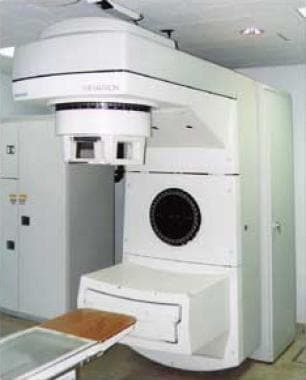
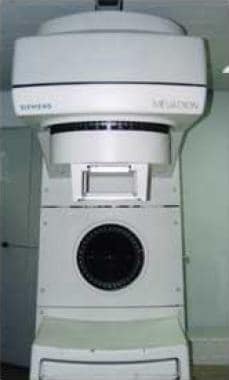
The image below depicts an EBRT unit.
Conventional EBRT
Conventional EBRT is typically delivered by means of a 4-field technique. The 4 fields (anteroposterior [AP], posteroanterior [PA], left lateral, and right lateral) are designed to include the prostate, the seminal vesicles, and the regional lymphatic vessels.
The morbidity of radiation treatment is intimately linked to the volume of normal tissue treated. Conventional radiotherapy includes irradiation of large volumes of tissue, including the skin, small bowel, bladder, large bowel, pelvic bones, and additional areas of soft tissue.
3-Dimensional conformal radiotherapy
In 3-dimensional conformal radiotherapy (3D-CRT), the radiation beam is shaped to include the 3D anatomic configuration of the prostate and any specified adjacent tissue (including the seminal vesicles and periprostatic adventitial tissues). This technique allows more precise delivery of therapy to the target organ or organs.
Intensity-modulated radiation therapy
Intensity-modulated radiation therapy (IMRT) can achieve tightly conformal dose distributions with the use of nonuniform radiation beams. The intent of this form of therapy is to create highly conformal fields by treating the patient with multiple static portals (so-called step-and-shoot IMRT) or dynamic fields. In dynamic IMRT, a series of arcs are administered through the area of interest. Multileaf collimators (MLCs) are reshaped many times as the machine performs a series of arc rotations around the target.
Image-guided radiotherapy
The term image-guided radiotherapy (IGRT) refers to the use of imaging techniques, including the following, in an attempt to ensure proper target localization during the course of radiotherapy:
-
Interfractional assessment (static) [1]
-
Portal imaging - The implantation of radiopaque fiducial markers into the prostatic target allows a soft tissue target to be localized with portal imaging technology
-
Ultrasonography - Ultrasonographic images of the prostate are obtained on a daily basis to identify the gland’s relative position
-
Computed tomography (CT) scanning - The radiation used for therapy can also be used to generate a CT image [2]
-
Radiofrequency localization - Small radio transponders can be implanted in the prostate to facilitate patient setup
Proton beam therapy
In contrast to photon beam therapy, the entrance radiation dose in proton beam therapy tends to be significantly less than the maximum energy of the clinical beam. Proton beams have a characteristic Bragg peak. Beyond this point, where energy is at a maximum intensity level, radiation energy rapidly falls off, which is important in the management of normal tissue toxicity.
Tomotherapy
Tomotherapy consists of helical radiotherapy using a computed tomography (CT)-like gantry and a rotating radiation beam that passes through the target area of interest; this modality has been used in the management of primary central nervous system tumors and viscera-based malignancies.
Hypofractionated radiotherapy
In this form of therapy, radiation is delivered from an accelerator, with the equipment mounted to a computer-guided robotic arm. Because the machine is capable of treating the target at angles that are not possible with conventional rotation-based equipment, it yields the theoretical advantage of conforming the dose more closely to the target organ’s shape
Radiotherapy and androgen ablation
Data from the Radiation Therapy Oncology Group (RTOG) have shown a clear improvement in biochemical control of disease when patients receive a combination of radiotherapy and androgen-suppressive treatment. The results of several phase III clinical trials suggest that the true benefit of combining radiotherapy with androgen blockade may lie in the potentially synergistic effects of the 2 treatments.
Adjunctive EBRT
Combined prostate implantation and EBRT
A comprehensive literature review indicated that for high-risk patients, combination therapies involving EBRT and brachytherapy with or without androgen deprivation therapy appear to be superior to more localized treatments, such as seed implant alone, surgery alone, or EBRT. [3]
EBRT after radical prostatectomy
Multi-institutional data from the American Society of Therapeutic Radiation Oncology (ASTRO) consensus conference suggest that in patients treated for a rising prostate-specific antigen (PSA) level, postoperative radiotherapy (typically in a dose range of 60-65 Gy) offers a PSA remission rate of 70%. Unfortunately, the durability of this response varies widely from center to center, with averages ranging from 25 to 67 months.
Overview
External beam radiation therapy (EBRT) remains one of the primary treatment modalities for patients with localized or locally advanced prostate cancer. The use of modern equipment has fostered greater interest in this form of treatment over the past 25 years; however, the origins of this therapy extend back to the early 20th century.
Radiation therapy for prostate cancer was first introduced to the United States in 1915 in the form of radium applicators that were positioned adjacent to the prostate (ie, in the urethra, bladder, or rectum). This form of therapy offered local treatment to the prostate but was associated with significant morbidity.
Unfortunately, the early therapy machines generated low-energy x-ray beams that lacked the ability to penetrate deep into the pelvis. As a result, treatments were often palliative and commonly caused significant skin morbidity. The technologic limitations of low-energy radiation beams greatly restricted the use of radiotherapy in advanced prostate cancer throughout the first half of the 20th century.
The role of radiotherapy in the palliative treatment of prostate cancer also was limited by discoveries regarding the endocrine-sensitive nature of this malignancy. As the androgen dependence of this tumor became increasingly clear, clinical interest turned toward eliminating the primary source of hormone production in patients with advanced disease. Accordingly, orchiectomy became an accepted form of therapy for advanced carcinoma of the prostate until estrogenic products were developed in the 1950s and 1960s.
The role of radiotherapy in the management of prostate carcinoma became clearer with technologic advancements that followed World War II. Megavoltage (> 1000 kV) radiation resulted in x-ray beams that penetrated more deeply and that were associated with significantly less skin and subcutaneous morbidity. This property prompted further investigation of the role of radiotherapy in more deeply seeded tumors.
During the 1950s and 1960s, megavoltage radiation was more commonly available from the decay of radioactive isotopes; however, in the following years, generation of high-energy x-rays became increasingly popular. Equipment names (eg, Betatron, Linear Accelerator, Proton Beam, and Neutron Beam) often indicated the mode by which the x-rays were created. By the early 1980s, the linear accelerator was established as the most common form of EBRT.
Clinical use of higher-energy radiation beams facilitated the development of radiation oncology as an accepted mode of therapy for both advanced and early-stage prostate carcinoma. This experience was largely due to the work of Bagshaw et al at Stanford University. [4] Besides allowing deeper beam penetration into tissue, linear accelerators generated a beam with more sharply delineated borders. This allowed higher doses of radiation to be directed at the clinical target (eg, prostate, seminal vesicles, or regional lymph nodes).
Improved technology, treatment planning, and dosimetry allowed localized therapy with curative intent. Heightened awareness of prostate cancer and an apparent increase in its incidence during the early 1990s served as a further stimulus for consideration of EBRT in the management of this disease.
With the evolution of improved computer-based treatment planning, conventional radiotherapy techniques have largely been supplanted by modern techniques such as 3-dimensional conformal radiotherapy (3D-CRT), intensity-modulated radiotherapy (IMRT), and image-guided radiotherapy (IGRT). The advantages of the modern approaches lie in their ability to escalate the tumor dose (thus enhancing disease control) while minimizing toxicity to normal tissue (thus improving patient compliance and satisfaction).
Nevertheless, a 2008 research summary by the Agency for Healthcare Research and Quality (AHRQ) reviewed 5 randomized controlled trials of EBRT and concluded that no regimen, whether conventional, high-dose conformal, dose fractionation, or hypofractionation, was superior in reducing overall or disease-specific mortality. [5]
For patient education information, see Prostate Cancer and Bladder Control Problems.
Guidelines
In 2013, for the first time in their history, the two medical organizations most responsible for the treatment of prostate cancer in the United States—the American Society for Radiation Oncology (ASTRO) and the American Urological Association (AUA)—issued a joint guideline, a series of 9 major statements on the use of radiation therapy (either adjuvant or salvage) after prostatectomy. The statements are categorized as follows [6, 7] :
-
Clinical principles - Wide agreement by urologists
-
Recommendations - Grade C; low-quality and certainty evidence
-
Standards - Grade A or B; high/moderate-quality and certainty evidence
-
Options – Nondirectives
The guideline statements are as follows:
-
If a patient is undergoing radical prostatectomy for localized prostate cancer, discuss the possibility of adverse pathologic findings indicating an increased cancer recurrence risk (clinical principle).
-
If adverse pathologic signs—such as seminal vesicle invasion, positive surgical margins, and extraprostatic extension—are found, inform the patient that the risk of biochemical (prostate-specific antigen [PSA]) recurrence, local recurrence, or clinical progression of cancer is lower following a combination of radical prostatectomy and adjuvant radiation therapy than it is after radical prostatectomy alone (clinical principle).
-
If adverse pathologic signs are found at prostatectomy, offer adjuvant radiation therapy to the patient (standard; evidence strength, grade A).
-
Inform patients that PSA recurrence after surgery is associated with a higher risk for metastatic prostate cancer and with an increased mortality risk (clinical principle).
-
Biochemical recurrence should be defined as a detectable or rising postsurgery PSA value of at least 0.2 ng/mL, with a second confirmatory level of at least 0.2 ng/mL (recommendation; evidence strength, grade C).
-
A restaging evaluation should be considered in patients with a PSA recurrence (option; evidence strength, grade C).
-
Offer salvage radiation therapy to patients who, after radical prostatectomy, demonstrate PSA or local recurrence but have no distant metastatic disease (recommendation; evidence strength, grade C).
-
Inform patients that radiation therapy is most effective against PSA recurrence when PSA levels are relatively low (clinical principle).
-
Inform patients that radiation therapy may cause short- or long-term urinary, bowel, and sexual adverse effects, but also discuss the treatment’s potential benefits as a means of controlling disease recurrence (clinical principle).
In 2019, the ASTRO and AUA updated their joint clinical guideline for radiation therapy after prostatectomy as follows [8] :
-
Inform patients with adverse pathologic findings that the impact of adjuvant radiotherapy on subsequent metastases and overall survival is less clear than its effect on reducing the risk of biochemical and local recurrence and clinical progression of cancer, compared with radical prostatectomy alone (clinical principle).
-
Offer hormonal therapy to patients who are treated with salvage radiotherapy and have a postoperative PSA level of >0.20 ng/mL or higher (standard; evidence strength: grade A).
Relevant anatomy
A normal prostate gland is approximately 20 g in volume, 3 cm in length, 4 cm wide, and 2 cm in depth. As men get older, the prostate gland is variable in size secondary to benign prostatic hyperplasia. The gland is located posterior to the pubic symphysis, superior to the perineal membrane, inferior to the bladder, and anterior to the rectum. The base of the prostate is in continuity with the bladder and the prostate ends at the apex before becoming the striated external urethral sphincter. The sphincter is a vertically oriented tubular sheath that surrounds the membranous urethra and prostate. For more information about the relevant anatomy, see Prostate Anatomy.
Indications and Contraindications
External beam radiation therapy (EBRT) is commonly used in the treatment of patients who have a greater likelihood of non–organ-confined disease. For patients who decide against surgical intervention, determining the presence of non–organ-confined disease is typically based on a review of tumor-related variables that have been shown to correlate with extraglandular disease extension (eg, high stage, high Gleason scores, and high prostate-specific antigen [PSA] levels). Certain patients may not be optimal candidates for EBRT, such as those with a history of bowel disorders (including ulcerative colitis and diverticular disease) and those with poorly controlled diabetes. [9] These patients are known to have preexisting conditions that may place them at risk for either more intense morbidity or more protracted morbidity.
Conventional EBRT
As biochemical endpoints of therapy replace clinical endpoints, the role of conventional external beam radiation therapy (EBRT) in the management of localized prostate cancer is likely to diminish. The reasons include an appreciation of the importance of dose escalation, the ability to offer patients more precise target localization, and the use of combined treatment strategies (ie, addition of hormonal manipulation or brachytherapy to the primary treatment).
The current literature supports the role of conventional EBRT in the management of localized prostate carcinoma. Countering this view is historical information reported by Paulson, [10] along with a meta-analysis by Wallis et al, [11] which showed that in early-stage disease, surgery offered better results than EBRT. However, the randomization schema and statistical analysis associated with these data make definitive conclusions difficult.
Although the Paulson study was a prospective trial, it was conducted before the routine use of prostate-specific antigen (PSA) testing and pelvic imaging. No similar study has been conducted in the PSA era. In fact, a trial at the US National Cancer Institutes (NCI), the Prostate Intervention versus Observation Trial (PIVOT), failed to include radiotherapy as a treatment arm.
The 2 forms of therapy are unlikely to be directly compared in the future. Retrospective comparisons, using PSA-based outcomes, suggest no significant difference between them. The results for patients with T1/T2 disease treated with conventional EBRT are similar to results achieved after radical prostatectomy. The 2 treatments offer comparable rates of disease control, and 10-year survival rates are similar (> 60%) in studies by both Bagshaw [12] and Perez [13] .
Despite the decreasing frequency of conventional EBRT in the management of prostate carcinoma, there is no significant body of data to indicate that this treatment should be abandoned. In fact, Radiation Therapy Oncology Group (RTOG) trial 94-13 suggests that whole-pelvis radiotherapy (ie, conventional treatment) may improve the local regional control of disease in patients with increased risk of non–organ-confined cancer (ie, high Gleason score and high prostate-specific antigen [PSA] level). [14]
Regardless of the current status of conventional EBRT in prostate cancer, a clear understanding of the former standard treatments will, at the least, allow a greater appreciation of more current techniques.
Simulation
Simulation is a process during which the patient is prepared for therapy. The patient is placed in the treatment position, and the radiation field or fields are marked. Simulation for conventional EBRT relies on fluoroscopy and plain radiography. These less sophisticated techniques are generally considered acceptable because the treatment borders (margins) are more inclusive than those used in 3-dimensional conformal therapy (3D-CRT) and intensity-modulated radiotherapy (IMRT). CT-based simulation is increasingly popular for 3D-CRT and IMRT.
Localization of the target and the adjacent normal tissue is critical in the planning of both conventional therapy and 3-dimensional treatment. For conventional therapy, the patient is placed in the supine position. Radiographs of the anteroposterior (AP), posteroanterior (PA), left lateral, and right lateral fields are obtained. Typically, treatment fields are shaped with corner blocks only, and borders are based on bony landmarks.
Normal tissue is delineated by using radiopaque contrast materials in the rectum and bladder. Retrograde urethrography is performed to establish the inferior margin of the prostate. Blocking of normal structures is indicated in most cases; however, custom blocking is not always necessary.
Technique
Conventional EBRT is typically delivered by means of a 4-field technique (see the image below). The 4 fields (AP, PA, left lateral, and right lateral) are designed to include the prostate, the seminal vesicles, and the regional lymphatic vessels. A cumulative dose of 45-50 Gy is delivered over a period of 5-5.5 weeks. An additional dose of approximately 20 Gy to a smaller field (ie, a boost) is delivered to the prostate and the periprostatic tissues.
Total doses of 66.6-70 Gy were once typically used; however, these doses are too low to provide the same rates of local and regional control achieved with currently used higher doses (72-80 Gy). [15, 16] The boost field is designed to limit treatment to the target volume (prostate, seminal vesicles, and 1- to 2-cm margin) and to offer additional shielding to the posterior wall of the rectum, the urethra, and the small bowel. The reduced volume of normal tissue included within the radiation field is associated with a reduction in morbidity.
Whole-pelvis radiotherapy (ie, superior border at L5-S1 junction) is rarely used, because of the increased bowel toxicity and lack of clear outcome improvement. Whole-pelvis radiotherapy is offered to some patients when extensive regional disease is either present or expected. RTOG trial 94-13 suggests that large-field radiation treatment may be beneficial in patients with higher Gleason scores and in those who receive adjuvant hormonal blockade. [14]
When regional lymph nodes are to be treated, the superior border of the pelvic field is at the level of the midsacroiliac joints, and the inferior border is usually 1-1.5 cm inferior to the junction of the membranous and prostatic urethra, as demonstrated on urethrography (ie, pencil point). The lateral borders on the AP and PA fields are 1.5-2 cm lateral to the pelvic brim.
The superior and inferior borders remain unchanged on the lateral fields. The posterior border of the lateral field is commonly placed at the S2-S3 interspace. The anterior border is established to include the anterior portion of the symphysis pubis. The field edges for cone-down, or boost-field treatments, share the same inferior border as the primary field.
Superiorly, the fields extend to the top of the acetabulum and laterally to include two thirds of the obturator foramen. Dose distributions for conventional treatment are typically generated in a single plane, and the dose is prescribed at the isocenter and normalized at the 100%-isodose line.
Outcome
One of the major outcome determinants for clinically localized prostate cancer is tumor stage. Staging systems have always acknowledged the significance of extraglandular disease. In both the Whitmore classification schema and the tumor-node-metastasis (TNM) system, the presence of disease beyond the prostate (ie, stage C and stage T3, respectively) indicates a poor prognosis. This observation predates the use of modern prognostic variables associated with increased risk of extracapsular disease (ie, elevated PSA level and high Gleason score).
Although radiation therapy was used in the management of locally advanced lesions, the long-term results, even in the pre-PSA era, were relatively poor. Patients diagnosed with locally advanced disease (ie, stage C or T3 disease) who were treated with EBRT had acceptable disease-free survival (DFS) rates at 5 years, approaching 60-65% in most series; however, 10-year DFS rates were less encouraging (30-40%). At 15 years, failure rates continued to increase (70-80%).
These results typify the experience gained through review of historical controls; however, they are significantly limited because of less sophisticated treatment techniques, lower total doses offered, and inadequacies of staging. Newer information continues to be generated by employing modern radiotherapy techniques and increasing therapy doses. Similarly, the rates of disease control are more carefully reported (eg, PSA-based outcome in lieu of clinical failure endpoints).
Patients undergoing definitive radiotherapy are typically considered to have achieved biochemical control of disease if the PSA level is not rising and the serum PSA level is below 0.5 ng/mL. Several definitions of biochemical failure have been proposed in the radiation oncology literature; however, 3 consecutive rises in the serum PSA level have been proposed as an accepted marker for failure in an American Society of Therapeutic Radiation Oncology (ASTRO) consensus conference.
Despite the increasing body of literature that considers PSA level a measurable determinant of treatment outcome, this tumor marker has been in wide use for a relatively short time. Before the adoption of PSA level as a marker, endpoints for clinical outcome were largely subjective and included prostate assessments based on clinical examination (eg, digital rectal examination [DRE]) and nonspecific serum markers (eg, alkaline phosphatase and acid phosphatase). The poor sensitivity of DRE has made this form of posttreatment assessment less and less useful.
As more studies use serum PSA level to assess treatment outcome, comparison with larger historic series (including those of the American College of Radiology’s Patterns of Care Studies) becomes harder. Several institutions have reported 5-year and 10-year PSA-based outcomes; however, the limited follow-up with posttreatment PSA studies and the protracted natural history of the disease make historical comparisons difficult. Unlike clinical outcomes, PSA-based outcomes suggest that long-term control can be difficult for locally advanced disease.
A study that examined the association between dose-escalated EBRT and overall survival among men with nonmetastatic prostate cancer reported that dose-escalated EBRT is associated with improved overall survival in men with intermediate- and high-risk prostate cancer but not low-risk prostate cancer. [17, 18]
A study that included 1,338 patients with lymph node metastasis after radical prostatectomy reported that adjuvant androgen deprivation therapy with external beam radiation therapy was associated with better overall survival that adjuvant androgen deprivation therapy or observation alone (hazard ratio [HR]: 0.46, 95% confidence interval [CI]: 0.32-0.66, p< 0.0001) or observation (HR: 0.41, 95% CI: 0.27-0.64, p< 0.0001). The study also found that ten-year mortality risk difference between adjuvant androgen deprivation therapy with external beam radiation therapy, observation, or adjuvant androgen deprivation therapy alone ranged from 5% in low-risk patients to 40% in high-risk patients. [19]
Complications
The morbidity of radiation treatment is intimately linked to the volume of normal tissue treated. Conventional radiotherapy includes irradiation of large volumes of tissue, including the skin, small bowel, bladder, large bowel, pelvic bones, and additional areas of soft tissue (including nerves). Each organ can experience irritation during and, potentially, after a course of radiotherapy.
A brief review of commonly encountered morbidities is appropriate. Patients are told that any of the following conditions may arise during the course of radiation therapy and may persist for up to 3 months afterward (ie, acute morbidity). A significantly smaller percentage of patients (3-5%) have symptoms that persist beyond 3 months or develop new symptoms after the completion of therapy.
Skin
Because radiation can cause perturbation of the epidermis and dermis, reddening of the skin may develop during therapy. This is relatively uncommon, given the frequent use of high-energy photon beams (≥ 10 MV). Skin irritation is common in patients with a fair complexion; however, early intervention can limit progression to either dry or moist desquamation. Hair loss in the irradiated field (epilation) is commonly seen.
Small intestine
Radiation therapy can cause changes to the epithelial lining of the small intestine. Absorption and transit can be impaired. Patients may experience more frequent bowel movements or less well formed stools. Minimizing the extent of the small bowel that is treated is the goal of treatment planning, but this is more difficult with conventional EBRT than with 3D-CRT or IMRT.
Patients undergoing radiotherapy are commonly advised to reduce their consumption of certain high-fiber foods (eg, fruits or uncooked vegetables), which can have a laxative effect. If dietary modifications do not correct frequent bowel movements, patients often receive prescriptions for antidiarrheal medications.
Bladder
During conventional radiotherapy, the entire bladder is usually treated, and this can cause adverse effects, including urinary frequency, urgency, and, less frequently, incontinence. Patients commonly experience dysuria, which can be lessened with increased fluid consumption. Hematuria occasionally occurs but should be carefully evaluated to ensure that an occult urinary tract infection is not present.
Whether other associated urinary tract symptoms (eg, frequency and urgency) are due to bladder or urethral irritations is often difficult to discern. Regardless, both symptoms occur quite frequently during EBRT and are seen throughout the continuum of radiotherapy (ie, from conventional therapy to IMRT). This observation supports the importance of urethral irritation to both urgency and frequency.
Large bowel
Toxicity, including proctitis, may develop in segments of the distal bowel included in the radiotherapy fields. This is observed more commonly with conventional radiotherapy than with conformal therapy. To minimize perianal inflammation, patients are encouraged to exercise good hygiene while receiving therapy.
Symptoms of proctitis can include fecal urgency, mucous discharge, and rectal bleeding. If rectal bleeding or mucous discharge is noted, treatment with steroid-containing suppositories may be necessary. Measurable changes in levels of hemoglobin and hematocrit may occur, albeit rarely. Blood should be tested weekly to assess this possibility.
Pelvic bones
A number of pelvic bones receive radiation during the treatment of prostate cancer. As expected, patients undergoing conventional EBRT have a large volume of bone tissue (ie, marrow reserve) irradiated. Weekly complete blood counts should be performed to review this possibility, which is typically noted in white blood cell and platelet counts. Cessation of therapy to allow for recovery of the blood count is rarely necessary.
Nerves
Pelvic irradiation exposes both sympathetic and parasympathetic nerves to radiation. Posttherapy defects with erectile and ejaculatory function are reported in 30-40% of patients undergoing a full course of EBRT (total doses ≥ 66.6 Gy). The etiology of this injury is not fully understood; however, current thinking suggests that the mechanism may be microvascular changes affecting the blood supply to the nerves.
Hormones
A study by Planas et al indicated that in the treatment of localized prostate cancer, EBRT has a greater effect on the hypothalamic-pituitary axis than does radical prostatectomy. The investigators found that at 3 months posttreatment, patients who underwent EBRT had significantly higher luteinizing and follicle-stimulating hormone levels and significantly lower total and free testosterone concentrations than did radical prostatectomy patients. At 12 months posttreatment, EBRT patients still demonstrated significantly higher follicle-stimulating hormone levels and significantly lower total testosterone levels than did the other patients. [20]
3-Dimensional Conformal Radiotherapy
In 3-dimensional conformal radiotherapy (3D-CRT), the radiation beam is shaped to include the 3-dimensional anatomic configuration of the prostate and any specified adjacent tissue (including the seminal vesicles and periprostatic adventitial tissues). 3D-CRT allows more precise delivery of therapy to the target organ or organs.
With increasing access to computed tomography (CT) and magnetic resonance imaging (MRI) simulation equipment, as well as more powerful treatment-planning equipment, the use of 3D-CRT has markedly increased over the past decade. Indeed, 3D-CRT has essentially replaced conventional external beam radiation therapy (EBRT) in the management of early-stage prostate cancer.
The transition to 3D-CRT has significantly reduced treatment-associated toxicity. More important, conforming the radiation dose to a limited target has resulted in several successful dose escalation trials. The success of 3D-CRT is the result of multiple factors, including favorable dose-response relations, increased ability to reduce radiation to neighboring normal tissue, the relative immobility of the organ (typically < 1 cm), and a high prevalence of disease.
Essential to the discussion of 3D-CRT is a working knowledge of the following terms:
-
Gross tumor volume (GTV) – This is the delineation of the visible target using common imaging modalities (eg, CT or MRI)
-
Clinical target volume (CTV) – This describes the structures that may be beyond the easily visible anatomic structures delineated by CT or MRI; specifically, the CTV encompasses the anticipated microscopic extent of tumor, as well as anticipated subclinical areas that may be at risk for disease involvement
-
Planning target volume (PTV) – The PTV acknowledges that daily patient positioning and setup vary and should be addressed in creating radiation portals
Although each of these terms is readily applied to single-organ structures (eg, prostate or seminal vesicles), it remains unclear how well the structures can be defined in the delineation of regional pelvic adenopathy.
Indications
Despite the successful completion of several prospective randomized trials evaluating 3D-CRT in the management of prostate cancer, several issues remain unclear. 3D-CRT is generally considered appropriate in the following circumstances:
-
The probability that tumor remains within the anticipated target portals (ie, delineation of the GTV, CTV, and PTV for organs at risk) can be adequately determined
-
The importance of regional adenopathy in varying clinical settings can be delineated
Technique
The process of 3D-CRT requires the acquisition of imaging data. The initial step is immobilization of the patient in either the supine or the prone position.
Theoretic advantages of supine immobilization include the ease of daily setup for the patient and staff, the ability to fuse treatment-planning images with previously obtained diagnostic images (ie, MRI), and the relative ease of use when performing daily setup localization with ultrasonographic assistance. However, many centers prefer prone positioning. Theoretic advantages of prone positioning relate to relative sparing of small bowel from the radiation portals and reproducibility of patient positioning on daily setup.
Once the patient has been appropriately positioned, fabrication of the immobilization device is performed. This step becomes increasingly critical as the margin around the target is decreased (eg, when a 1.5-cm margin around the prostate is decreased to a 0.5-cm margin).
Numerous materials have been used to immobilize patients before CT or MRI data are acquired for treatment planning. Commercially available products include thermoplastic casts, vacuum-shaping bags, and self-contained thermochemicals. Regardless of which type of device is chosen, the goal of immobilization is to reproduce the position in which the patient is treated each day.
Upon fabrication of the device or devices, axial images of the area of interest are obtained. Consecutive CT scans or MRIs are obtained, starting from 3 cm below the prostate and extending superiorly to 3 cm above the superior tip of the seminal vesicles. Additional CT imaging or MRI data can be obtained above or below the areas of interest. However, this information has minimal impact on patient treatment or dose computation.
The targets (CTV, PTV, or both) are identified on each relevant axial CT slice. Similarly, normal structures, including the bladder wall, rectum, small bowel, bony structures, and skin surface, are outlined on each relevant CT slice.
The target volume and normal structures are then digitally reconstructed in 3 dimensions and displayed with the beam’s-eye view (BEV) technique. The adequacy of target coverage and normal tissue doses can be viewed by using dose-volume histograms (DVHs) or serial 2-dimensional images with isodose curves superimposed on them.
Compared with conventional EBRT, 3D-CRT techniques implement a larger number of beams daily to improve the tumor–to–normal tissue dose ratio. Implementation of 3D-CRT requires the use of newer treatment machines capable of rapidly delivering a large number of precisely shaped fields under automated computer control (ie, multileaf collimators [MLCs]; see the image below).
 Conformal radiation therapy. A linear accelerator equipped with a multileaf collimator is a device that can decrease the time a patient spends in the treatment room and one that improves treatment accuracy.
MLCs are capable of automatically shaping the apertures of each treatment field in rapid succession under computer control. Treatment times are shortened, individual treatment blocks need not be fabricated, and more complex beam shaping can be attempted.
Results
The results of 3D-CRT demonstrate superior bNED (biochemical, no evidence of disease) control rates, largely because of the ability to escalate the dose with less concern over the toxicity to normal tissue. 3D-CRT allows delivery of higher doses of radiation to the prostate without significant complications to the normal tissue.
Even small degrees of dose escalation have been shown to improve the biochemical outcome in patients diagnosed with prostate cancer. Comparison studies have shown superior outcomes with doses of 78 or 79 Gy versus doses of 70 Gy. [15, 16]
The 5- and 10-year follow-up results with 3D-CRT indicate increased rates of bNED control, especially in patients with intermediate prognostic factors (ie, Gleason score of 7 and PSA level of 10-20 ng/mL). The bNED rates in patients with pretreatment PSA levels of 10-20 ng/mL are approximately 30% better than those in patients treated with conventional radiotherapy at 5 years.
Patients with more favorable prognostic factors (ie, Gleason score ≤ 6 and PSA level ≤ 10 ng/mL) may not benefit from dose escalation, although this issue remains highly controversial. Similarly, the bNED rates in patients at high risk for locally or regionally advanced disease (ie, Gleason scores of 8-10 and PSA ≥ 20 ng/mL) may not markedly improve after dose escalation, probably because this group of patients is ultimately at higher risk for distant metastasis.
Intensity-Modulated Radiation Therapy
Intensity-modulated radiation therapy (IMRT) can achieve tightly conformal dose distributions with the use of nonuniform radiation beams. The intent of this form of therapy is to create highly conformal fields by treating the patient with multiple static portals (so-called step-and-shoot IMRT) or dynamic fields. In dynamic IMRT, a series of arcs are administered through the area of interest. Multileaf collimators (MLCs) are reshaped many times as the machine performs a series of arc rotations around the target (see the image below).
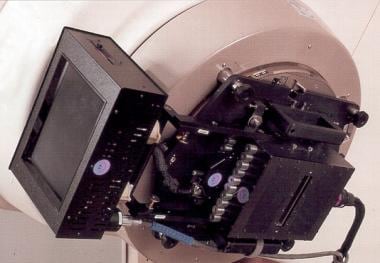 Multileaf intensity-modulating collimator (MIMiC) unit. This is used to deliver intensity-modulated radiotherapy.
Complex treatment-planning software algorithms allow exceedingly high doses of radiation to be delivered to the target while significantly smaller doses of radiation are delivered to the adjacent normal tissue. In contrast to the traditional method of radiation planning, inverse treatment planning is commonly used for the calculation of doses during IMRT.
IMRT establishes a treatment plan after the establishment of acceptable doses to regional (normal) anatomy. For instance, the maximum tolerable dose to be delivered to the involved segments of the bladder, bowel, and rectum is specified. The desired target dose is then prescribed to the planning target volume (PTV).
The computer, through a series of complex iterations, designs a treatment that maximizes the dose delivered to the target and minimizes the dose delivered to adjacent normal tissue. Implicit in the name of this form of therapy is the concept that the intensity of the radiation beam changes throughout the course of therapy.
IMRT has been successfully used to treat tumors when the target area is readily identifiable at the initiation of daily treatments and the desired dose for optimum tumor control is significantly higher than the acceptable dose limits for adjacent normal tissue. Tumors of the head and neck and tumors of the breast are clinical sites where this treatment has been successfully used.
IMRT is no longer considered an investigational technique in the management of prostate cancer. Rather, it has rapidly become a highly precise method of delivering increasing doses of radiotherapy to the prostate and the immediate periprostatic tissues.
To date, however, no multicenter, phase III, prospective, randomized trial has been performed to establish whether IMRT is superior to well-designed 3-dimensional conformal radiotherapy (3D-CRT). Data from the Memorial Sloan-Kettering Cancer Center have demonstrated that doses of more than 80 Gy can be safely delivered by means of IMRT. The value of dose escalation when additional adjuvant treatments are being considered (eg, hormonal blockade, chemotherapy) remains unclear.
IMRT in the treatment of prostate cancer continues to evolve; however, reproducible identification of the target (on daily treatments) remains challenging. The use of implantable fiducial markers and ultrasonographic localization devices has become increasingly popular. Both techniques allow the treating therapists to identify the desired target immediately before each day’s treatment. Without such specificity, the logic of using IMRT is questionable.
Image-Guided Radiotherapy
The term image-guided radiotherapy (IGRT) refers to the use of additional verification tools in an attempt to ensure proper target localization during the course of radiotherapy. This term has been used for a wide range of imaging techniques, from those as simple as daily port films to those as complicated as computer-assisted patient repositioning devices. Regardless, as highly conformal radiotherapy is administered with increasing frequency (eg, intensity-modulated radiotherapy [IMRT]), accurate target localization becomes mandatory.
Historically, attempts to identify the target’s position focused on imaging performed before each daily treatment, or fraction. Subsequent efforts have attempted to address the significance of organ movement during the daily treatment fraction. [2]
Interfractional assessment (static)
As radiotherapy for prostate cancer has become increasingly conformal, dose escalation has become a standard approach to the management of early-stage disease, [21] and accurate target and normal tissue localization has become increasingly important. Proper target identification becomes necessary in 3 distinct phases of treatment planning and treatment execution.
When patients are selected for primary radiotherapy treatment, simulation is performed. Computed tomography (CT)-based imaging is most commonly used; however, some centers prefer magnetic resonance imaging (MRI). [1]
Accurate delineation of normal tissues in relation to the prostatic target is equally important in this phase of patient care. Physician review of axial CT images allows delineation of the prostate. Because most radiation oncologists lack formal training in the interpretation of radiographic imaging, appropriate assessment requires either review with a diagnostic radiologist or extensive clinical experience.
After identification of the prostatic target (ie, the gross target volume [GTV]), the clinical target volume (CTV) and the planning target volume (PTV) are created. Although this phase of treatment planning allows accurate target localization on the basis of the gland’s location at the time of CT imaging, it does not address organ motion subsequent to that date.
Portal imaging
Early attempts to identify the location of the prostate before each daily treatment resulted in suboptimal target identification. A commonly used approach includes obtaining daily port films before therapy. This strategy provides limited value to the image guidance process.
Megavoltage imaging (portal imaging) of pelvic anatomy provides reasonable confidence that the patient’s fixed pelvic structures (bones) are in the same position as that observed during CT simulation; however, it does not provide information regarding the prostate’s position in reference to these bony structures. Nevertheless, it remains an accepted method for approximating target localization in the absence of more sophisticated imaging.
An evolution from this simple form of therapy occurred with the implantation of radiopaque fiducial markers into the prostatic target, which allows a soft tissue target to be localized with portal imaging technology. Imaging of the implanted fiducial markers can be obtained with either megavoltage imaging (ie, port films) or kilovoltage imaging. In the latter setting, low-energy x-ray equipment and detectors are mounted on the treatment machine.
Ultrasonography
Transabdominal ultrasonography is an accepted noninvasive method for obtaining detailed imaging of pelvic contents. It is regularly used by gynecologists and urologists in the daily assessment of their patients and has been adapted to the management of target localization in patients undergoing radiation-based treatments.
In an attempt to displace normal tissue(s) from the radiation treatment portals and to facilitate the immobilization of the gland, patients are asked to undergo their daily treatments with a full urinary bladder and a relatively empty distal bowel. The increased volume within the bladder provides a good window for prostate target localization using ultrasonography.
Several commercially available systems allow the operator to identify the target’s localization on the basis of CT data. Ultrasonographic images of the prostate are obtained on a daily basis to identify the gland’s relative position. If the gland is not identified in the anticipated location, a series of calculated shifts are proposed to correct the image. After repositioning of the patient, repeat imaging can confirm that the target is correctly localized. [22]
Limitations of this technology include the need to train staff to interpret ultrasonographic images, day-to-day variations in image quality, and the potential for organ movement while the ultrasound transducer is applied to the patient’s lower abdominal wall. Although this added procedure is still used in many clinical centers, it increases the time patients are required to remain in the treatment position.
CT-based imaging
As noted (see above), kilovoltage imaging devices have been successfully added to linear accelerators. They are normally mounted at a 90° angle to the treatment head of the machine and are opposed by digital image detectors. These x-ray cameras can be used to generate axial images representative of the target area of interest, which are then compared with CT scans previously obtained during treatment planning.
Coordinated shifts can be made if positioning inaccuracies are detected in the patient setup. Alternatively, and more simply, the radiation used for therapy (megavoltage) can be used to generate a CT image. Megavoltage images can be used to perform the requisite shift to ensure that the patient’s position matches that obtained during initial simulation. [2]
Radiofrequency localization
Newer forms of target tracking have become clinically available. Like the radiopaque fiducial markers used in portal imaging, small radio transponders can be implanted into the prostate for facilitating patient setup. These transponders emit radiofrequency waves that can be detected and used to reposition the target before execution of therapy.
Intrafractional assessment (dynamic)
As increasing information regarding the potential for interfractional organ movement has become available, concerns about this possibility have grown accordingly. This issue is particularly important because many intensity-modulated radiation treatments can require 20-30 minutes or longer.
Of the devices listed above, only radiofrequency-based localization has been regularly used to obtain information about target localization during the course of linear accelerator–based radiotherapy. Newer generations of radiation-based equipment (eg, robotic arm linear accelerator–based therapy and the Accuray CyberKnife) have unique treatment repositioning techniques that are not found in association with linear accelerator–based therapy.
The use of radiofrequency transponders can be valuable as each treatment fraction is administered. After initial patient positioning using this image-guided technique, any potential target movement can be accurately tracked during treatment. In the simplest of models, when a discordance is detected between the expected location and the actual location, the machine can be stopped from delivering further radiotherapy.
In a more complicated system, the patient can be automatically repositioned to correct for the measured change in target localization. Because this technology is relatively new, phase 3, multi-institutional studies of overall benefit are still under way. [23, 24]
Proton Beam Therapy
Conventional external beam radiation therapy (EBRT) is delivered via photon beams. Data from Loma Linda and Harvard suggest that prostate cancer can be effectively managed with conformal proton beam therapy. [25] What is less clear is whether the overall cost-benefit ratio will make this form of therapy a continued source of clinical interest. [26]
Photons are packets of energy that are capable of entering the body to a depth that is proportional to their energy (ie, higher energy equals deeper penetration). Photons are generated for clinical use in linear accelerators by accelerating electrons to varying potentials (speeds) and aiming them at a tungsten target.
The resultant collision of the electron with the target generates a photon beam whose energy is proportional to the accelerating potential of the electron. Energies of commonly used clinical beams range from 4 to 25 MV.
The electron is a relatively small particle with essentially no weight. Consequently, the equipment needed to accelerate the electron before it collides with the target is comparatively small. In contrast, other charged particles (eg, protons) have as much as 2000 times the mass of an electron. Accordingly, the equipment needed to accelerate these particles is significantly larger; in fact, it can require up to several thousand square feet of space.
A unique feature of proton beam therapy is the way in which it deposits its most concentrated radiation dose. Unlike photon beam therapy, the entrance radiation dose tends to be significantly less than the maximum energy of the clinical beam. Proton beams have a characteristic Bragg peak. Beyond this point, where energy is at a maximum intensity level, radiation energy rapidly falls off, which is important in the management of normal tissue toxicity.
Technologic advances in proton beam therapy have resulted in the construction of several centers throughout the United States. Although costs remain in excess of $100 million per unit, this technology has generated significant interest in the management of prostate carcinoma.
Proton beam therapy has been successfully used in the management of prostate cancer. Early work from the cyclotron center at Harvard formed an important basis for current clinical trials. [27]
Tomotherapy and Hypofractionated Radiotherapy
The radiotherapeutic approach to carcinoma has been relatively consistent for more than 3 decades. Although refinements in existing treatment technique have allowed higher doses of radiation treatment to be offered to patients with limited disease, there are 2 unique forms of external beam radiotherapy (EBRT) that have garnered increasing interest over the past few years.
The first of these is helical radiotherapy using a computed tomography (CT)-like gantry and a rotating radiation beam that passes through the target area of interest. This modality, termed tomotherapy, has been used in the management of primary central nervous system (CNS) tumors and viscera-based malignancies. Although the CT-like gantry generates megavoltage radiation, its design allows ready acquisition of CT-like images, which have been successfully used in optimizing patient positioning on a daily basis. [28]
The second distinct modality is hypofractionated radiotherapy. In this form of therapy, radiation is delivered from an accelerator; however, the equipment is mounted to a computer-guided robotic arm. Because the machine is capable of treating the target at angles that are not possible with conventional rotation-based equipment, it yields the theoretical advantage of conforming the dose more closely to the target organ’s shape.
Unlike conventional forms of radiotherapy, this treatment process is administered over the course of approximately 1 week. This duration is substantially shorter than that of conventional intensity-modulated radiotherapy (IMRT) or image-guided radiotherapy (IGRT), which entails daily (Monday through Friday) treatment for 6-8 weeks.
The Accuray CyberKnife is used in several clinical centers throughout the United States. Early clinical data suggest that the hypofractionated regimens may allow adequate dose delivery while causing similar (and, potentially, reduced) toxicity to normal tissue. [29]
Accelerated hypofractionated radiotherapy may offer great promise in the management of prostate carcinoma. To date, however, the clinical use of this form of therapy has been insufficient to support its routine use in all patients. [30, 31, 32]
Radiotherapy and Androgen Ablation
The primary purpose of combining hormonal blockade with radiotherapy continues to evolve. Initial attempts at combined treatment approaches centered on the obvious results of hormonal blockade (ie, prostate downsizing). The results of several phase III clinical trials suggest that the true benefit of combining radiotherapy with androgen blockade may lie in the potentially synergistic effects of the 2 treatments.
Data from the Radiation Therapy Oncology Group (RTOG) have shown a clear improvement in biochemical control of disease when patients are treated with a combination of radiotherapy and androgen-suppressive therapy. Initial data suggest that patients with a Gleason score of 7 or higher may have an improved survival rate.
The mechanism by which hormonal blockade enhances the effects of radiotherapy is unclear; however, induction of apoptosis may be an important component of its action. Others have proposed that shifting of the cell cycle (ie, to a more sensitive component of the cycle) may underlie the benefit of combined therapy.
The 5-year results of the RTOG 86-10 trial in patients with bilobar and more severe prostate carcinoma demonstrated that 4 months of total androgen blockade (TAB) in conjunction with conventional external beam radiotherapy (EBRT) was more beneficial than radiotherapy alone. Specifically, improved prostate-specific antigen (PSA) relapse-free survival, disease-free survival (DFS), and local control were observed. Overall survival rate was not better in the neoadjuvant androgen ablation arm; however, this may be because of the lack of protracted follow-up.
The 5-year results from the European Organization for Research and Treatment of Cancer (EORTC) trial that compared radiotherapy alone for locally advanced disease (T1, T2 grade 3 disease, any T-T4 without pelvic lymph node involvement) with radiotherapy followed by adjuvant androgen ablation for 3 years demonstrated improved outcome, including a survival advantage for the combined modality arm.
RTOG trial 92-02, which evaluated the role of continued androgen blockade for 2 years, found that PSA relapse-free survival, DFS, and local control were better with combined therapy (ie, TAB and radiotherapy) than with radiotherapy alone. However, it did not prove that combined therapy conferred an overall survival advantage. As the data mature, a demonstrable survival advantage to combined therapy may be anticipated.
Preliminary analysis of a trial by Laverdiere et al showed a potential for marked improvement in outcome when patients with early-stage prostate cancer are offered combined therapy. [33] In this prospective study, more than 120 patients with early-stage prostate carcinoma were divided into the following 3 treatment arms:
-
Radiotherapy alone
-
3 months of neoadjuvant antiandrogen therapy, followed by radiotherapy
-
3 months of neoadjuvant antiandrogen therapy, followed by combined therapy (ie, radiotherapy and TAB) and then by 6 months of TAB (post radiotherapy)
The study followed both posttreatment PSA and gland biopsy. [33] As expected, patients receiving TAB reached lower PSA nadirs. In addition, at 1 year posttherapy, the rate of repeat prostate biopsy was twice as high in patients not receiving TAB.
The RTOG 94-08 trial demonstrated definitively that the addition of short-term TAB to radiation therapy does not improve survival in patients with low-risk prostate cancer (T1/T2a). However, a subgroup analysis of the trial, presented at the 2010 Genitourinary Cancers Symposium in San Francisco, showed that patients with intermediate-risk prostate cancer obtained a significant survival benefit from TAB. [34]
Nevertheless, full determination of the benefits of TAB for this group of patients may require more study. For example, RTOG 94-08 employed lower doses of radiation than are currently used.
At present, TAB is used primarily for volume reduction in early-stage disease. This can be of great importance for patients undergoing either brachytherapy or 3-dimensional conformal radiotherapy (3D-CRT).
Although the results of combined therapy continue to offer encouraging results, each component of therapy (TAB and EBRT) is associated with its own potential morbidity. Adverse effects of androgen ablation include anemia, decreased muscle tone, gynecomastia, hepatotoxicity, hot flashes, impotence, osteoporosis, and loss of libido. These effects rarely necessitate termination of therapy.
Radiation and adjuvant androgen ablation for T3 disease
Clinical trials have shown significant greater freedom from relapse in patients with advanced local-regional prostate cancer treated with radiation and adjuvant androgen ablation than in those treated with radiation alone. Improved disease control, specifically in patients with stage III disease treated with combined radiation and estrogen, has been noted. Even though treated with both modalities have considerably less favorable disease characteristics, their outcomes are significantly better than those of patients treated with radiation alone.
Adjuvant androgen ablation leads to suppression of the postirradiation rising PSA profile. Patients treated with both radiation and androgen ablation have a significantly decreased incidence of positive findings on postirradiation prostate biopsy samples than patients treated with androgen alone. Therefore, the combination of androgen ablation and radiation probably achieves greater local tumor cell killing than radiation alone does. The incidence of metastatic relapse is low in combined-therapy patients, as is the incidence of distant metastases.
Conventional EBRT as the sole treatment has limited curative potential for patients with clinical stage III prostate cancer. Radiation doses lower than 68 Gy appear to be relatively ineffective. However, patients with pretreatment PSA levels higher than 10 ng/mL have little chance for long-term freedom from PSA relapse, even when treated with the conventional dose limit of 70 Gy. Such patients are best treated with combined radiation and adjuvant androgen ablation or with 3D-CRT dose-escalation protocols.
Patients with PSA levels lower than 10 ng/mL fare relatively better with conventional radiation to a dose-equivalent of 70 Gy in 7 weeks; however, their PSA outcome is also improved by adjuvant androgen ablation.
Publication of results from RTOG 94-13 has given rise to additional debate regarding the use of antiandrogen therapy in conjunction with EBRT. RTOG 94-13 was a phase III, prospective, randomized, clinical trial that compared the sequencing of androgen blockade (ie, before therapy, during therapy, and after therapy) and the size of the radiation field (ie, large field, conventional EBRT vs limited field, and 3D-CRT).
Initial analysis of this trial demonstrated 2 trends. First, in patients presenting with high risk of non–organ-confined disease (ie, Gleason scores of 8-10), larger-field radiotherapy seems to carry a disease-control advantage (ie, treatment of regional pelvic lymph nodes). Second, in patients treated with limited-field radiotherapy (prostate only), sequencing of hormonal therapy (ie, neoadjuvant vs adjuvant) may not be a critical outcome variable.
Note that this trial has not had sufficient time for thorough maturation. Continued follow-up may alter these early results. More specifically, the initial observations may become increasingly significant after longer follow-up periods.
As noted (see above), patients with early-stage prostate cancer, a low Gleason score, and a low PSA level may also benefit from highly localized conformal EBRT. This requires 3-dimensional CT-guided planning with specialized block cutting or multileaf collimation of the external beam in order to deliver the highest possible dose to the prostate gland and to protect the surrounding normal tissues. Results of treatment with this method of delivery are encouraging.
Adjunctive EBRT
Combined prostate implantation and EBRT
Current data suggest that the clinical outcomes of patients treated with external beam radiotherapy (EBRT) and those of patients treated with permanent prostate brachytherapy are comparable if properly selection criteria are used (typically, T1 or T2 disease, prostate-specific antigen [PSA] level lower than 10 ng/mL, and Gleason score of 6 or less).
An ongoing Radiation Therapy Oncology Group (RTOG) trial should clarify the difference between the 2 forms of therapy. Although permanent brachytherapy is widely accepted for patients with low risk of extracapsular disease, its role in other patients is less clear.
A well-performed prostate implant is perhaps the most conformal type of radiation treatment available. To this end, brachytherapy has been used more frequently for boost purposes in patients with more advanced disease (eg, T3a). Prospective data demonstrating an improved outcome in comparison with EBRT alone are scant; however, the logic of combined treatment is irrefutable.
Using brachytherapy alone is reasonable if one wishes to treat only the prostate and immediate periglandular tissue. However, if the patient is believed to be at increased risk for extracapsular disease, brachytherapy may not adequately address all sites of potential disease. In this instance, many clinicians consider supplementing the brachytherapy dose with a shortened course of EBRT.
The proper sequence of the 2 treatments is uncertain; however, the authors believe that the biologic effectiveness of the therapy is improved when brachytherapy is provided first. In this setting, EBRT is offered 5-6 weeks after permanent prostate implantation. Although by then, the implanted radioisotopes have begun to decay, they contribute to the daily radiation dose from EBRT. This increased daily dose may improve the effectiveness of the radiotherapy in controlling the cancer.
It should be kept in mind that when EBRT is offered in conjunction with brachytherapy, neither form of treatment employs maximal doses. Conventional iodine (I)-125 prostate implants offer doses in the range of 144 Gy. Likewise, palladium (Pd)-103 implants offer doses of 110 Gy. Trials predict similar biologic outcome with cesium (Cs)-131, which is given in a dose of 115 Gy.
When implantation is followed by EBRT, the brachytherapy doses with I-125, Pd-103, and Cs-131 are reduced to 108 Gy, 90 Gy, and 90 Gy, respectively. When EBRT follows permanent prostate implantation, the dose usually is limited to 45 Gy per 5 weeks.
Selection of the isotope has varied widely in different practice settings. One of the strongest potential advantages to Cs-131 for combination radiotherapy is its markedly short half-life. This should allow successful integration with EBRT when this modality is initiated several weeks after implantation.
A large-scale comprehensive review of the literature by the Prostate Cancer Results Study Group, which compared risk-stratified patients by treatment option and included long-term follow-up, came to the following conclusions [3] :
-
In terms of biochemical-free progression, brachytherapy alone provides superior outcome in patients with low-risk disease
-
For intermediate-risk disease, the combination of EBRT and brachytherapy appears equivalent to brachytherapy alone
-
For high-risk patients, combination therapies involving EBRT and brachytherapy plus or minus androgen deprivation therapy appear superior to more localized treatments such as seed implant alone, surgery alone, or EBRT
EBRT after radical prostatectomy
Radiation therapy has been used as an adjuvant after surgical treatment of prostate cancer (ie, radical prostatectomy). Selection of candidates for this approach is increasingly difficult. Prognostic variables that predict extracapsular extension can be used before the selection of surgical candidates. Unfortunately, many of the clinical trials that attempted to answer the role of postsurgical radiotherapy were conducted before the era of PSA testing.
Multi-institutional data from the American Society of Therapeutic Radiation Oncology (ASTRO) consensus conference suggest that in patients treated for a rising PSA level, postoperative radiotherapy (typically in a dose range of 60-65 Gy) offers a PSA remission rate of 70%. Unfortunately, the durability of this response varies widely from center to center, with averages ranging from 25 to 67 months. The ASTRO panel also noted that the data support initiation of therapy when the PSA level is below 1.5 ng/mL.
Subsequently reported data further support the use of adjuvant radiotherapy, indicating improved biochemical control of disease with immediate postoperative therapy as compared with benefits seen when treatment is started for a rising PSA level. Finally, the ASTRO panel noted that the use of hormonal therapy in this setting is investigational.
Guidelines
Guidelines on external-beam radiation therapy (EBRT) for prostate cancer were released by the American Society for Radiation Oncology, American Society of Clinical Oncology, and American Urological Association on October 11, 2018. [35, 36]
The following definitions are used in the guidelines:
-
Hypofractionation is subdivided into moderate hypofractionation and ultrahypofractionation.
-
Conventional fractionation is defined as external-beam radiation therapy (EBRT) with a fraction size of 180-200 cGy.
-
Moderate hypofractionation is defined as EBRT with a fraction size between 240 cGy and 340 cGy.
-
Ultrahypofractionation is defined as EBRT with a fraction size ≥500 cGy.
-
The fraction size gap (>340 cGy but < 500 cGy) is a relatively little studied and little used intermediate range outside of the scope of these guidelines.
The guidelines contain the following recommendations:
-
Moderate hypofractionation (between 240 cGy and 340 cGy) should be offered to men with low-risk prostate cancer who decline active surveillance and receive EBRT to the prostate with or without radiation to the seminal vesicles.
-
Moderate hypofractionation should be offered to men with intermediate-risk prostate cancer receiving EBRT to the prostate with or without radiation to the seminal vesicles.
-
Moderate hypofractionation should be offered to men with high-risk prostate cancer receiving EBRT to the prostate but not including pelvic lymph nodes.
-
Moderate hypofractionation should be offered regardless of patient age, comorbidity, anatomy, or urinary function to patients who are candidates for EBRT, but physicians should discuss the limited follow-up beyond 5 years for most randomized clinical trials that have evaluated moderate hypofractionation.
-
In patients with localized prostate cancer who are candidates for EBRT, a regimen of 6,000 cGy delivered in 20 fractions of 300 cGy and a regimen of 7,000 cGy delivered in 28 fractions of 250 cGy are suggested, because they are supported by the greatest evidence.
-
Ultrahypofractionated EBRT (≥500 cGy per fraction) may be offered as an alternative to conventional fractionation (180-200 cGy per fraction) to men with low-risk prostate cancer who decline active surveillance and choose to receive active treatment with EBRT.
-
Ultrahypofractionation may be offered as an alternative to conventional fractionation to men with intermediate-risk prostate cancer receiving EBRT. The task force strongly encourages that these patients be treated as part of a clinical trial or multi-institutional registry.
-
In men with high-risk prostate cancer receiving EBRT, the task force does not suggest offering ultrahypofractionation outside of a clinical trial or multi-institutional registry because of insufficient comparative evidence.
-
Ultrahypofractionated prostate EBRT of 3,500 to 3,625 cGy in 5 fractions of 700 to 725 cGy to the planning target volume may be offered to low-risk and intermediate-risk patients whose prostate size is < 100 cm 3.
-
Five-fraction prostate ultrahypofractionation at doses greater than 3,625 cGy to the planning target volume is not suggested outside of a clinical trial or multi-institutional registry because of the risk of late toxicity.
-
Five-fraction prostate ultrahypofractionation using consecutive daily treatments is not suggested because of the potential increased risk of late urinary and rectal toxicity.
-
Image-guided radiotherapy (IGRT) is universally recommended when delivering moderately or ultrahypofractionated EBRT.
Future of EBRT
Technical developments have significantly enhanced the ability of external beam radiation therapy (EBRT) to deliver high-dose radiation to the prostate while minimizing the dose delivered to the surrounding structures.
A current trend in well-localized radiotherapy, intensity-modulated radiotherapy (IMRT), has enhanced the precision of conformal therapy. IMRT further minimizes the toxicities associated with conventional EBRT. In addition, careful target assessment both before and during each fraction of therapy (ie, image-guided radiotherapy [IGRT]) has become a current standard of care. Whether image guidance will allow more successful dose escalation is uncertain.
Doses previously considered unsafe for clinical use have become current standards. Patients with early-stage prostate carcinoma typically receive doses in the range of 72-78 Gy. Those with more advanced disease are commonly offered doses that approach 80-82 Gy.
Prospective data suggest that in certain groups of patients, increasing the dose yields improved disease control; however, dose escalation can be safely accomplished only with the use of modern technology (eg, IMRT and IGRT) and the assistance of technically proficient staff (including physicists, dosimetrists, and therapists). The role of chemotherapy in patients with locally advanced disease continues to evolve.
Current clinical trials are attempting to address the potential advantages of adding taxane-based therapy to EBRT and androgen blockade. Chemotherapy in conjunction with radiotherapy should not be considered a current standard; rather, it is a potential alternative that is still undergoing clinical investigation.
Patients with early-stage disease (ie, T1c/T2a) who are at a low risk of extracapsular disease extension (ie, prostate-specific antigen [PSA] level of 10 ng/mL or lower and Gleason score of 6 or lower) may opt for either EBRT or permanent prostate implants (brachytherapy) if they do not wish to undergo surgery.
Current literature suggests that conformal radiotherapy reduces the morbidity associated with EBRT; this may also be particularly true of IMRT. An additional benefit of this form of therapy is the ability to offer an increased dose to the primary target. In turn, this should improve bNED (biochemical, no evidence of disease) control rates.
Patients with early-stage disease who have either an increased PSA level (≥ 10 ng/mL) or an elevated Gleason score (≥ 7) likely require aggressive therapy. Dose-escalation studies suggest that many of these patients may benefit from higher doses of local therapy. Data that assess the role of androgen blockade in this setting should be available within the next few years.
Patients with more advanced disease (T2b and higher) appear to benefit from combined treatment including total androgen blockade (TAB) and radiotherapy. Several prospective studies have shown improved bNED control rates when both forms of therapy are offered. Protracted use of TAB after radiotherapy may also confer a survival advantage. This issue also awaits further clarification from the follow-up of completed clinical trials.
Patients who are at high risk and have poor prognostic features may be treated more effectively by adding systemic therapies and androgen ablation.
The advent of new techniques such as patient immobilization devices, computed tomography (CT) planning, beam’s-eye view (BEV) visualization and planning, 3-dimensional dose calculation, multileaf collimation, and electronic portal imaging has significantly improved the management of prostate cancer, resulting in increased radiation doses to the prostate. At the same time, these technologies have enabled a reduction in the normal tissue volume that receives clinically significant doses of radiation, thus minimizing complication rates.
Radiation oncology will continue to offer great benefit to patients with carcinoma of the prostate. IGRT appears especially promising in the management of this disease. Ongoing clinical trials should help solidify the role of this approach in the treatment of patients with prostate cancer.
Combined use of antiandrogen therapy and radiotherapy has been shown to markedly affect bNED control rates in patients with locally advanced disease. In Radiation Therapy Oncology Group (RTOG) trial 94-08, patients with intermediate-risk prostate cancer obtained a significant survival benefit from antiandrogen therapy; survival was not improved in patients with low-risk disease. [34]
Lee et al analyzed prostate cancer-specific mortality (PCSM) according to treatment-specific nomogram-predicted risk of biochemical recurrence (BCR) for men treated by radical prostatectomy (RP), external-beam radiation therapy (EBRT), and brachytherapy. [37] The authors studied more than 13,800 men who underwent RP, EBRT, or brachytherapy in an 18 year period at two academic medical centers. The 5-yr progression-free probability (5Y-PFP) was calculated for each patient based on the treatment received using a validated treatment-specific nomogram to predict biochemical recurrence. Men receiving EBRT had higher 10-yr PCSM compared with those treated by RP across the range of nomogram-predicted risks of BCR. After adjusting for nomogram-predicted 5Y-PFP, EBRT was associated with a significantly increased PCSM risk compared with RP. EBRT patients with similar nomogram-predicted 5Y-PFP appear to have a significantly increased risk of PCSM compared with those treated by RP. [37]
Questions & Answers
Overview
What is the role of external beam radiotherapy (EBRT) in the treatment of prostate cancer?
What is external beam radiotherapy (EBRT)?
What is conventional external beam radiotherapy (EBRT)?
What is 3-dimensional conformal radiotherapy (3D-CRT)?
What is intensity-modulated radiation therapy (IMRT)?
What is image-guided radiotherapy (IGRT)?
What is hypofractionated radiotherapy?
What is the role of radiotherapy and androgen ablation in the treatment of prostate cancer?
What are indications and contraindications of external beam radiotherapy (EBRT) in prostate cancer?
What is simulation prior to conventional external beam radiotherapy (EBRT)?
How is 3-dimensional conformal radiotherapy (3D-CRT) delivered for the treatment of prostate cancer?
What is the role of image-guided radiotherapy (IGRT) in the treatment of prostate cancer?
What is the role of proton beams in the treatment of prostate cancer?
-
Adenocarcinoma of the prostate. Note the atypical hyperchromatic epithelium lining the glands and the pleomorphic nature of the malignant cells.
-
Conformal radiation therapy. A linear accelerator equipped with a multileaf collimator is a device that can decrease the time a patient spends in the treatment room and one that improves treatment accuracy.
-
Unit used to deliver conventional external beam radiotherapy.
-
Multileaf intensity-modulating collimator (MIMiC) unit. This is used to deliver intensity-modulated radiotherapy.
Tables

What would you like to print?
- Practice Essentials
- Overview
- Indications and Contraindications
- Conventional EBRT
- 3-Dimensional Conformal Radiotherapy
- Intensity-Modulated Radiation Therapy
- Image-Guided Radiotherapy
- Proton Beam Therapy
- Tomotherapy and Hypofractionated Radiotherapy
- Radiotherapy and Androgen Ablation
- Adjunctive EBRT
- Guidelines
- Future of EBRT
- Questions & Answers
- Show All
- Media Gallery
- References


