Practice Essentials
Definitions
Sepsis is a life-threatening syndrome usually caused by bacterial infection. Sepsis is a response of the body's immune system that results in organ dysfunction or failure. The systemic inflammatory response syndrome (SIRS) criteria were recently replaced by the quick Sequential Organ Failure Assessment (qSOFA) in 2016, allowing for quick bedside analysis of organ dysfunction in patients with suspected or documented infection. The qSOFA score includes a respiratory rate of 22 breaths/minute or more, systolic blood pressure of 100 mm Hg or less, and altered level of consciousness. A score >2 is associated with poor outcomes. However, due to conflicting data on diagnosing sepsis it has fallen out of favor in the 2021 Sepsis guidelines and may be deemed a predictive tool rather than a diagnostic one. [1, 2, 3] For completeness, severe sepsis is defined as sepsis complicated by organ dysfunction. The definitions of sepsis and septic shock were revised in 2001 and 2021 by the Society of Critical Care Medicine and European Society of Intensive Care Medicine [1, 4] ; qSOFA and NEWS (National Early Warning Score) are the most used. NEWS is derived from measurement of respiration rate, oxygen saturation, systolic blood pressure, pulse rate, level of consciousness or confusion, and temperature. A score between 0-4 is low risk, 5-6 is medium risk and 7 or higher is associated with high risk for sepsis-associated mortality. [1] Early identification of sepsis scores including NEWS (National Early Warning Score), MEWS (Modified Early Warning Score) and SIRS were found to be more reliable and predictive than qSOFA.
Multiple organ dysfunction syndrome (MODS) is characterized by progressive organ dysfunction in a severely ill patient, with failure to maintain homeostasis without intervention such as pressors or IV fluids. It is the end stage in infectious conditions (sepsis, septic shock) and noninfectious conditions (eg, SIRS due to pancreatitis). The greater the number of organ failures, the higher the mortality risk, with the greatest risk associated with respiratory failure requiring mechanical ventilation.
MODS can be classified as primary or secondary. [5] Primary MODS is the direct result of identifiable injury or insult with early organ dysfunction (eg, renal failure due to a nephrotoxic agent or liver failure due to a hepatotoxic agent).
Secondary MODS is organ failure that has no attributable cause and is a consequence of the host's response (eg, acute respiratory distress syndrome [ARDS] in individuals with pancreatitis).
The following parameters are used to assess individual organ dysfunction:
-
Respiratory system: Partial pressure of arterial oxygen (PaO 2)/fraction of inspired oxygen (FiO 2) ratio
-
Hematology: Platelet count, coagulation panel (prothrombin time and partial thromboplastin time)
-
Liver: Serum bilirubin
-
Renal: Serum creatinine (or urine output)
-
Brain: Glasgow coma score
-
Cardiovascular: Hypotension and vasopressor requirement
Septic shock is defined as sepsis with hypotension requiring vasopressor therapy to maintain a mean blood pressure of more than 65 mm Hg and a serum lactate level exceeding 2 mmol/L (18 mg/dL) after adequate fluid resuscitation. [2] This has a greater risk for mortality and long-term morbidity.
Pseudosepsis is defined as fever, leukocytosis, and hypotension due to causes other than sepsis. Examples might include the clinical picture seen with salicylate intoxication, methamphetamine overdose, or bilateral adrenal hemorrhage.
Etiology
Sepsis can be caused by an obvious injury or infection or a more complicated etiology such as perforation, compromise, or rupture of an intra-abdominal or pelvic structure. [6] Other etiologies can include meningitis, head and neck infections, deep neck space infections, pyelonephritis, renal abscess (intrarenal or extrarenal), acute prostatitis/prostatic abscess, severe skin or skin structure infections (eg, necrotizing fasciitis), postsurgical infections, or systemic infections such as rickettsial infection. A more detailed discussion of sepsis etiology in various organ systems is provided in Etiology.
Clinical Presentation
Individuals with sepsis may present with localizing symptoms related to a specific site or source of infection or may present with nonspecific symptoms. Individuals with nonspecific symptoms are usually acutely ill with fever and may present with or without shaking chills. Mental status may be impaired in the setting of fever or hypotension. Patients with bacteremia from any source often display an increased breathing rate resulting in respiratory alkalosis. The skin of patients with sepsis may be warm or cold, depending on the adequacy of organ and skin perfusion. A detailed history and physical examination is essential in determining the likely source of the septic process. This helps the clinician to determine the appropriate treatment and antimicrobial therapy.
See Clinical Presentation, History and Physical Examination, and Treatment for more detail.
Diagnosis
A diagnosis of sepsis is based on a detailed history, physical examination, laboratory and microbiology testing, and imaging studies.
Laboratory studies that may be considered include the following:
-
Complete blood count (CBC) - May show elevated or low white blood cell count, anemia, and/or thrombocytopenia
-
Chemistry studies, such as markers of liver or kidney injury - May suggest organ dysfunction
-
Bacterial cultures - Blood cultures and site-specific cultures based on clinical suspicion (eg, wound culture, sputum culture, or urine culture)
-
Stained buffy coat smears or Gram staining of peripheral blood - May be helpful in certain infections
-
Urine studies (urinalysis, microscopy, urine culture)
-
Certain biomarkers, such as procalcitonin [7, 8, 9] and presepsin [10] - May be useful in diagnosing early sepsis and in determining prognosis
Imaging modalities should be focused on areas of clinical concern, based on the history and physical examination, and may include the following:
-
Chest radiography (to rule out pneumonia and diagnose other causes of pulmonary infiltrates)
-
Chest CT scanning (to further evaluate for pneumonia or other lung pathology)
-
Abdominal ultrasonography (for suspected biliary tract obstruction)
-
Abdominal CT scanning or MRI (for assessing a suspected non-biliary intra-abdominal source of infection or delineating intrarenal and extrarenal pathology)
-
Site-specific soft tissue imaging, including ultrasonography, CT scanning, or MRI (to assess for possible abscess, fluid collection, or necrotizing skin infection)
-
Contrast-enhanced CT scanning or MRI of the brain/neck (to assess for possible masses, abscess, fluid collection, or necrotizing infection)
The following cardiac studies may be useful if cardiac involvement or disease is suspected as a cause or complication of infection:
-
Electrocardiography (ECG) to evaluate for conduction abnormalities or delays or arrhythmias; pericarditis may be a cause of “pseudosepsis”
-
Cardiac enzyme levels
-
Echocardiography to evaluate for structural heart disease
Invasive diagnostic procedures that may be considered include the following:
-
Thoracentesis (in patients with pleural effusion)
-
Paracentesis (in patients with ascites)
-
Drainage of fluid collections/abscesses
-
Bronchoscopy with washing, lavage, or other invasive sampling (in patients with suspected pneumonia)
See Workup for more detail.
Management
Initial management may include the following [1] :
-
Inpatient admission or ICU admission for monitoring and treatment
-
Initiation of empiric antibiotic therapy, to be followed by focused treatment based on culture, laboratory, and imaging data
-
Supportive therapy as necessary to maintain organ perfusion and respiration; timely intervention with infection source control, hemodynamic stabilization, and ventilatory support
-
Transfer if requisite facilities are not available at the admitting hospital
Appropriate empiric antimicrobial therapy depends on adequate coverage of the presumed pathogen(s) responsible for the septic process, potential antimicrobial resistance patterns, and patient-specific issues such as drug allergies or chronic medical conditions. Tying sites of infection to specific pathogens should occur, as follows:
-
Intravenous line infections: Consider broad-spectrum coverage for gram-positive organisms, especially methicillin-resistant Staphylococcus aureus (MRSA) (linezolid, vancomycin, or daptomycin) and gram-negative nosocomial pathogens (especially Pseudomonas species and other Enterobacteriaceae [piperacillin-tazobactam, carbapenems, or cefepime]), and line removal. Some of these may be Candida infections.
-
Biliary tract infections: Typical bacterial agents include Enterobacteriaceae, gut-associated anaerobes, and Enterococcus. Consider carbapenems, piperacillin-tazobactam, cephalosporins, or quinolones in combination with an anaerobic agent such as metronidazole.
-
Intra-abdominal and pelvic infections: Typically Enterobacteriaceae, gut-associated anaerobes, or Enterococcus (carbapenems, piperacillin-tazobactam, or cephalosporins or quinolones in combination with an anaerobic agent such as metronidazole)
-
Urosepsis: Typically Enterobacteriaceae or Enterococcus (carbapenems, piperacillin-tazobactam, cephalosporins, quinolones, or aminoglycosides)
-
Pneumococcal sepsis: Third-generation cephalosporins, respiratory quinolone (levofloxacin or moxifloxacin), carbapenem, or vancomycin if resistance is suspected
-
Sepsis of unknown origin: Meropenem, imipenem, piperacillin-tazobactam, or tigecycline; metronidazole plus levofloxacin, cefepime, or ceftriaxone may be alternatives
Early surgical evaluation for presumed intra-abdominal or pelvic sepsis is essential. Procedures that may be warranted depend on the source of the infection, the severity of sepsis, and the patient’s clinical status, among other factors.
Once an etiologic pathogen is identified, typically via culture, narrowed antibiotic therapy against the identified pathogen is appropriate (eg, penicillin for penicillin-susceptible Streptococcus pneumoniae).
See Treatment and Medication for more detail.
Background
Hippocrates, in the fourth century BCE, used the term sepsis denoting decomposition. Avicenna, in the eleventh century, called diseases causing purulence as blood rot. In the nineteenth century, the term sepsis was widely used to describe severe systemic toxicity. A closely derived term of septicemia was used for bacterial infection in the blood, which has been replaced by the term bacteremia. In the last 2 centuries, the processes underlying infections have been better studied and elucidated. The role of microorganisms in causing infections and the intricate mechanisms of various intrinsic and extrinsic toxins in damaging body tissues that result in fever and shock has been discovered with painstaking research. At the beginning of the twentieth century, the term endotoxin was devised by Pfeiffer to explain the causative agent in infection with cholera. It was later linked to other gram-negative bacterial pathogenicity. [11]
The initial sepsis guidelines were published in 2004 and revised in 2008 and 2012. The current clinical practice guidelines are a revision of the 2012 Surviving Sepsis Campaign (SSC) guidelines for the management of severe sepsis and septic shock. See Guidelines for more detail.
Etiology
The etiology of sepsis is diverse, and clinical clues to various organ systems aid in appropriate workup and diagnosis. It is pertinent to be able to distinguish between the infectious and noninfectious causes of fever in a septic patient. The following are organ system–specific etiologies of possible sepsis:
-
Skin/soft tissue: Necrotizing fasciitis, cellulitis, myonecrosis, or gas gangrene, among others, with erythema, edema, lymphangitis and positive skin biopsy result
-
Wound infection: Inflammation, edema, erythema, discharge of pus, with positive Gram stain and culture results from incision and drainage or deep cultures
-
Upper respiratory tract: Pharyngitis, tonsillitis, or sinusitis, among others, with inflammation, exudate with or without swelling, and lymphadenopathy or positive throat swab culture or rapid test result
-
Lower respiratory tract: Pneumonia, empyema, or lung abscess, among others, with productive cough, pleuritic chest pain, consolidation on auscultation, positive sputum culture result, positive blood culture result, rapid viral testing, urinary antigen testing (eg, Pneumococcus, Legionella), quantitative culture of protected brush, or bronchoalveolar lavage
-
Central nervous system: Meningitis, brain abscess, or infected hematoma, among others, with signs of meningeal irritation, elevated CSF cell count and protein level, reduced CSF glucose level, positive Gram stain and culture results
-
Cerebrovascular system: Myocardial infarction, acute valvular dysfunction, myocarditis, pericarditis, ruptured aortic aneurysm, aortitis, or septic emboli, among others, with elevated levels of cardiac enzymes, and imaging (ultrasonography, CT scanning, or MRI) of the chest, abdomen, and/or pelvis showing vascular involvement
-
Vascular catheters (arterial, venous): Redness or drainage at insertion site, positive blood culture result (from the catheter and a peripheral site), and catheter tip culture after sterile removal
-
Gastrointestinal: Colitis, infectious diarrhea, ischemic bowel, or appendicitis, among others, with abdominal pain, distension, diarrhea, and vomiting; positive stool culture result and testing for toxigenic Escherichia coli, Salmonella, Shigella, Campylobacter, or Clostridium difficile
-
Intra-abdominal: Renal abscess, pyelonephritis, pancreatitis, cholecystitis, liver abscess, intra-abdominal abscesses, or perforation, compromise, or rupture of an intra-abdominal or pelvic structure, among others, with specific symptoms and signs4; aerobic and anaerobic culture of drained abdominal fluid collections; peritoneal dialysis (PD) catheter infection with cloudy PD fluid, abdominal pain, deranged cell count, and positive PD fluid culture result
-
Urinary tract: Cystitis, pyelonephritis, urethritis, or renal abscess, among others, with urgency, dysuria, pelvic, suprapubic, or back pain; urine microscopy showing pyuria or a positive urine culture result; urosepsis has been reported after prostatic biopsy [12]
-
Female genital tract: Pelvic inflammatory disease, cervicitis, or salpingitis, among others, with lower abdominal pain, vaginal discharge, positive results on endocervical and high vaginal swabs
-
Male genital tract: Orchitis, epididymitis, acute prostatitis, balanitis, or prostatic abscess, among others, with dysuria, frequency, urgency, urge incontinence, cloudy urine, prostatic tenderness, and positive urine Gram stain and culture results
-
Bone: Osteomyelitis presenting with pain, warmth, swelling, decreased range of motion, positive blood and/or bone culture results, and MRI changes
-
Joint: Septic arthritis presenting with pain, warmth, swelling, decreased range of motion, positive arthrocentesis with cell counts, and positive Gram stain and culture results
-
Nonspecific systemic febrile syndromes: Babesiosis, rickettsial diseases, lyme disease, typhus, or typhoid fever, among others, with multiorgan involvement, specific travel and epidemiologic exposures, and associated rashes or other symptoms
There are numerous noninfectious causes of fever and organ dysfunction that can mimic sepsis [13] :
-
Alcohol/drug withdrawal
-
Postoperative fever (48 hours postoperatively)
-
Transfusion reaction
-
Drug fever
-
Allergic reaction
-
Cerebral infarction/hemorrhage
-
Adrenal insufficiency/adrenal hemorrhage
-
Myocardial infarction
-
Pancreatitis
-
Acalculous cholecystitis
-
Ischemic bowel
-
Aspiration pneumonitis
-
ARDS (both acute and late fibroproliferative phase)
-
Subarachnoid hemorrhage
-
Fat emboli
-
Transplant rejection
-
Deep venous thrombosis
-
Pulmonary emboli
-
Gout/pseudogout
-
Hematoma
-
Cirrhosis (without primary peritonitis)
-
Gastrointestinal hemorrhage
-
Phlebitis/thrombophlebitis
-
IV contrast reaction
-
Neoplastic fevers
-
Decubitus ulcers
Table 1. Infectious and Noninfectious Causes of Fever [14] (Open Table in a new window)
System |
Infectious Causes |
Noninfectious Causes |
|---|---|---|
Central nervous |
Meningitis, encephalitis |
Posterior fossa syndrome, central fever, seizures, cerebral infraction, hemorrhage, cerebrovascular accident |
Cardiovascular |
Central line, infected pacemaker, endocarditis, sternal osteomyelitis, viral pericarditis, myocardial/perivalvular abscess |
Myocardial infarction, balloon pump syndrome, Dressler syndrome |
Pulmonary |
Ventilator-associated pneumonia, mediastinitis, tracheobronchitis, empyema |
Pulmonary emboli, ARDS, atelectasis (without pneumonia), cryptogenic organizing pneumonia, bronchogenic carcinoma without postobstructive pneumonia, systemic lupus erythematosus, pneumonitis, vasculitis |
Gastrointestinal |
Intra-abdominal abscess, cholangitis, cholecystitis, viral hepatitis, peritonitis, diarrhea (Clostridium difficile) |
Pancreatitis, acalculous cholecystitis, ischemia of the bowel/colon, bleeding, cirrhosis, irritable bowel syndrome |
Urinary tract |
Catheter-associated bacteremia, urosepsis, pyelonephritis, cystitis |
Allergic interstitial nephritis |
Skin/soft tissue |
Decubitus ulcers, cellulitis, wound infection |
Vascular ulcers |
Bone/joint |
Chronic osteomyelitis, septic arthritis |
Acute gout |
Other |
Transient bacteremia, sinusitis |
Adrenal insufficiency, phlebitis/thrombophlebitis, neoplastic fever, alcohol/drug withdrawal, delirium tremens, drug fever, fat emboli, deep venous thrombosis, postoperative fever (48 h), fever after transfusion |
An abdominal wall abscess is depicted on the CT scan below.
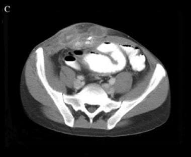 A right lower quadrant abdominal wall abscess and enteric fistula are observed and confirmed by the presence of enteral contrast in the abdominal wall.
Organisms can be introduced via various mechanisms, including direct inoculation of microbes into the body or body site, such as in skin or soft tissue infections or bloodstream infections associated with indwelling venous catheters. Inhalational acquisition is a mode of infection in the setting of respiratory infection, as is aspiration of oral/gastric content. Ascending urinary tract infection can cause systemic infection. The gastrointestinal tract also can be a source of infection if contents macroscopically rupture or seed the intra-abdominal compartment or if organisms translocate through the mucosal barrier. Other mucosal surfaces can serve as entry points, including the conjunctiva, the upper respiratory tract, and the genitourinary tract. External disease-transmitting vectors, such as arthropods, also can cause infection. [6, 15]
The pathophysiology of sepsis is complex and results from the effects of circulating bacterial products, mediated by cytokine release, caused by sustained bacteremia. Cytokines are responsible for the clinically observable effects of bacteremia in the host. [15, 16, 17, 18] Impaired pulmonary, hepatic, or renal function may result from excessive cytokine release during the septic process.
Prognosis
Sepsis is a common cause of mortality and morbidity worldwide. The prognosis depends on underlying health status and host defenses, prompt and adequate surgical drainage of abscesses, relief of any obstruction of the intestinal or urinary tract, and appropriate and early empiric antimicrobial therapy. [19]
The prognosis of sepsis treated in a timely manner and with appropriate therapy is usually good, except in those with intra-abdominal or pelvic abscesses due to organ perforation. When timely and appropriate therapy has been delivered, the underlying physiologic condition of the patient determines outcome.
A systematic review by Winters et al suggested that beyond the standard 28-day in-hospital mortality endpoint, ongoing mortality in patients with sepsis remains elevated up to 2 years and beyond. [20] In addition, survivors consistently demonstrate impaired quality of life. [21]
Clinical characteristics that affect the severity of sepsis and, therefore, the outcome include the host's response to infection, the site and type of infection, and the timing and type of antimicrobial therapy.
Host-related
Abnormal host immune responses may increase susceptibility to severe disease and mortality. For example, extremes of temperature and the presence of leukopenia and/or thrombocytopenia, advanced age, presence of co-morbid conditions, hyperglycemia, bleeding diatheses, and failure of procalcitonin levels to fall are associated with worsened outcome. [22]
Important risk factors for mortality include the patient's comorbidities, functional health status, newly onset atrial fibrillation, hypercoagulability state, hyperglycemia on admission, AIDS, liver disease, cancer, alcohol dependence, and immune suppression.
Age older than 40 years is associated with comorbid illnesses, impaired immunologic responses, malnutrition, increased exposure to potentially resistant pathogens in nursing homes, and increased use of medical devices, such as indwelling catheters and central venous lines. [23, 24, 25, 26]
Infection site
Sepsis due to urinary tract infection has the lowest mortality rate, whereas mortality rates are higher with unknown sources of infection, gastrointestinal sources (highest in ischemic bowel), and pulmonary sources. [27, 28, 29]
Infection type
Sepsis due to nosocomial pathogens has a higher mortality rate than sepsis caused by community-acquired pathogens. Increased mortality is associated with bloodstream infections due to Staphylococcus aureus, fungi, and Pseudomonas, as well as polymicrobial infections. When bloodstream infections become severe (ie, septic shock), the outcome may be similar regardless of whether the pathogenic bacteria are gram-negative or gram-positive.
Antimicrobial therapy
Studies have shown that the early administration of appropriate antibiotic therapy (ie, antibiotics to which the pathogen is sensitive) is beneficial in septic patients demonstrating bacteremia. Previous antibiotic therapy (ie, antibiotics within the prior 90 days) may be associated with increased mortality risk, at least among patients with gram-negative sepsis. Patients who have received prior antibiotic therapy are more likely to have higher rates of antibiotic resistance, reducing the likelihood that appropriate antibiotic therapy will be chosen empirically. [30, 31, 32, 33]
Restoration of perfusion
Failure to attempt aggressive restoration of perfusion early may be associated with an increased mortality risk. A severely elevated lactate level (>4 mmol/L) is associated with a poor prognosis in patients with sepsis.
Epidemiology
Incidence
The incidence of sepsis and the number of sepsis-related deaths are increasing because of an increased use of immunosuppressive medications. The incidence varies by race and sex. The highest incidence is among Black males. The incidence also shows seasonal variation, with the highest number of cases in winter, probably because of the increased prevalence of respiratory infections during this season. Older patients (≥65 years) account for most (60-85%) sepsis cases, attributable to multiple comorbidities and frequent hospitalizations. [19]
Pathogens
The predominant infectious organisms that cause sepsis have changed over the years. Gram-positive bacteria are the most common etiologic pathogens, although the incidence of gram-negative sepsis remains substantial. The incidence of fungal sepsis has been rising with more patients on immunosuppressive therapies and more cases of HIV infection. In approximately half of sepsis cases, the organism is not identified (culture-negative sepsis).
Risk Factors
Risk factors for sepsis and septic shock include the following:
-
ICU admission with subsequent nosocomial infection
-
Bacteremia
-
Advanced age (≥65 years)
-
Immunosuppression - Conditions that impair host defenses such as seen with neoplasms, renal failure, hepatic failure, AIDS, asplenism, diabetes, autoimmune diseases, organ transplant, alcoholism, and the use of immunosuppressant medications and immunomodulators
-
Community-acquired pneumonia
-
Previous hospitalization and antibiotic therapy in the preceding 90 days
-
Genetic factors - Defects of cellular and humoral immunity (low or absent antibody production, T cells, phagocytes, natural killer cells, complement)
-
Urosepsis due to benign prostatic hypertrophy (BPH) in older males or complicated UTI
-
Major trauma and burn injuries
-
A right lower quadrant abdominal wall abscess and enteric fistula are observed and confirmed by the presence of enteral contrast in the abdominal wall.
Tables
- Table 1. Infectious and Noninfectious Causes of Fever [14]
- Table 2. Clinical Conditions Associated With Sepsis
- Table 3. Noninfectious Conditions Mimicking Clinical and Hemodynamic Parameters of Sepsis
- Table 4. Characteristics of Pseudosepsis and Sepsis
- Table 1. Table of Current Recommendations and Changes From Previous 2016 Recommendations [1]
System |
Infectious Causes |
Noninfectious Causes |
|---|---|---|
Central nervous |
Meningitis, encephalitis |
Posterior fossa syndrome, central fever, seizures, cerebral infraction, hemorrhage, cerebrovascular accident |
Cardiovascular |
Central line, infected pacemaker, endocarditis, sternal osteomyelitis, viral pericarditis, myocardial/perivalvular abscess |
Myocardial infarction, balloon pump syndrome, Dressler syndrome |
Pulmonary |
Ventilator-associated pneumonia, mediastinitis, tracheobronchitis, empyema |
Pulmonary emboli, ARDS, atelectasis (without pneumonia), cryptogenic organizing pneumonia, bronchogenic carcinoma without postobstructive pneumonia, systemic lupus erythematosus, pneumonitis, vasculitis |
Gastrointestinal |
Intra-abdominal abscess, cholangitis, cholecystitis, viral hepatitis, peritonitis, diarrhea (Clostridium difficile) |
Pancreatitis, acalculous cholecystitis, ischemia of the bowel/colon, bleeding, cirrhosis, irritable bowel syndrome |
Urinary tract |
Catheter-associated bacteremia, urosepsis, pyelonephritis, cystitis |
Allergic interstitial nephritis |
Skin/soft tissue |
Decubitus ulcers, cellulitis, wound infection |
Vascular ulcers |
Bone/joint |
Chronic osteomyelitis, septic arthritis |
Acute gout |
Other |
Transient bacteremia, sinusitis |
Adrenal insufficiency, phlebitis/thrombophlebitis, neoplastic fever, alcohol/drug withdrawal, delirium tremens, drug fever, fat emboli, deep venous thrombosis, postoperative fever (48 h), fever after transfusion |
| System | Associated With Sepsis |
Not Typically Associated With Sepsis |
| GI tract | Liver Gallbladder Colon Abscess Intestinal obstruction Instrumentation |
Esophagitis Gastritis Pancreatitis (may have multiorgan dysfunction but not infectious in origin) Small bowel disorders GI bleeding |
| GU tract | Pyelonephritis Intra- or perinephric abscess Renal calculi Urinary tract obstruction Acute prostatitis/abscess Renal insufficiency Instrumentation in patients with bacteriuria |
Urethritis Cystitis Cervicitis Vaginitis Catheter-associated bacteriuria (in otherwise healthy hosts without genitourinary tract disease) |
| Pelvis | Peritonitis Abscess |
|
| Upper respiratory tract | Deep neck space infection Abscess |
Pharyngitis Sinusitis Bronchitis Otitis |
| Lower respiratory tract | Community-acquired pneumonia (with asplenia) Empyema Lung abscess |
Community-acquired pneumonia (in otherwise healthy host)
|
| Intravascular | IV line sepsis Infected prosthetic device Acute bacterial endocarditis |
|
| Cardiovascular | Acute bacterial endocarditis Myocardial/perivalvular ring abscess |
Subacute bacterial endocarditis
|
| CNS | Bacterial meningitis |
Aseptic meningitis |
Skin/soft-tissue
|
Necrotizing fasciitis
|
Osteomyelitis Uncomplicated wound infections |
CNS = central nervous system; GI = gastrointestinal; GU = genitourinary; IV = intravenous. Adapted from: Cunha BA, Shea KW. Fever in the intensive care unit. Infect Dis Clin North Am. Mar 1996;10(1):185-209. [41] |
||
Clinical Presentations Mimicking Sepsis |
Hemodynamic Parameters Mimicking Sepsis |
Myocardial infarction |
Spinal cord injury |
Pancreatitis |
Adrenal insufficiency |
Diabetic ketoacidosis |
Acute pancreatitis |
Systemic lupus erythematosus flare with abdominal crisis |
Hemorrhage |
Ventricular pseudoaneurysm |
Pulmonary embolism |
Massive aspiration/atelectasis |
Anaphylaxis |
Systemic vasculitis |
|
Hypovolemia (eg, due to diuretics, dehydration) |
|
Parameters |
Pseudosepsis |
Sepsis |
Microbiologic |
No definite source PLUS ≥1 abnormalities Negative blood cultures excluding contaminants |
Proper identification/process/source PLUS ≥1 microbiologic abnormalities Positive buffy coat smear result OR several positive blood culture results with a pathogenic organism |
Hemodynamic |
⇓ PVR ⇑ CO |
⇓ PVR ⇑ CO Left ventricular dilatation |
Laboratory |
⇑ WBC count (with left shift) Normal platelet count ⇑ FSP ⇑ Lactate ⇑ D-dimers ⇑ PT/PTT ⇓ Albumin ⇓ Fibrinogen ⇓ Globulins |
⇑ WBC count (with left shift) ⇓ Platelets ⇑ FSP ⇑ Lactate ⇑ D-dimers ⇑ PT/PTT ⇓ Albumin |
Clinical |
≤102°F ± Tachycardia ± Respiratory alkalosis ± Hypotension |
≥102°F OR Hypothermia ± Mental status changes ± Hypotension |
CO = cardiac output; FSP = fibrin split products; GI = gastrointestinal; GU = genitourinary; PT/PTT = prothrombin time/partial thromboplastin time; PVR = peripheral vascular resistance; WBC = white blood cell. |
||
| Recommendations 2021 | Recommendation Strength and Quality of Evidence | Changes From 2016 Recommendations |
|---|---|---|
| 1. For hospitals and health systems, we recommend using a performance improvement program for sepsis, including sepsis screening for acutely ill, high-risk patients and standard operating procedures for treatment. | Strong; moderate-quality evidence (for screening) | Changed from Best practice statement |
| “We recommend that hospitals and hospital systems have a performance improvement program for sepsis including sepsis screening for acutely ill, high-risk patients.” | ||
| Strong; very low-quality evidence (for standard operating procedures) | ||
| 2. We recommend against using qSOFA compared with SIRS, NEWS, or MEWS as a single-screening tool for sepsis or septic shock. | Strong; moderate-quality evidence | NEW |
| 3. For adults suspected of having sepsis, we suggest measuring blood lactate. | Weak; low quality of evidence | |
| INITIAL RESUSCITATION | ||
| 4. Sepsis and septic shock are medical emergencies, and we recommend that treatment and resuscitation begin immediately. | Best practice statement | |
| 5. For patients with sepsis induced hypoperfusion or septic shock we suggest that at least 30 mL/kg of IV crystalloid fluid should be given within the first 3 hr of resuscitation. | Weak; low quality of evidence | DOWNGRADE from Strong; low quality of evidence |
| “We recommend that in the initial resuscitation from sepsis-induced hypoperfusion, at least 30 mL/kg of IV crystalloid fluid be given within the first 3 hr” | ||
| 6. For adults with sepsis or septic shock, we suggest using dynamic measures to guide fluid resuscitation, over physical examination or static parameters alone. | Weak; very low quality of evidence | |
| 7. For adults with sepsis or septic shock, we suggest guiding resuscitation to decrease serum lactate in patients with elevated lactate level, over not using serum lactate. | Weak; low quality of evidence | |
| 8. For adults with septic shock, we suggest using capillary refill time to guide resuscitation as an adjunct to other measures of perfusion. | Weak; low quality of evidence | NEW |
| MEAN ARTERIAL PRESSURE | ||
| 9. For adults with septic shock on vasopressors, we recommend an initial target mean arterial pressure (MAP) of 65 mm Hg over higher MAP targets. | Strong; moderate-quality evidence | |
| ADMISSION TO INTENSIVE CARE | ||
| 10. For adults with sepsis or septic shock who require ICU admission, we suggest admitting the patients to the ICU within 6 hr. | Weak; low quality of evidence | |
| INFECTION | ||
| 11. For adults with suspected sepsis or septic shock but unconfirmed infection, we recommend continuously re-evaluating and searching for alternative diagnoses and discontinuing empiric antimicrobials if an alternative cause of illness is demonstrated or strongly suspected. | Best practice statement | |
| 12. For adults with possible septic shock or a high likelihood for sepsis, we recommend administering antimicrobials immediately, ideally within 1 hr of recognition. | Strong; low quality of evidence (Septic shock) | CHANGED from previous: “We recommend that administration of intravenous antimicrobials should be initiated as soon as possible after recognition and within one hour for both a) septic shock and b) sepsis without shock” |
| Strong; very low quality of evidence (Sepsis without shock) | ||
| Strong recommendation; moderate quality of evidence | ||
| 13. For adults with possible sepsis without shock, we recommend rapid assessment of the likelihood of infectious versus noninfectious causes of acute illness. | Best practice statement | |
| 14. For adults with possible sepsis without shock, we suggest a time-limited course of rapid investigation and if concern for infection persists, the administration of antimicrobials within 3 hr from the time when sepsis was first recognized. | Weak; very low quality of evidence | NEW from previous: |
| “We recommend that administration of IV antimicrobials should be initiated as soon as possible after recognition and within 1 hr for both a) septic shock and b) sepsis without shock” | ||
| Strong recommendation; moderate quality of evidence | ||
| 15. For adults with a low likelihood of infection and without shock, we suggest deferring antimicrobials while continuing to closely monitor the patient. | Weak; very low quality of evidence | NEW from previous: |
| “We recommend that administration of IV antimicrobials should be initiated as soon as possible after recognition and within 1 hr for both a) septic shock and b) sepsis without shock“ | ||
| Strong recommendation; moderate quality of evidence | ||
| 16. For adults with suspected sepsis or septic shock, we suggest against using procalcitonin plus clinical evaluation to decide when to start antimicrobials, as compared with clinical evaluation alone. | Weak; very low quality of evidence | |
| 17. For adults with sepsis or septic shock at high risk for MRSA, we recommend using empiric antimicrobials with MRSA coverage over using antimicrobials without MRSA coverage. | Best practice statement | NEW from previous: |
| “We recommend empiric broad-spectrum therapy with one or more antimicrobials for patients presenting with sepsis or septic shock to cover all likely pathogens (including bacterial and potentially fungal or viral coverage.” | ||
| Strong recommendation; moderate quality of evidence | ||
| 18. For adults with sepsis or septic shock at low risk for MRSA, we suggest against using empiric antimicrobials with MRSA coverage, as compared with using antimicrobials without MRSA coverage. | Weak; low quality of evidence | NEW from previous: |
| “We recommend empiric broad-spectrum therapy with one or more antimicrobials for patients presenting with sepsis or septic shock to cover all likely pathogens (including bacterial and potentially fungal or viral coverage.” | ||
| Strong recommendation; moderate quality of evidence | ||
| 19. For adults with sepsis or septic shock and high risk for multidrug resistant (MDR) organisms, we suggest using two antimicrobials with gram-negative coverage for empiric treatment over one gram-negative agent. | Weak; very low quality of evidence | |
| 20. For adults with sepsis or septic shock and low risk for multidrug resistant (MDR) organisms, we suggest against using two gram-negative agents for empiric treatment, as compared to one gram-negative agent. | Weak; very low quality of evidence | |
| 21. For adults with sepsis or septic shock, we suggest against using double gram-negative coverage once the causative pathogen and the susceptibilities are known. | Weak; very low quality of evidence | |
| 22. For adults with sepsis or septic shock at high risk for fungal infection, we suggest using empiric antifungal therapy over no antifungal therapy. | Weak; low quality of evidence | NEW from previous: |
| “We recommend empiric broad-spectrum therapy with one or more antimicrobials for patients presenting with sepsis or septic shock to cover all likely pathogens (including bacterial and potentially fungal or viral coverage.” | ||
| Strong recommendation; moderate quality of evidence | ||
| 23. For adults with sepsis or septic shock at low risk of fungal infection, we suggest against empiric use of antifungal therapy | Weak; low quality of evidence | NEW from previous: |
| “We recommend empiric broad-spectrum therapy with one or more antimicrobials for patients presenting with sepsis or septic shock to cover all likely pathogens (including bacterial and potentially fungal or viral coverage.“ | ||
| Strong recommendation; moderate quality of evidence | ||
| 24. We make no recommendation on the use of antiviral agents. | No recommendation | |
| 25. For adults with sepsis or septic shock, we suggest using prolonged infusion of beta-lactams for maintenance (after an initial bolus) over conventional bolus infusion. | Weak; moderate-quality evidence | |
| 26. For adults with sepsis or septic shock, we recommend optimising dosing strategies of antimicrobials based on accepted pharmacokinetic/pharmacodynamic (PK/PD) principles and specific drug properties. | Best practice statement | |
| 27. For adults with sepsis or septic shock, we recommend rapidly identifying or excluding a specific anatomical diagnosis of infection that requires emergent source control and implementing any required source control intervention as soon as medically and logistically practical. | Best practice statement | |
| 28. For adults with sepsis or septic shock, we recommend prompt removal of intravascular access devices that are a possible source of sepsis or septic shock after other vascular access has been established. | Best practice statement | |
| 29. For adults with sepsis or septic shock, we suggest daily assessment for de-escalation of antimicrobials over using fixed durations of therapy without daily reassessment for de-escalation. | Weak; very low quality of evidence | |
| 30. For adults with an initial diagnosis of sepsis or septic shock and adequate source control, we suggest using shorter over longer duration of antimicrobial therapy. | Weak; very low quality of evidence | |
| 31. For adults with an initial diagnosis of sepsis or septic shock and adequate source control where optimal duration of therapy is unclear, we suggest using procalcitonin AND clinical evaluation to decide when to discontinue antimicrobials over clinical evaluation alone. | Weak; low quality of evidence | |
| HEMODYNAMIC MANAGEMENT | ||
| 32. For adults with sepsis or septic shock, we recommend using crystalloids as first-line fluid for resuscitation. | Strong; moderate-quality evidence | |
| 33. For adults with sepsis or septic shock, we suggest using balanced crystalloids instead of normal saline for resuscitation. | Weak; low quality of evidence | CHANGED from weak recommendation; low quality of evidence. |
| “We suggest using either balanced crystalloids or saline for fluid resuscitation of patients with sepsis or septic shock” | ||
| 34. For adults with sepsis or septic shock, we suggest using albumin in patients who received large volumes of crystalloids. | Weak; moderate-quality evidence | |
| 35. For adults with sepsis or septic shock, we recommend against using starches for resuscitation. | Strong; high-quality evidence | |
| 36. For adults with sepsis and septic shock, we suggest against using gelatin for resuscitation. | Weak; moderate-quality evidence | UPGRADE from weak recommendation; low quality of evidence |
| “We suggest using crystalloids over gelatins when resuscitating patients with sepsis or septic shock.” | ||
| 37. For adults with septic shock, we recommend using norepinephrine as the first-line agent over other vasopressors. | Strong | |
| Dopamine. High-quality evidence | ||
| Vasopressin. Moderate-quality evidence | ||
| Epinephrine. Low quality of evidence | ||
| Selepressin. Low quality of evidence | ||
| Angiotensin II. Very low-quality evidence | ||
| 38. For adults with septic shock on norepinephrine with inadequate mean arterial pressure levels, we suggest adding vasopressin instead of escalating the dose of norepinephrine. | Weak; moderate quality evidence | |
| 39. For adults with septic shock and inadequate mean arterial pressure levels despite norepinephrine and vasopressin, we suggest adding epinephrine. | Weak; low quality of evidence | |
| 40. For adults with septic shock, we suggest against using terlipressin. | Weak; low quality of evidence | |
| 41. For adults with septic shock and cardiac dysfunction with persistent hypoperfusion despite adequate volume status and arterial blood pressure, we suggest either adding dobutamine to norepinephrine or using epinephrine alone. | Weak; low quality of evidence | |
| 42. For adults with septic shock and cardiac dysfunction with persistent hypoperfusion despite adequate volume status and arterial blood pressure, we suggest against using levosimendan. | Weak; low quality of evidence | NEW |
| 43. For adults with septic shock, we suggest invasive monitoring of arterial blood pressure over noninvasive monitoring, as soon as practical and if resources are available. | Weak; very low quality of evidence | |
| 44. For adults with septic shock, we suggest starting vasopressors peripherally to restore mean arterial pressure rather than delaying initiation until a central venous access is secured. | Weak; very low quality of evidence | NEW |
| 45. There is insufficient evidence to make a recommendation on the use of restrictive versus liberal fluid strategies in the first 24 hr of resuscitation in patients with sepsis and septic shock who still have signs of hypoperfusion and volume depletion after the initial resuscitation. | No recommendation | NEW |
| “We suggest using either balanced crystalloids or saline for fluid resuscitation of patients with sepsis or septic shock” | ||
| Weak recommendation; low quality of evidence | ||
| “We suggest using crystalloids over gelatins when resuscitating patients with sepsis or septic shock.” | ||
| Weak recommendation; low quality of evidence | ||
| VENTILATION | ||
| 46.There is insufficient evidence to make a recommendation on the use of conservative oxygen targets in adults with sepsis-induced hypoxemic respiratory failure. | No recommendation | |
| 47. For adults with sepsis-induced hypoxemic respiratory failure, we suggest the use of high flow nasal oxygen over noninvasive ventilation. | Weak; low quality of evidence | NEW |
| 48. There is insufficient evidence to make a recommendation on the use of noninvasive ventilation in comparison to invasive ventilation for adults with sepsis-induced hypoxemic respiratory failure. | No recommendation | |
| 49. For adults with sepsis-induced ARDS, we recommend using a low tidal volume ventilation strategy (6 mL/kg), over a high tidal volume strategy (> 10 mL/kg). | Strong; high-quality evidence | |
| 50. For adults with sepsis-induced severe ARDS, we recommend using an upper limit goal for plateau pressures of 30 cm H2O, over higher plateau pressures. | Strong; moderate-quality evidence | |
| 51. For adults with moderate to severe sepsis-induced ARDS, we suggest using higher PEEP over lower PEEP. | Weak; moderate-quality evidence | |
| 52. For adults with sepsis-induced respiratory failure (without ARDS), we suggest using low tidal volume as compared with high tidal volume ventilation. | Weak; low quality of evidence | |
| 53. For adults with sepsis-induced moderate-severe ARDS, we suggest using traditional recruitment maneuvers. | Weak; moderate-quality evidence | |
| 54. When using recruitment maneuvers, we recommend against using incremental PEEP titration/strategy. | Strong; moderate-quality evidence | |
| 55. For adults with sepsis-induced moderate-severe ARDS, we recommend using prone ventilation for greater than 12 hr daily. | Strong; moderate-quality evidence | |
| 56. For adults with sepsis induced moderate-severe ARDS, we suggest using intermittent NMBA boluses, over NMBA continuous infusion. | Weak; moderate-quality evidence | |
| 57. For adults with sepsis-induced severe ARDS, we suggest using Veno-venous (VV) ECMO when conventional mechanical ventilation fails in experienced centers with the infrastructure in place to support its use. | Weak; low quality of evidence | NEW |
| ADDITIONAL THERAPIES | ||
| 58. For adults with septic shock and an ongoing requirement for vasopressor therapy we suggest using IV corticosteroids. | Weak; moderate-quality evidence | UPGRADE from Weak recommendation , low quality of evidence |
| “We suggest against using IV hydrocortisone to treat septic shock patients if adequate fluid resuscitation and vasopressor therapy are able to restore hemodynamic stability (see goals for Initial Resuscitation). If this is not achievable, we suggest IV hydrocortisone at a dose of 200 mg/day.” | ||
| 59. For adults with sepsis or septic shock we suggest against using polymyxin B hemoperfusion. | Weak; low quality of evidence | NEW from previous: “We make no recommendation regarding the use of blood purification techniques” |
| 60. There is insufficient evidence to make a recommendation on the use of other blood purification techniques. | No recommendation | |
| 61. For adults with sepsis or septic shock we recommend using a restrictive (over liberal) transfusion strategy. | Strong; moderate-quality evidence | |
| 62. For adults with sepsis or septic shock we suggest against using IV immunoglobulins. | Weak; low quality of evidence | |
| 63. For adults with sepsis or septic shock, and who have risk factors for gastrointestinal (GI) bleeding, we suggest using stress ulcer prophylaxis. | Weak; moderate-quality evidence | |
| 64. For adults with sepsis or septic shock, we recommend using pharmacologic venous thromboembolism (VTE) prophylaxis unless a contraindication to such therapy exists. | Strong; moderate-quality evidence | |
| 65. For adults with sepsis or septic shock, we recommend using low molecular weight heparin over unfractionated heparin for VTE prophylaxis | Strong; moderate-quality evidence | |
| 66. For adults with sepsis or septic shock, we suggest against using mechanical VTE prophylaxis, in addition to pharmacological prophylaxis, over pharmacologic prophylaxis alone. | Weak; low quality of evidence | |
| 67. In adults with sepsis or septic shock and AKI, we suggest using either continuous or intermittent renal replacement therapy. | Weak; low quality of evidence | |
| 68. In adults with sepsis or septic shock and AKI, with no definitive indications for renal replacement therapy, we suggest against using renal replacement therapy. | Weak; moderate-quality evidence | |
| 69. For adults with sepsis or septic shock, we recommend initiating insulin therapy at a glucose level of ≥ 180mg/dL (10 mmol/L). | Strong; moderate-quality evidence | |
| 70. For adults with sepsis or septic shock we suggest against using IV vitamin C. | Weak; low quality of evidence | NEW |
| 71. For adults with septic shock and hypoperfusion-induced lactic acidemia, we suggest against using sodium bicarbonate therapy to improve hemodynamics or to reduce vasopressor requirements. | Weak; low quality of evidence | |
| 72. For adults with septic shock and severe metabolic acidemia (pH ≤ 7.2) and acute kidney injury (AKIN score 2 or 3), we suggest using sodium bicarbonate therapy | Weak; low quality of evidence | |
| 73. For adult patients with sepsis or septic shock who can be fed enterally, we suggest early (within 72 hr) initiation of enteral nutrition. | Weak; very low quality of evidence | |
| LONG-TERM OUTCOMES AND GOALS OF CARE | ||
| 74. For adults with sepsis or septic shock, we recommend discussing goals of care and prognosis with patients and families over no such discussion. | Best practice statement | |
| 75. For adults with sepsis or septic shock, we suggest addressing goals of care early (within 72 hr) over late (72 hr or later). | Weak; low quality of evidence | |
| 76. For adults with sepsis or septic shock, there is insufficient evidence to make a recommendation on any specific standardized criterion to trigger goals of care discussion. | No recommendation | |
| 77. For adults with sepsis or septic shock, we recommend that the principles of palliative care (which may include palliative care consultation based on clinician judgement) be integrated into the treatment plan, when appropriate, to address patient and family symptoms and suffering. | Best practice statement | |
| 78. For adults with sepsis or septic shock, we suggest against routine formal palliative care consultation for all patients over palliative care consultation based on clinician judgement. | Weak; low quality of evidence | |
| 79. For adult survivors of sepsis or septic shock and their families, we suggest referral to peer support groups over no such referral. | Weak; very low quality of evidence | |
| 80. For adults with sepsis or septic shock, we suggest using a handoff process of critically important information at transitions of care over no such handoff process. | Weak; very low quality of evidence | |
| 81. For adults with sepsis or septic shock, there is insufficient evidence to make a recommendation on the use of any specific structured handoff tool over usual handoff processes. | No recommendation | |
| 82. For adults with sepsis or septic shock and their families, we recommend screening for economic and social support (including housing, nutritional, financial, and spiritual support), and make referrals where available to meet these needs. | Best practice statement | |
| 83. For adults with sepsis or septic shock and their families, we suggest offering written and verbal sepsis education (diagnosis, treatment, and post-ICU/post-sepsis syndrome) prior to hospital discharge and in the follow-up setting. | Weak; very low quality of evidence | |
| 84. For adults with sepsis or septic shock and their families, we recommend the clinical team provide the opportunity to participate in shared decision making in post-ICU and hospital discharge planning to ensure discharge plans are acceptable and feasible. | Best practice statement | |
| 85. For adults with sepsis and septic shock and their families, we suggest using a critical care transition program, compared with usual care, upon transfer to the floor. | Weak; very low quality of evidence | |
| 86. For adults with sepsis and septic shock, we recommend reconciling medications at both ICU and hospital discharge. | Best practice statement | |
| 87. For adult survivors of sepsis and septic shock and their families, we recommend including information about the ICU stay, sepsis and related diagnoses, treatments, and common impairments after sepsis in the written and verbal hospital discharge summary. | Best practice statement | |
| 88. For adults with sepsis or septic shock who developed new impairments, we recommend hospital discharge plans include follow-up with clinicians able to support and manage new and long-term sequelae. | Best practice statement | |
| 89. For adults with sepsis or septic shock and their families, there is insufficient evidence to make a recommendation on early post-hospital discharge follow-up compared with routine post-hospital discharge follow-up. | No recommendation | |
| 90. For adults with sepsis or septic shock, there is insufficient evidence to make a recommendation for or against early cognitive therapy. | No recommendation | |
| 91. For adult survivors of sepsis or septic shock, we recommend assessment and follow-up for physical, cognitive, and emotional problems after hospital discharge. | Best practice statement | |
| 92. For adult survivors of sepsis or septic shock, we suggest referral to a post-critical illness follow-up program if available. | Weak; very low quality of evidence | |
| 93. For adult survivors of sepsis or septic shock receiving mechanical ventilation for > 48hr or an ICU stay of > 72 hr, we suggest referral to a post-hospital rehabilitation program. | Weak; very low quality of evidence | |