Continually Updated Clinical Reference
 
 
  All Sources     eMedicine     Medscape     Drug Reference     MEDLINE
 
eMedicine - Transureteroureterostomy : Article by

Quick Find
Authors & Editors
Introduction
Indications
Relevant Anatomy
Contraindications
Workup
Treatment
Complications
Outcome And Prognosis
Future And Controversies
References


Patient Education
Kidneys and Urinary System Center

Intravenous Pyelogram Introduction

Intravenous Pyelogram Preparation


Author: Bradley Fields Schwartz, DO, FACS, Associate Professor of Urology, Director, Center for Laparoscopy and Endourology, Department of Surgery, Southern Illinois University School of Medicine

Bradley Fields Schwartz is a member of the following medical societies: American College of Surgeons, American Urological Association, Association of Military Osteopathic Physicians and Surgeons, Endourological Society, Society of Laparoendoscopic Surgeons, and Society of University Urologists

Editors: Michael Grasso, MD, Chairman, Department of Urology, Saint Vincent's Medical Center; Professor and Vice Chairman, Department of Urology, New York Medical College; Francisco Talavera, PharmD, PhD, Senior Pharmacy Editor, eMedicine; Shlomo Raz, MD, Professor, Department of Surgery, Division of Urology, University of California at Los Angeles School of Medicine; J Stuart Wolf, Jr, MD, FACS, David A Bloom Professor of Urology, Director, Division of Minimally Invasive Urology, Department of Urology, University of Michigan Medical Center; William J Cromie, MD, MBA, President and Chief Executive Officer, Health Care, Capital District Physicians' Health Plan

Author and Editor Disclosure

Synonyms and related keywords: transureteroureterostomy, TUU, cross ureteroureterostomy, urinary reconstruction, ureters, ureteral obstruction, distal ureteral obstruction, cutaneous ureterostomy, pelvic malignancies, vesicoureteral reflux, exstrophy amyloidosis, malakoplakia, leukoplakia, solitary kidney, intrinsic ureter obstruction, extrinsic ureteral obstruction, bilateral hydronephrosis, unilateral hydronephrosis

Transureteroureterostomy (TUU) is a urinary reconstruction technique that joins one ureter to the other across the midline. It offers patients with distal ureteral obstruction an option to live without external urostomy appliances or internal urinary stents. TUU is also used in undiversion procedures when the surgeon wants to avoid the pelvis because of previous trauma, surgery, or radiation therapy. It can be combined with other procedures, such as cutaneous ureterostomy, in extreme cases.
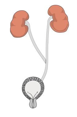
Requirements for performing a successful TUU include a salvageable ipsilateral kidney with a normal ureter proximal to the diseased portion. The accepting ureter must have unobstructed drainage and must not be affected by any disease process that will put both kidneys at risk postoperatively. Indications for TUU include trauma, pelvic malignancies, vesicoureteral reflux, exstrophy, and rare conditions such as amyloidosis, malakoplakia, and leukoplakia involving large segments of ureter. This procedure is seldom used if ureteral reimplantation using the psoas hitch or Boari flap is possible. Other options include ileal ureter, autotransplant, or nephrectomy.

The concept of the procedure is to reestablish ureteral continuity by bringing the ureter across the midline and anastomosing (connecting) it to the contralateral ureter. This is generally performed in an end-to-side or side-to-side fashion. The accepting ureter must be normal because, after the TUU is performed, any disease process that affects one ureter or kidney puts the contralateral ureter and kidney at risk. TUU can also be performed in cases of a solitary kidney with a normal contralateral ureteral stump.

History of the Procedure

The procedure was first attempted in animals in 1906, and Higgins applied it to humans in 1935. Since then, TUU has been performed in children and adults for benign and malignant diseases that cause ureteral obstruction.

Problem

TUU is a surgical procedure used in patients who have no other option of regaining ureteral continuity. It is used in only patients who cannot undergo reimplantation with psoas hitch or Boari flap. These patients are very challenging, and proper patient selection is important.

Frequency

A literature search using Medline revealed 6 articles that reported fewer than 600 cases performed worldwide from 1975 to present. This number is most likely much higher considering the fact that most procedures are not reported in scientific literature.

The largest modern series is from England, where 253 procedures were performed for both benign and malignant diseases that affected the distal ureter (Noble, 1997). The authors experienced 5 complications, all of which involved the common ureter distal to the TUU. Transient leak occurred in 16 (6%) patients. This emphasizes the need to ensure a normal accepting ureter distal to the TUU anastomosis. In addition, any disease processes that may risk both upper units, such as stone disease, medicorenal disease, or chronic renal insufficiency, are contraindications to this procedure.

Etiology

Medical conditions that may necessitate TUU include trauma, pelvic malignancies, vesicoureteral reflux, exstrophy, and rare conditions such as amyloidosis, malakoplakia, and leukoplakia involving large segments of ureter.

Clinical

Patients who require TUU present with either intrinsic or extrinsic ureteral obstruction. This may manifest as flank pain, fever, malaise, or sepsis or may be completely asymptomatic. Sonograms and CT scans usually demonstrate unilateral or bilateral hydronephrosis.


Indications for transureteroureterostomy (TUU) include trauma, pelvic malignancies, vesicoureteral reflux, exstrophy, and rare conditions such as amyloidosis, malakoplakia, and leukoplakia involving large segments of ureter. This procedure is seldom used if ureteral reimplantation using the psoas hitch or Boari flap is possible. Other options include ileal ureter, autotransplant, or nephrectomy.


The ureters are roughly 20-30 cm long and lie completely in the retroperitoneum. They have a rich blood supply, making them relatively forgiving organs on which to operate. However, meticulous surgical technique is required in diseased ureters because they are prone to ischemia with resulting stricture formation or necrosis.

Intraoperative complications include injury to adjacent structures such as bowel or vascular structures. Care must be taken when the bowel is retracted because lacerations or mesentery injuries may occur. The aorta, inferior vena cava, and iliac vessels are in the operative field and must be identified and spared throughout the procedure. Previous surgery and/or radiation therapy increase the risk of injury to these organs.


Contraindications to transureteroureterostomy (TUU) generally include conditions that affect both mid ureters or proximal ureters and/or kidneys. These include genitourinary tuberculosis, a long history of severe stone disease, retroperitoneal fibrosis, transitional cell carcinoma of the renal pelvis or ureter (except in cases of palliation), vesicoureteral reflux in the accepting ureter, and large luminal discrepancy in the 2 ureters. If a procedure such as a psoas hitch or Boari flap can be performed, TUU may not be the best long-term alternative for the patient. In poorly functioning kidneys (<15%), nephrectomy may be the best option.


Lab Studies

  • Obtain baseline renal function tests, including BUN and creatinine, CBC counts, and electrolytes. Generally, patients with normal or slightly low hemoglobin levels do not require transfusion, and physicians do not routinely type and screen or type and crossmatch patients. Urine cultures identify urinary pathogens preoperatively, so directed therapy can be administered.

Imaging Studies

  • The necessary evaluation prior to a transureteroureterostomy (TUU) should ensure overall patient stability. The surgery ranges from 2-6 hours, depending on the tissue encountered in the retroperitoneum, the health of the ureters, and the experience of the surgeon.
  • Obtain a thorough cardiopulmonary history to ascertain whether preoperative pulmonary function tests, graded exercise stress test, Persantine thallium stress test, or angiography needs to be performed. Consultation with a cardiologist or internist is recommended if the perioperative risk is significant.
  • Imaging studies of the ureters and renal pelvis bilaterally are important. They characterize the ureters and potentially define the distal limits of dissection necessary to obtain adequate ureteral length. Multiple modalities are available to opacify the urothelium. The quality, character, and useable length of the ureters can be defined using any of the following modalities:
    • Intravenous pyelography (IVP)
    • Retrograde pyelography (RPG)
    • Antegrade nephrostography
    • CT scan with reformatting and 3-dimensional reconstruction
    • MRI with intravenous gadolinium
    • Pouch-o-gram or loop-o-gram if reflux is present

Histologic Findings

Histology may play a role in patients with malignancy or in patients with idiopathic ureteral obstruction due to an unknown cause. Recurrence of a malignant process requires involvement with multiple specialists, including a medical oncologist and radiation oncologist. Frozen section at the time of TUU may be used to diagnose rare or uncommon disease such as retroperitoneal fibrosis, amyloidosis, or malakoplakia.


Medical therapy

No medical substitute for transureteroureterostomy (TUU) exists. This is strictly a surgical issue and is simply one technique available to reconstitute ureteral integrity.

Preoperative details

Similar to most urologic procedures, TUU may be performed in various ways. The most common involves a transperitoneal midline approach with the patient in the supine position. Flexion of the table, placement of a lumbar roll, or Trendelenburg positioning may aid in exposure and bowel retraction. Standard prophylactic antibiotics are administered 1-2 hours preoperatively; in the case of infected urine, culture-directed therapy is used. Bowel preparation is not necessary unless an ileal ureter is contemplated in the event a TUU cannot be performed. Cystoscopy and stent placement prior to the incision may aid in ureter identification and intraoperative stent positioning.

Intraoperative details

The retroperitoneum is exposed after the bowel is either packed out of the way or placed in a bowel bag. The ureters are best identified where they cross the iliac vessels anteriorly. The posterior peritoneum is incised using either 2 separate longitudinal incisions or 1 large transverse incision.

The 2 ureters are examined and assessed for mobility and length. If the disease process is bilateral, the most amenable ureter is chosen to implant. The ureter is then mobilized above the diseased portion to the ipsilateral ureteropelvic junction (UPJ) while the periureteric tissue is maintained to preserve its blood supply. Stripping the ureter of this rich source of vascularity places the ureter at great risk of ischemia and subsequent stricture or necrosis. The recipient ureter needs to be mobilized only a short distance (1-4 cm) to limit periureteral trauma.

When the left ureter is transposed, it is passed inferior to the inferior mesenteric artery (IMA) without kinking or obstruction, or it is passed superior to the IMA in a more oblique fashion. The latter route causes the anastomosis to be more proximal on the recipient ureter. Transposing the ureter caudad to the IMA predisposes the ureter to obstruction by the vascular structures. Either method is acceptable as long as the surgeon is aware of the advantages and disadvantages of both techniques.

Once the ureter is transected and brought across the midline in a retroperitoneal tunnel, tension absolutely must be avoided. To gain more distance, the ureter can be mobilized up to the kidney. The recipient ureter can also be mobilized if necessary, taking care to preserve the periureteral tissue. As a final resort, the receiving kidney may be mobilized and nephropexy performed. This maneuver can provide an additional 2-4 cm of length.

When a tension-free anastomosis is ensured, the donor ureter is spatulated 15-20 mm. If an end-to-end procedure is performed, both ureters are spatulated. A 15-mm ureterotomy into the receiving ureter is made so that the donor ureter will drain in a dependent position, usually at a 45º angle.

The ureteroureteral anastomosis may be performed in various methods. A watertight, tension-free anastomosis using absorbable sutures should be performed. Use two 4-0 running absorbable sutures, such as Vicryl or Dexon, starting at each apex and finishing at the contralateral apex. Stenting is decided by the surgeon. The author's practice has been to place 4.8F double pigtail stents up each proximal ureter, through the common distal ureter, and into the bladder. This can be accomplished intraoperatively, similar to that performed during an ileal conduit.

Retroperitonealization of the ureters may limit urine leak into the peritoneal cavity. Caution must be taken not to injure the ureters when this is performed. A drain is placed in the area of the anastomosis and brought out to the abdominal wall through a separate stab incision.

Postoperative details

The Foley catheter remains for 3-5 days to limit reflux up the stented ureter. The drain is removed a day after the Foley comes out or when drainage significantly diminishes. The stents remain for 4-6 weeks, at which time retrograde ureterography can be performed. If extravasation at the anastomosis occurs, the stents stay in for another 2 weeks. Otherwise, the stents are removed. IVP is performed 4-6 weeks after the stents are removed to rule out silent obstruction and to check the patency of the repair.

Follow-up

IVP is repeated at 6 months, 1 year postoperatively, and then yearly. Renal ultrasonography may be substituted for yearly surveillance, although urine leak may not demonstrate hydronephrosis.

For excellent patient education resources, visit eMedicine's Kidneys and Urinary System Center. Also, see eMedicine's patient education article Intravenous Pyelogram.


Assuming meticulous surgical technique and proper patient selection, the complication rate should be less than 5-10%. Complications are classified based on their time of occurrence—intraoperative, postoperative, and long-term. Only complications specifically related to urologic aspects are discussed.

The surgeon must keep in mind the potential for coronary events, pulmonary complications, deep venous thrombosis, and pulmonary embolism. Intraoperative complications include injury to adjacent structures such as bowel or vascular structures. Care must be taken when the bowel is retracted because lacerations or mesentery injuries may occur. The aorta, inferior vena cava, and iliac vessels are in the operative field and must be identified and spared throughout the procedure. Inadequate ureteral length may be encountered. This may lead to ureteral kinking and subsequent obstruction. Stent malposition can lead to obstruction and eventual breakdown of the anastomosis.

Lastly, a tension-free watertight anastomosis prevents prolonged urinary leakage. Anastomotic leak is the most frequently encountered complication and manifests as increased drain output. The fluid obtained can be measured for creatinine; an elevated level indicates urine. Use of abdominal radiography (kidneys, ureters, and bladder [KUB]) ensures proper stent placement. Prolonged urinary drainage usually resolves within 3-5 days with observation and patience. If anastomotic breakdown has occurred or distal obstruction is present, the drainage persists and evaluation with IVP or CT scan is indicated. Bilateral percutaneous nephrostomy tube drainage may be necessary if the urine leak is prolonged.

The possibility of delayed complications makes long-term follow-up of these patients' cases imperative. Late anastomotic leak may manifest as urinoma formation, fever, ureterocutaneous fistula formation, small bowel obstruction, or ureterointestinal fistula. Occurrence of any of these problems mandates an evaluation for distal ureteral obstruction. This may be because of stones, recurrent pelvic disease such as malignancy, inflammatory disease, abscess, or radiation injury. Small bowel obstruction may occur many years after surgery and usually responds to conservative management. Urinary fistulae are treated initially with ureteral stenting and Foley catheterization. Place percutaneous nephrostomy tubes bilaterally if stenting is not successful. Open repair and, finally, nephrectomy are used as last resorts.


The outcome and prognosis of patients undergoing transureteroureterostomy (TUU) depends on numerous factors. If the underlying disease process is progressive, then involvement of the common ureter may occur. Patients who have pelvic malignancies and/or a history of radiation therapy have the most potential for failure. As mentioned above, poor surgical technique that leads to prolonged leak, fistula, or stricture is the leading cause of failure in these patients.

A group from France monitored the cases of 69 patients for a minimum of 1 year at a median of 6 years after TUU was performed (Mure, 2000). They reported complications in 4 (6.3%) patients, including a urinoma that required open drainage, prolonged urinary leak, common ureteral necrosis, and progressive renal failure in the donor kidney. Long-term follow-up is stressed because 2 of these complications occurred as late as 3 and 4 years postoperatively. In a recent study that compared the performance of 34 TUUs with 67 ileal conduit urinary diversions, TUU favored well in terms of reoperation and early and late complications (Kilciler, 2006).

The National Cancer Institute performed TUU in 10 patients for pelvic malignancy (Strup, 1996). Mean follow-up was 77.9 months (range, 2-191 mo). One patient experienced common ureteral stricture, 1 patient underwent pyelolithotomy and subsequent nephrectomy, recurrence of disease with ureteral obstruction occurred in 1 patient, and 1 patient experienced unilateral disease progression in a case of vasculitis/fibrosis. One patient died of sepsis due to urine leak at the anastomosis, 1 patient died from myocardial infarction, and 3 patients died from metastatic disease of their primary process. While these results may not appear promising, one must remember the patient population with which these physicians were working. All 10 patients had extensive pelvic malignancy, with 4 patients receiving radiation therapy and 5 patients receiving chemotherapy.

TUU is one more tool in the urologists' armamentarium that, when performed well, is a successful operation. Patient selection, meticulous attention to detail in the operating room, and long-term follow-up improve the results of this challenging procedure.


The turn of the new century brings an abundance of new technology, with the advent of minimally invasive surgery and laparoscopy. Laparoscopic transureteroureterostomy (TUU) has been performed in the animal model by several centers. The literature contains no reports regarding laparoscopic TUU in the human; however, it will be attempted eventually. Significant barriers to laparoscopy in this setting are the dense pelvic and abdominal adhesions. The hand-assisted approach may play a role in the application of minimally invasive TUU.


  • Dechet CB, Young MM, Segura JW. Laparoscopic transureteroureterostomy: demonstration of its feasibility in swine. J Endourol. Sep 1999;13(7):487-93. [Medline].
  • Ehrlich RM, Skinner DG. Complications of transureteroureterostomy. J Urol. Apr 1975;113(4):467-73. [Medline].
  • Elliott SP, McAninch JW. Ureteral injuries from external violence: the 25-year experience at San Francisco General Hospital. Journal of Urology. 2003;170 (4 pt 1):1213-1216. [Medline].
  • Hendren WH, Hensle TW. Transureteroureterostomy: experience with 75 cases. J Urol. Jun 1980;123(6):826-33. [Medline].
  • Higgins C. Transuretero-ureteral anastomosis. Report of a clinical case. J Urol. 1935;34:349.
  • Hodges CV, Barry JM, Fuchs EF. Transureteroureterostomy: 25-year experience with 100 patients. J Urol. Jun 1980;123(6):834-8. [Medline].
  • Kilciler M, Bedir S, Erdemir F, et al. Comparison of ileal conduit and transureteroureterostomy with ureterocutaneostomy urinary diversion. Urol Int. 2006;77(3):245-50.
  • Maxwell KL, McDougall EM, Shalhav AL. Laparoscopic ureteroureterostomy using vascular closure staples in porcine model. J Endourol. Jun 1998;12(3):265-8. [Medline].
  • Mure PY, Mollard P, Mouriquand P. Transureteroureterostomy in childhood and adolescence: long-term results in 69 cases. J Urol. Mar 2000;163(3):946-8. [Medline].
  • Noble IG, Lee KT, Mundy AR. Transuretero-ureterostomy: a review of 253 cases. Br J Urol. Jan 1997;79(1):20-3. [Medline].
  • Rushton HG, Parrott TS, Woodard JR. The expanded role of transureteroureterostomy in pediatric urology. J Urol. Aug 1987;138(2):357-63. [Medline].
  • Sandoz IL, Paull DP, MacFarlane CA. Complications with transureteroureterostomy. J Urol. Jan 1977;117(1):39-42. [Medline].
  • Schwartz BF, Stoller ML. Endourologic management of urinary fistulae. Tech Urol. Sep 2000;6(3):193-5. [Medline].
  • Sharpe NW. Transuretero-ureteral anastomosis. Ann Surg. 1906;44:687.
  • Strup SE, Sindelar WF, Walther MM. The use of transureteroureterostomy in the management of complex ureteral problems. J Urol. May 1996;155(5):1572-4. [Medline].
  • Sugarbaker PH, Gutman M, Verghese M. Transureteroureterostomy: an adjunct to the management of advanced primary and recurrent pelvic malignancy. International Journal of Colorectal Disease. 2003;18(1):40-44. [Medline].

Transureteroureterostomy excerpt

Article Last Updated: Feb 9, 2007