Continually Updated Clinical Reference
 
 
  All Sources     eMedicine     Medscape     Drug Reference     MEDLINE
 
eMedicine - Cystectomy, Radical : Article by

Quick Find
Authors & Editors
Introduction
Indications
Relevant Anatomy
Contraindications
Workup
Treatment
Complications
Outcome and Prognosis
Future and Controversies
Further Reading
Multimedia
References


Patient Education
Men's Health Center

Cancer and Tumors Center

Kidneys and Urinary System Center

Bladder Cancer Overview

Bladder Cancer Causes

Bladder Cancer Symptoms

Bladder Cancer Treatment

Blood in Urine Overview


Author: Scott E Eggener, MD, Fellow in Urologic Oncology, Department of Urology, Memorial Sloan Kettering Cancer Center

Scott E Eggener is a member of the following medical societies: American Urological Association

Coauthor(s): Steven C Campbell, MD, PhD, Professor of Surgery, Division of Urologic Oncology, Cleveland Clinic

Editors: Richard A Santucci, MD, FACS, Chief of Urology, Detroit Receiving Hospital; Specialist-in-Chief of Urology, Detroit Medical Center; Chief of Urologic Trauma Surgery, Sinai Grace Hospital; Director, The Center for Urologic Reconstruction; Francisco Talavera, PharmD, PhD, Senior Pharmacy Editor, eMedicine; J Stuart Wolf, Jr, MD, FACS, David A Bloom Professor of Urology, Director, Division of Minimally Invasive Urology, Department of Urology, University of Michigan Medical Center; Bradley Fields Schwartz, DO, FACS, Associate Professor of Urology, Director, Center for Laparoscopy and Endourology, Department of Surgery, Southern Illinois University School of Medicine

Author and Editor Disclosure

Synonyms and related keywords: radical cystectomy, radical cystoprostatectomy, anterior pelvic exenteration, bladder cancer, superficial bladder cancer, muscle-invasive bladder cancer, muscle-invasive transitional cell carcinoma, TCC, squamous cell carcinoma, SCC, Schistosoma haematobium, S haematobium, carcinoma in situ, CIS, bladder barbotage, transurethral resection of bladder tumor, TURBT, cystoprostatectomy, urethrectomy, anterior pelvic exenteration, urinary diversion/reconstruction, orthotopic neobladder, Indiana pouch, ileal conduit, muscularis propria, bladder-sparing multimodality therapy, pelvic lymph node dissection, nerve-sparing cystectomy, proto-oncogenes, total gross painless hematuria, cystoscopic resection, diverticulectomy, prostatic urethral involvement, bilateral pelvic lymphadenectomy, continent cutaneous urinary diversions, incontinent cutaneous urinary diversion, Studer pouch, prostate-sparing approach, cyclophosphamide, phenacetin, schistosomiasis, indwelling catheter


In the United States, bladder cancer is the fifth most common cancer (following cancers of the lung, colon, prostate, and breast) and the fourth most common among men. More than 90% of bladder cancers are transitional cell in origin, while, in countries with high endemic schistosomiasis rates (eg, Egypt and the Nile River Valley), squamous cell carcinoma of the bladder is more common.

Lesions limited to the urothelium [pCIS], mucosa [pTa], or lamina propria [pT1] represent 70-80% of all newly diagnosed bladder cancer cases. Although prone to recurrences and, less commonly, progression to higher-stage disease, these lesions are typically managed with transurethral resection and selectively with intravesical chemotherapy, such as bacille Calmette-Guérin (BCG), mitomycin, or thiotepa. Patients with pT1 disease, particularly those with high-risk features (eg, multifocality, recurrence after intravesical therapy, extensive lamina propria invasion, concomitant carcinoma in situ [CIS]) are at considerable risk of disease progression and may benefit from early radical cystoprostatectomy.

Muscle-invasive bladder cancer, defined as tumors that invade the muscularis propria (pT2 or higher), requires more intensive therapy. To date, surgical resection via radical cystoprostatectomy (bladder and prostate) and pelvic lymph node dissection remains the criterion standard for determining accurate pathologic staging, optimizing curative potential, and minimizing the risk of tumor recurrence.

History of the Procedure

The first record of a radical cystectomy dates to the late 1800s. In 1949, Marshall and Whitmore described the basic surgical principles of radical cystoprostatectomy. In 1987, following the neuroanatomic mapping of the pelvic plexus by Schlegel and Walsh, nerve-sparing cystectomy became a surgical option that allowed for preservation of sexual function.1

For many years, radical cystectomy carried a significant perioperative mortality rate (5-10%). However, presumably because of improvements in surgical technique, the evolution of intensive care medicine, and the availability of new antibiotics, radical cystectomy is now a common procedure in major medical centers and carries a perioperative mortality rate of approximately 2-3%.2 At high-volume centers with postoperative pathway care programs, an ICU stay is no longer routine and the median hospital stay is 7 days.

Problem

Bladder cancer can be axiomatically subdivided into non–muscle invasive and muscle-invasive disease. This article focuses primarily on the management of muscle-invasive transitional cell carcinoma (TCC) and the role of radical cystectomy. For a more in-depth review of the management of non–muscle invasive bladder cancer, see Bladder Cancer.

Frequency

United States

  • More than 90% of bladder cancers are TCCs.
  • Bladder cancer diagnoses increased by 36% from 1984-1993.
  • In the United States, up to 600,000 people have bladder cancer. In 2006, an estimated 60,000 new cases of bladder cancer were diagnosed, and 12,000 persons died of the disease.
  • The male-to-female incidence ratio is 2.9:1, and the male-to-female mortality ratio is 2.2:1.
  • Bladder cancer is more common in whites than in African Americans.
  • The average age at diagnosis is 65 years.

International

  • In 1996, an estimated 310,000 new cases of bladder cancer were diagnosed worldwide.
  • The incidence rate in Western Europe and North America is higher than in East Asian countries.
  • In developing countries, many bladder cancers are squamous cell carcinomas (SCCs) caused by the parasite Schistosoma haematobium. In high-prevalence regions, such as the Nile River Valley, SCC of the bladder has enormous health implications (eg, SCC is the most common solid tumor in Egyptian men).

Etiology

Environmental risk factors

  • Tobacco accounts for 50% of all bladder cancer cases; people who smoke heavily quintuple their risk. The carcinogenic components of tobacco include 4-aminobiphenyl, nitrosamine, 2-naphthylamine, and oxygen-derived free radicals.
  • Exposure to aromatic amines found in some dyes, paints, solvents, leather dust, inks, combustion products, rubbers, and textiles is a risk factor.
  • Prior radiation therapy is a risk factor; women who have undergone pelvic radiation (eg, for cervical cancer) have a 2- to 4-fold increased incidence rate.
  • Treatment with cyclophosphamide (Cytoxan, Neosar) and ifosfamide (Ifex) may lead to the development of bladder cancer through their metabolite acrolein. Following high-dose cyclophosphamide treatment, the 12-year prevalence of bladder cancer is as high as 11%.
  • Low daily fluid intake may be a contributing factor; the relative risk in persons who drink 6 cups of water per day is 0.49 compared with that in persons who drink one cup of water per day.
  • Schistosomiasis caused by the parasite S haematobium can cause SCC; this is common in Egypt and the Nile River Valley.
  • Long-term phenacetin use is a risk factor; this agent is no longer approved for use in the United States.
  • Long-term placement of indwelling catheters is a risk factor; patients who have indwelling catheters for longer than 10 years should undergo bladder surveillance via cytology and cystoscopy.
  • Artificial sweeteners (saccharin, cyclamate), when administered in high doses to laboratory animals, are risk factors for bladder cancer; no similar evidence has been shown in humans.

Pathophysiology

As with most neoplasms, bladder carcinogenesis is a complex multistep process that is not fully understood. Activation of proto-oncogenes, loss or inactivation of tumor suppressor genes, and abnormal growth factor or receptor expression have been implicated.

Multiple mutations of chromosome 9 have been identified in bladder cancer cells, and 68% of advanced tumors exhibit such mutations. Mutations of the TP53 and retinoblastoma tumor suppressor genes are common in patients with advanced bladder cancer. Mutations and nuclear accumulation of TP53 have been correlated with an increased grade, stage, and recurrence risk.

The risk of progression to muscle-invasive disease is associated with tumor grade, stage (Ta vs T1), size, number of lesions (solitary vs multiple lesions), previous tumor recurrence, and presence of CIS.

Clinical

Gross or microscopic hematuria is the initial presenting sign in 80-90% of patients. Approximately 20% of patients have irritative symptoms such as urinary urgency, dysuria, or frequency. This presentation is typical in patients with diffuse CIS, which can be confused with a urinary tract infection and can result in a delayed diagnosis. With the more routine use of cross-sectional imaging, many bladder lesions are incidentally diagnosed. Patients with muscle-invasive disease can present with incidental or symptomatic obstructive hydroureteronephrosis or, less commonly, with metastatic deposits.


Indications for radical cystectomy include the following:

  • Infiltrating muscle-invasive bladder cancer without evidence of metastasis or with low-volume, resectable locoregional metastases (stage T2-T3b)
  • Superficial bladder tumors characterized by any of the following:
    • Refractory to cystoscopic resection and intravesical chemotherapy or immunotherapy
    • Extensive disease not amenable to cystoscopic resection
    • Invasive prostatic urethral involvement
  • Stage-pT1, grade-3 tumors unresponsive to intravesical BCG vaccine therapy
  • CIS refractory to intravesical immunotherapy or chemotherapy
  • Palliation for pain, bleeding, or urinary frequency
  • Primary adenocarcinoma, SCC, or sarcoma

Indications for urethrectomy include the following:

  • Tumor in the anterior urethra
  • Prostatic stromal invasion that is noncontiguous with the primary
  • Positive urethral margin during radical cystectomy
  • Diffuse CIS of bladder, prostatic ducts, or prostatic urethra (a relative indication)

Rarely, radical cystoprostatectomy is indicated for salvage treatment for recurrent prostate cancer following primary therapy with radiation.


The bladder is an extraperitoneal muscular urine reservoir that lies behind the pubis symphysis in the pelvis. At the dome of the bladder lies the median umbilical ligament, a fibrous cord that is anchored to the umbilicus and that represents the obliterated urachus. This ligament contains vessels that must be ligated when divided. The ureters approach the bladder obliquely and posterosuperiorly and enter the bladder at the trigone. The intravesical ureteral orifices are roughly 2-3 cm apart and form the superolateral borders of the trigone. The trigone consists of the area between the interureteric ridge and the bladder neck. The bladder neck serves as an internal sphincter, which is sacrificed during a radical cystectomy.

In males, the seminal vesicles, vas deferens, ureters, and rectum border the inferoposterior aspect of the bladder. Anterior to the bladder is the space of Retzius, which is composed of fibroadipose tissue and the prevesical fascia. The dome and posterior surface of the bladder are covered by parietal peritoneum, which reflects superiorly to the seminal vesicles and is continuous with the anterior rectal peritoneum. In females, the posterior peritoneal reflection is continuous with the uterus and vagina.

The vascular supply to the bladder arrives primarily via the internal iliac (hypogastric) arteries, branching into the superior, middle, and inferior vesical arteries, which are often recognizable as lateral and posterior pedicles. The arterial supply also arrives via the obturator and inferior gluteal artery and, in females, via the uterine and vaginal arteries. Bladder venous drainage is a rich network that often parallels the named arterial vessels, most of which ultimately drain into the internal iliac vein.

Recent extensive anatomic pathology studies have determined that initial lymphatic drainage from the bladder is primarily into the external iliac, obturator, internal iliac (hypogastric), and common iliac nodes. Following the drainage to these sentinel pelvic regions, spread may continue to the presacral, paracaval, interaortocaval, and paraaortic lymph node chains. For a more detailed explanation of lymphatic drainage, see Treatment.


Contraindications to radical cystectomy include (1) bleeding diathesis, (2) evidence of gross, unresectable metastatic disease (unless performed for palliation), and (3) medical comorbidities that preclude operative intervention (eg, advanced heart disease, poor pulmonary mechanics, advanced age).


Lab Studies

  • Urinalysis
    • Urinalysis is used to identify or to confirm microscopic or gross hematuria. When hematuria is present along with bacteruria or pyuria, obtain a urine culture to rule out urinary tract infection.
    • Urinalysis may yield false-negative results because hematuria associated with bladder cancer is often intermittent.
  • Urine cytology
    • Exfoliated urothelial cells are viewed using microscopy. In some urothelial cancers, cellular clumping, a high nuclear-to-cytoplasmic ratio, nucleoli, and atypia are seen.
    • Avoid the first morning sample because of cellular degeneration.
    • Cytology is useful for identifying CIS and high-grade tumors but is less useful for low-grade tumors.
    • The sensitivity of cytology for grades 1, 2, and 3 disease is 20%, 50%, and 90%, respectively.
    • Tests that are used to assess for protein (NMP22) or chromosomal abnormalities (chromosomes 3, 7, 9, 17) are emerging as possible urine-based adjuncts for the diagnosis of bladder cancer.
  • Bladder barbotage
    • Bladder barbotage consists of repeated washings of bladder urothelium with normal saline.
    • This test has better sensitivity than urine cytology because the total yield of cells is higher.
  • Liver function tests and bony fraction of alkaline phosphatase: These tests are used to evaluate for metastatic spread to liver and bone.

Imaging Studies

  • An appropriate evaluation of hematuria includes radiographic (CT scanning, ultrasonography, retrograde pyelography, intravenous pyelography [IVP]) or direct imaging (cystoscopy, ureteroscopy) of the entire urinary tract. A standard hematuria evaluation includes (1) CT scanning of the abdomen and pelvis, with and without intravenous contrast, often termed CT urography and (2) flexible cystoscopy. If a patient cannot receive intravenous contrast, the evaluation commonly consists of renal ultrasonography, cystoscopy, and retrograde ureteropyelography.
  • CT scanning and ultrasonography can reveal filling defects or masses in the urinary tract; however, they are frequently unable to demonstrate small urothelial tumors.
  • A bone scan is indicated if the patient has symptomatic bone pain, elevated calcium levels, or elevated alkaline phosphatase levels.
  • MRI is used in some centers for evaluation of both local and metastatic disease; however, its staging accuracy is unknown. Therefore, the use of MRI is currently under investigation.

Other Tests

  • Upon recognition of disease that may prove amenable to radical cystectomy, preoperative staging is essential for stratifying the chance of disease eradication and recurrence risk, as well as for identifying patients with metastatic foci who are not candidates for surgery. Preoperative staging involves the following:
    • The patient is examined under anesthesia to determine if the mass is fixed (pT4). In women, this includes a bimanual pelvic examination; in men, this includes a bimanual examination with one hand per rectum and the other on the lower abdominal wall.
    • CT scanning of the abdomen and pelvis is performed. Both overstaging (20%) and understaging (50%) are common if only one study is performed.
    • Chest radiography is performed to assess for metastatic deposits.

Diagnostic Procedures

  • Biopsy - Transurethral resection of bladder tumor
    • The biopsy consists of cystoscopic resection of tumors with biopsy forceps or a resectoscope. Superficial-appearing papillary tumors, as assessed by an experienced urologist, are virtually always noninvasive lesions. As a rule, any lesion that may extend into the lamina propria or deeper should include deep resection specimens that incorporate muscularis propria.
    • The biopsy can be performed in an operating room under general or spinal anesthesia.
    • Document the site, size, and number of tumors and whether papillary tumors are sessile, solid, nodular, or pedunculated.
    • Consider obtaining a biopsy specimen of the prostatic urethra in patients at high risk for urethral disease (extensive CIS, multifocal high-grade disease, disease at or near the bladder neck, an abnormal-appearing prostate); 10-40% of patients with bladder cancer have prostatic urethral involvement.

Histologic Findings

In the United States, 90% of bladder cancers are TCC, 5% are SCC, and 2% are adenocarcinoma. Grades 1, 2, and 3 refer to well, moderately, and poorly differentiated microscopic classifications, respectively.

Staging

Upon diagnosis of bladder cancer, accurately staging the cancer is essential for instituting appropriate therapy.

Table 1. Staging of Bladder Cancer

Disease TypeStageCharacteristics
Superficial diseaseTaConfined to mucosa
T1Involving lamina propria and muscularis mucosa
CISMalignant cells still confined to the flat urothelial layer
Muscle-invasive diseaseT2Invasion of muscularis propria
T3Extension into perivesical fat
T4Invasion of pelvic sidewall or adjacent organs or metastatic disease

Lymph node stages include the following:

  • NX - Unknown lymph node status
  • N0 - No lymph node involvement
  • N1 - One lymph node smaller than 2 cm with disease
  • N2 - One lymph node 2-5 cm with disease or multiple nodes, none larger than 5 cm
  • N3 - Any lymph node larger than 5 cm with disease


Medical therapy

Alternative therapy for muscle-invasive disease

  • Transurethral resection of bladder tumor (TURBT) alone: Risks include incomplete resection, a high rate of disease recurrence, and the potential for disease progression.
  • Systemic chemotherapy in combination with TURBT
    • This regimen has historically included methotrexate, vinblastine, doxorubicin (Adriamycin), and cisplatin (MVAC).
    • A doublet of gemcitabine and cisplatin has shown similar response rates to MVAC in the metastatic setting and is easier to tolerate; therefore, this regimen is often considered the first-line therapy.3, 4
  • Primary radiation therapy: This therapy is more commonly used in countries outside the United States for patients with T2 and T3 cancer. The 5-year survival rate is 20-40% (less than radical cystectomy).
  • Bladder-sparing multimodality therapy
    • Transurethral resection plus radiation therapy and concomitant cisplatin-based chemotherapy carries a 3- to 5-year estimated survival rate of 45-64%. Delayed cystectomy is often required for palliation of symptoms or for recurrent disease. At 5 years following treatment, approximately 40% of patients are disease-free with their native bladder.
    • No significant improvement over up-front cystectomy has been shown, and the burden of therapy is often greater in patients who undergo multimodal therapy.
    • This therapy is considered only in patients who are highly motivated to preserve their bladder and reliably adhere to the rigorous surveillance protocol required.
  • Partial cystectomy: Partial cystectomy is for highly selected patients with a single tumor at a single point in time in a surgically amenable location who have no associated CIS and a bladder volume capable of tolerating a partial resection. Patients must be willing to accept the risk of local recurrence within the retained bladder and the risk of disease progression.

Surgical therapy

The criterion standard for the treatment of patients with stage T2-T4 disease is radical cystoprostatectomy for men and anterior pelvic exenteration for women. Additionally, all patients should undergo bilateral pelvic lymphadenectomy.

Patients who undergo radical cystectomy may benefit from a cancer-specific survival advantage when neoadjuvant chemotherapy is given prior to surgery. The rationale of preoperative chemotherapy includes treatment of micrometastatic disease and pathologic downstaging. However, some patients with node-negative disease are needlessly treated with chemotherapy; in addition, surgery is significantly delayed. A recent meta-analysis of 11 trials showed an overall survival rate benefit of 6.5% among patients who received neoadjuvant chemotherapy. If locally advanced TCC is suspected based on clinical staging, the rationale for neoadjuvant chemotherapy prior to cystectomy is even stronger.5

Emerging retrospective data from multiple institutions suggest that an increased interval from the time of the diagnosis to radical cystectomy can adversely affect pathologic stage and survival. For example, at the University of Pennsylvania, patients who underwent radical cystectomy within 12 weeks of the diagnosis had a lower incidence of advanced pathologic stage (42% vs 84% with extravesical disease), lower incidence of positive lymph nodes, and an increased 3-year survival rate (62% vs 35%).6 Although all studies have been retrospective, sufficient data support the concept of prompt surgical intervention.

Survival data based on pathologic stage and nodal status are detailed in Outcome and Prognosis.

Cystoprostatectomy

Cystoprostatectomy involves a bilateral pelvic lymphadenectomy and removal of the bladder, peritoneal covering, perivesical fat, distal ureters, prostate, seminal vesicles, vas deferentia, and, sometimes, the membranous or entire urethra. At centers with advanced laparoscopic experience, cystoprostatectomies and urinary diversion can be performed laparoscopically.

Anterior pelvic exenteration

This consists of a bilateral pelvic lymphadenectomy, cystectomy, urethrectomy, hysterectomy, salpingo-oophorectomy, and partial anterior vaginectomy.

Bilateral pelvic lymphadenectomy

A lymph node dissection must be bilateral and should include either a standard or extended template. A standard pelvic lymph node dissection generally includes removal of the bilateral external iliac, obturator, internal iliac (hypogastric), and common iliac lymph node chains. An extended dissection includes all nodes from the standard template plus paracaval, interaortocaval, para-aortic, and presacral lymph nodes.

Two recent studies have detailed the pattern of lymphatic spread for bladder cancer. A group from Mansoura, Egypt performed pelvic lymph node dissections (extending cranially to the inferior mesenteric artery) in 200 consecutive patients and harvested a mean of 50 lymph nodes per patient. Twenty-three of the patients had only one positive lymph node. Twenty-two of the positive lymph nodes were located in the obturator or hypogastric region. Based on these data, the obturator and hypogastric region may represent sentinel regions. Thus, the authors suggest that a pelvic lymph node dissection should include the obturator and hypogastric regions and that, if the frozen sections are negative for metastatic deposits, extending the lymphadenectomy to other regions may be of limited utility.

A separate analysis of 290 lymphadenectomies from a European multicenter trial identified 7% of patients with metastases in only the external iliac/internal iliac/obturator region, 7% of patients with metastases in only the common iliac region, and no patient with metastases more proximal to the common iliac region without also having more caudal regions of metastases.7

In concert with the previously detailed study, these findings suggest that the lymph node regions caudal to the aortic bifurcation may represent the initial areas of metastatic spread. Because some patients have a solitary positive lymph node in the common iliac region, this area should be included in the pelvic lymphadenectomy. However, these 2 well-done studies suggest that, if the common iliac, external iliac, obturator, and internal iliac lymph node packets are grossly and microscopically uninvolved with tumor, further cranial resection may have little benefit. Nonetheless, if any of these sentinel regions harbors metastases, a lymphadenectomy that extends more cranially up to the inferior mesenteric artery is appropriate with inclusion of the presacral lymph nodes.

Always attempt a thorough lymph node dissection; however, dissection cannot be safely performed in some rare circumstances, such as the following:

  • Extensive radiation changes
  • Prior pelvic surgery
  • Large arterial aneurysms
  • Severe patient comorbidities that limit the length of surgery
  • Large volume, fixed lymphadenopathy

Urinary diversion and reconstruction

Many methods of urinary diversion following radical cystectomy are possible. These methods can be classified into 3 categories: incontinent urinary diversions, continent cutaneous urinary diversions, and orthotopic ileal neobladders.

For a more detailed description of these 3 categories, see Intraoperative details.

An ileal conduit is, technically, the easiest and quickest method of urinary diversion to perform. Continent cutaneous urinary diversions require intermittent catheterization of a small periumbilical stoma and obviate the need for a stoma bag. The major advantages of neobladders include improved body image and the lack of a stomal device; however, they are contraindicated in some patients, including women with tumor involvement of the bladder neck and most men with prostatic stromal or urethral involvement. Relative contraindications to neobladder include a radiated pelvis, advanced age (lower continence rates), azotemia, and liver dysfunction. Complications of all types of urinary diversion include the following:

  • Hyperchloremic metabolic acidosis that may require treatment with an alkalinizing agent (eg, sodium bicarbonate, potassium citrate, nicotinamide)
  • Urinary tract infections from bacterial colonization, noncompliant catheterization, and intestinal mucous production serving as a nidus
  • Stomal-peristomal inflammation, parastomal hernia, stomal stenosis, or urinary leaks from the ureterointestinal anastomosis
  • Urinary calculi (20-30% incidence rate) from acidic urine, increased calciuria, urinary tract infections, or foreign body reactions
  • Vitamin B-12 deficiency with bile salt and fat malabsorption that occurs when a significant portion of terminal ileum is used for the urinary reconstruction
  • Ureterointestinal stenosis leading to hydronephrosis and renal dysfunction

Preoperative details

Prehospital

  • Consideration of neoadjuvant chemotherapy for stage pT2 disease or higher: Adequate renal function (estimated glomerular filtration rate >60 mL/min) is required to administer cisplatin-based regimens (MVAC or gemcitabine/cisplatin). Cisplatin-based neoadjuvant chemotherapy is the preferred standard when possible, as it has been shown to improve survival rates. Neoadjuvant chemotherapy is preferred over adjuvant chemotherapy because of improved patient tolerance.
  • Evaluation and optimization of cardiopulmonary disease
  • Stoma marking and counseling, if necessary
  • Autologous blood donation, if desired
  • Bowel preparation, with surgeon's choice of clear-liquid diet, magnesium citrate, Phospho-soda, GoLYTELY, or enemas
  • Mechanical bowel preparation with or without oral nonabsorbable antibiotics (eg, neomycin and erythromycin in 3 separate doses 1 day prior to surgery)
  • Confirmation of urine sterility

In-hospital

  • Adequate intravenous hydration
  • Nasogastric tube placed intraoperatively (Some centers remove gastric suction at the end of surgery.)
  • Antibiotics with bowel flora coverage
  • Subcutaneous heparin or pneumatic compression stockings

Operating room

  • Paralytic anesthetic agent
  • Long, curved, and angled instruments; self-retaining retractor
  • Supine position in men (For concomitant urethrectomy, lithotomy is needed.)
  • Modified lithotomy position with Allen or Lloyd-Davies stirrups in women
  • Sterile preparation and drape from mid chest to mid thigh, including the genitalia and perineum
  • Bladder catheter

Intraoperative details

Exposure

Make a vertical midline incision from symphysis pubis to a few centimeters above the umbilicus. Enter the peritoneum above the umbilicus and incorporate the urachal remnant into the specimen. Palpate the pelvic and abdominal viscera and collect frozen section biopsy samples of any suspicious sites. Expose the ventral bladder and prostate. Identify, ligate, and divide the vas deferentia or round ligaments. Continue the peritoneal incision laterally to expose the aortic bifurcation, common iliac arteries, and external iliac arteries.

Lymphadenectomy

Dissect the adventitia from the external and common iliac vessels and isolate all of the perivascular lymphatic tissue. Enter the obturator space, being cognizant of accessory obturator vessels and the obturator nerve. Clip or tie all lymphatics to limit the incidence and size of lymphoceles. Standard lymphadenectomy template borders are typically as follows:

  • Lateral - Genitofemoral nerve on the psoas muscle
  • Superior - Aortic bifurcation
  • Inferior - Inguinal ligament, including the node of Cloquet
  • Medial - Perivesical tissue

As detailed in Surgical therapy, if the standard template is without gross or microscopic disease, stopping the lymph node dissection is reasonable; however, if these regions harbor metastases, extend the lymphadenectomy to include the paracaval, interaortocaval, paraaortic, and presacral regions.

If grossly positive lymph node disease is encountered, consider whether the lymph nodes can be safely and completely resected. If this can be fully achieved, proceed with the lymphadenectomy. Up to 25% of patients with lymph node–positive disease survive long-term, as detailed in Outcome and Prognosis.

Studies have shown that the survival rate is related to the total number of lymph nodes removed, regardless of the number of positive lymph nodes (for further detail, see Outcome and Prognosis).8 The survival rate is likely related to more accurate pathologic staging and the removal of lymph nodes with micrometastatic disease that the pathologist does not identify; therefore, a more extensive lymph node dissection is often advocated. Another rationale for an extended lymph node dissection is that more limited templates (caudal to the bifurcation of the iliac vessels) are associated with pelvic recurrence rates as high as 30%.

Cystoprostatectomy

The distal ureters are mobilized, taking care to preserve the periureteral tissue that contains the vasculature; they are ligated at the ureterovesical junction; margins may be sent for frozen section. Some groups forego frozen section analysis, as it has not been shown to alter disease recurrence or systemic progression rates. Bluntly enter Denonvilliers space anterior to the rectum and posterior to the bladder, prostate, and seminal vesicles. For lateral pedicles, the first branch of the anterior division of the internal iliac artery (superior vesical artery) is ligated and divided bilaterally; repeat with the inferior vesical artery.

For the posterior pedicles, in multiple steps, divide the tissue that lies laterally to the bladder, seminal vesicles, and prostate. This can be accomplished with clamps and ties or with the gastrointestinal anastomosis (GIA) stapler. At this point, the bladder and proximal prostate should be mobile. Bluntly open the endopelvic fascia on the lateral edge of the prostate. Turn attention to the apex of the prostate. Partially release the puboprostatic ligaments. Ligate the dorsal venous complex (DVC) proximally and distally. Transect the DVC and control any remaining bleeding with suture ligatures. Transect the urethra, divide the rectourethralis muscle, and remove the bladder, seminal vesicles, and prostate en bloc. If an orthotopic neobladder is planned, frozen sections of the prostatic urethra and prostatic apex are mandatory to exclude disease. Confirm hemostasis. Closed suction drainage of the pelvis is suggested.

Urethrectomy

Place the patient in the dorsal lithotomy position. A midline perineal incision is made. The urethra and accompanying corpus spongiosum are isolated from the corpora cavernosum to the fossa navicularis. A ventral urethral meatotomy is made, and the urethra is dissected from the proximal and distal aspects and removed en bloc with the bladder and the prostate.

Incontinent cutaneous urinary diversion

Bricker popularized the ileal conduit in the 1950s. The conduit drains to a cutaneous stoma and requires the use of an external appliance. Ureteral anastomoses are freely refluxing. Approximately 12-15 cm of ileum is mobilized, and the proximal end is closed with a staple line or sutures. The left ureter is tunneled through the sigmoid mesocolon and both ureters are spatulated and sutured to the ileal segment with interrupted absorbable sutures (eg, 4-0 Vicryl). Temporary stenting of the ureterointestinal anastomoses is traditionally performed, although some groups no longer routinely practice this. The Wallace technique is a variation, with a distal ureteroureteral anastomosis prior to the ileal anastomosis. This provides a wider lumen anastomosis; however, in the rare occurrence of an anastomotic recurrence, it places both kidneys at risk of obstruction.

A circular 2.5-cm diameter incision is made on the skin in a premarked position and carried down through the Scarpa fascia. A cruciate incision large enough for 2 fingers is made in the anterior and posterior rectus sheath. The stoma is "rosebudded" with eversion of the end segment of ileum.

Absorbable sutures are placed from the stomal skin edge to the serosa of the conduit, approximately 3 cm proximal to its distal end, and are continued as a full-thickness bite through the distal end of the conduit. Then, tailor the posterior rectus fascial sheath to an appropriate fit to prevent intestinal herniation.

A colonic conduit can be used if the ileum has been irradiated. Every segment of colon has been used, with the most popular being the transverse colon because it is outside the field of any previous pelvic radiation.

Continent cutaneous urinary diversions

In 1987, Rowland and associates introduced the Indiana pouch, which is the most commonly used continent cutaneous diversion technique. Approximately 30-40 cm of cecum and ascending colon are isolated with approximately 10 cm of terminal ileum. The colon is detubularized. The terminal ileum is plicated to the size of a 14F catheter with a GIA stapler. The ileocecal valve is buttressed and imbricated as a continence mechanism. The cecum is folded down, shaped into a spherical reservoir, and closed with 2-0 running absorbable suture. The ureterointestinal anastomoses are placed on the posterior colonic wall and stented perioperatively. A cecostomy tube is placed to drain the pouch and to provide for postoperative irrigation, as needed. The estimated rate of daytime continence is 93% and nocturnal continence, 76%.

Orthotopic neobladders

Orthotopic neobladders are constructed in the anatomic position and anastomosed to the native urethra. Volitional voiding is achieved by increasing the abdominal pressure and relaxing the external sphincter. The neobladders can be fashioned from ileum, ileocolonic tissue, or sigmoid colon. During dissection, special attention must be given to protect the urethra, periurethral musculature, and sphincter. Complications include daytime incontinence (approximately 10% of patients), nocturnal incontinence (20-30%), hypercontinence requiring catheterization (in women), urinary retention (due to obstruction caused by stricture, residual prostate tissue, disease recurrence, or mechanical kinking of the urethra or neobladder dysfunction), and, rarely, ureterointestinal anastomotic stenosis or fistula formation.

Numerous variations of the orthotopic neobladder have been introduced, but the Studer-type pouch, because of its versatility, is currently used most often. The pouch is particularly useful with short ureters because the proximal limb can be configured to reach cephalad, if necessary. A 50- to 60-cm ileal segment is isolated approximately 15-20 cm proximally to the ileocecal valve. The distal 40-45 cm are detubularized, folded, and fashioned into a pouch with 2-0 absorbable sutures. The ureteroileal anastomoses are placed in the unopened, isoperistaltic afferent segment. A small opening is placed in a dependent portion of the pouch and anastomosed to the urethral stump with interrupted absorbable sutures.

Other neobladders include the Camey, a 60-cm segment of ileum fashioned into a U shape; the Hautmann, which is similar to other neobladders, but W-shaped to increase capacity; and the Mainz, LeBag, and UCLA pouches, which all use ileocecal segments.

Table 2. Advantages and Disadvantages of Intestinal Segments Used for Urinary Diversion

SegmentAdvantagesDisadvantages
StomachCan be used in patients with renal failure, hepatic failure, acidosis, and pelvic radiation; no mucus productionHypokalemic hypochloremic metabolic alkalosis, hematuria dysuria syndrome due to acid irritation of the urothelium
JejunumN/AHyperkalemic hypochloremic metabolic acidosis, hyponatremia, osteomalacia (Avoid if at all possible.)
IleumFamiliarity to urologistsHypokalemic hyperchloremic metabolic acidosis, vitamin B-12 deficiency, fat malabsorption, diarrhea, osteomalacia (not a good option following pelvic radiation)
ColonTransverse colon can be used in patients who have had pelvic radiation.Hypokalemic hyperchloremic metabolic acidosis, osteomalacia; most mucus production of all intestinal segments

Other considerations

Nerve-sparing radical cystectomy is a technique that can be used to preserve erectile function. Postoperative potency is 40-50% in optimally selected patients, namely men without tumors at the bladder base or prostate.

Most of the dissection is performed in a retrograde manner. The apex of the prostate is addressed first, using a technique virtually identical to that used for a nerve-sparing radical prostatectomy. The nerves are most commonly damaged at the apex and at the tips of the seminal vesicle—an important point to remember during dissection. The best candidates for this procedure are patients who have superficial TCC or invasive disease that does not involve the base of the bladder posteriorly because they have a lower risk of disease extension posterolaterally. An appropriate cancer surgery takes utmost precedence; abandon nerve sparing if the patient's cancer status posterolaterally is of any concern.

Survival rates are comparable with those of the traditional approach in appropriately

selected patients. Nerve-sparing cystectomy has also been associated with improved neobladder continence rates, although the exact mechanism is unclear.

Alternative surgical approaches

  • Perineal access may be used for mobilization of the posterior plane between the rectum and the prostate following radiation therapy to the pelvis (salvage cystectomy).
  • Laparoscopic (or robot-assisted laparoscopic) cystectomy with miniature-laparotomy urinary diversion or neobladder (performed at specialized centers) is an emerging treatment option with early perioperative outcomes that suggest less blood loss, potentially earlier return of bowel function, and apparently similar pathologic outcomes. However, given the small series and early experience, further data are mandatory, and this approach should be considered experimental.9, 10.
  • Radical cystectomy in women is often technically easier because women have a larger pelvic cavity. Warn patients of a smaller vaginal cavity and the possibility of dyspareunia. Unique technical considerations in females include the following:
    • When the superior vesical artery is ligated, the uterine arteries should also be addressed.
    • The broad ligament is incised on the posterior side down to the posterior fornix of the vagina.
    • The round ligament is ligated and divided.
    • Sacrifice the gonadal vessels above the ovaries.
    • The fallopian tubes and ovaries are removed, along with the uterus and bladder.
    • The vagina is mobilized and incised at the posterior fornix along the lateral vaginal wall to the bladder neck at the 2-o'clock and 10-o'clock positions.
    • Anterior vaginectomy is a U-shaped anterior vaginal wall incision on both sides of the bladder neck. Labia can be retracted laterally with suture ligatures. The dorsal venous plexus anterior to the urethra is controlled with suture ligature. The anterior vaginal wall is then divided and removed en bloc with the entire specimen.
    • The vagina is reconstructed by suturing the lateral walls together or by flipping the posterior wall forward in a clam-shell fashion. A vaginal pack soaked with Betadine can be left in the vagina postoperatively for 1-2 days.
    • Vagina-sparing cystectomy has undergone feasibility studies; however, adequate long-term cancer control has not been established.11

Postoperative details

  • Optional use of nasogastric suction and stress gastritis prophylaxis
  • Bowel-spectrum antibiotics for 1-2 days
  • Deep vein thrombosis (DVT) prophylaxis - Includes serial compression devices, subcutaneous unfractionated heparin, or low molecular weight heparin
  • Incentive spirometry and chest physiotherapy
  • Removal of Jackson-Pratt drains when less than 150 mL per day accumulates

Follow-up

Because recurrence is a significant risk following radical cystectomy, frequent and appropriate surveillance is essential.

A group from the University of Texas MD Anderson Cancer Center retrospectively reviewed their post–radical cystectomy surveillance protocol for 382 patients and concluded that a stage-specific approach was most appropriate.12 With a median follow-up of 38 months, 97 of 382 (25%) patients experienced recurrences, with a median time to recurrence of 12 months. The 4 most common sites of recurrence (in decreasing order of incidence) included the lung, pelvis, bone, and liver. Seventy-four percent of recurrences were asymptomatic, and 43 of the 72 asymptomatic recurrences were detected with chest radiography or liver function serum tests.

Only 5% of patients with pT1 disease had subsequent metastases, and all were identified with chest radiography or liver function tests. Among 10 patients who were found to have asymptomatic intra-abdominal recurrences based on CT scan findings, 9 had pT3 disease. Patients with pT2 and pT3 disease had recurrence rates of 20% and 40%, respectively. All recurrences in patients with pT2 or pT3 disease occurred within 24 months.

Based on these findings, the group recommends that patients with pT1 disease should undergo an annual history, physical examination, chest radiography, liver function tests, and alkaline phosphatase assessment. Patients with pT2 disease should undergo the same studies, but they should be performed every 6 months for 3 years, then annually. Patients with pT3 disease should be observed similarly to those with pT2 disease, except surveillance should start at 3 months, with CT scanning performed at 6, 12, and 24 months. All patients with TCC, particularly those at higher risk of recurrence (distal ureteral involvement at cystectomy, multiple recurrent bladder tumors, CIS), should undergo upper tract radiographic studies every 1-2 years.

For excellent patient education resources, visit eMedicine's Men's Health Center, Cancer and Tumors Center, and Kidneys and Urinary System Center. Also, see eMedicine's patient education articles Bladder Cancer and Blood in the Urine.


A complication rate of 28% and a mortality rate of 2% (primarily due to cardiovascular events, sepsis, and pulmonary embolism) were found in a series of 1,359 patients who underwent radical cystectomy at the University of Southern California.2 Other series estimate similar complication rates of 25-35% and mortality rates of 1-3%.

Common complications include ileus, atelectasis, DVT, and wound infection. Less common complications include rectal injury, ureteroileal anastomotic leaks, and bowel obstruction.

Lymphadenectomy typically carries a low morbidity rate. In a large series that detailed complication rates directly attributable to the lymph node dissection, about 5% of patients had prolonged lymphatic drainage via an externalized tube; however, all tubes were postoperatively removed within 10 days. Although an extended lymph node dissection typically takes approximately 60 minutes longer than a standard dissection, perioperative morbidity and mortality rates were not higher in a small series of 92 patients from Austria.

Rectal injury results from undue excessive traction of the specimen, which can cause tenting and avulsion of rectal tissue or direct incision into the rectum. If the rectum is injured, a meticulous multilayered repair using the Lembert technique is necessary. Suture a flap of peritoneum or omentum over the repaired injury and copiously irrigate. Perirectal drainage, length of nothing-by-mouth status, and duration of antibiotic therapy are case- and surgeon-dependent.


Cure rates among patients with stage pT2-pT3b TCC following radical cystectomy are equal or superior to those of any bladder-salvage technique. Despite radical cystectomy, 50% of patients die from their disease (18-35% of patients with stage pT2). The local recurrence rate among patients with pT3 and pT4 disease is 5-10% and 15-25%, respectively.

Radical cystectomy plus pelvic lymph node dissection and negative nodes

The following is the pathologic stage with the corresponding 5-year survival rate for radical cystectomy plus pelvic lymph node dissection and negative nodes:

  • pTa-pT1 - 85-100%
  • P2 - 63-83%
  • P3 - 17-58%
  • P4 - 0-59%
  • Node positive - 10-30%

Radical cystectomy plus pelvic lymph node dissection with nodal disease

In most large series, the incidence of lymph node metastases at the time of cystectomy ranged from 20-30%; patients with positive nodes have a 5-year survival rate of 20-35%. Of patients with nodal disease, survival depends on numerous factors, such as the following:

  • Extent of the primary bladder disease
  • Extent of nodal metastases (N1, N2, N3)
  • Total number of lymph nodes (diseased plus disease-free) removed8
  • Lymph node density (total number of positive lymph nodes divided by total lymph nodes removed)

A cure rate of up to 45% can be achieved in patients with nodal disease and a tumor confined to bladder (pT2 or less), whereas lower cure rates (15-30%) are achieved in patients with nodal disease and an extravesical primary tumor.

N1 disease carries a 5-year survival rate of 26-44%, N2 disease carries a 5-year survival rate of 22-26%, and those with N3 disease rarely survive longer than 5 years.

The survival rate has also been correlated to the total number of lymph nodes removed, regardless of the number of diseased nodes; this is likely a result of more accurate pathologic staging and the removal of micrometastases not visualized pathologically. In one large study, patients with both node-negative and node-positive disease had an increased survival rate if the total number of lymph nodes removed was greater than 9.8 Another recent study that analyzed lymph node dissection data from 1,121 patients undergoing cystectomy showed that, regardless of lymph node status, as more lymph nodes were resected, overall survival improved. Survival probability never plateaued at a specific lymph node threshold but continued to improve as more nodes were removed. This finding implies that more extensive lymph node dissections may result in improved long-term survival.13

Another prognostic tool is the lymph node ratio, defined as the total number of positive lymph nodes divided by total lymph nodes removed. A cutpoint of 20% has been used to risk-stratify patients with positive lymph nodes. In large Memorial Sloan Kettering Cancer Center and University of Southern California series, patients with a lymph node ratio of less than 20% had 5-year survival rates of 45-64%, whereas those with a lymph node ratio of more than 20% had 5-year survival rates of 6-18%.

When disease is limited to the prostatic urethra or prostatic ducts, the prognosis is similar to that in patients without prostatic involvement. Patients with urethral CIS, ductal prostatic involvement, and stromal prostatic involvement have 5-year survival rates of 74%, 67%, and 36%, respectively.

Other prognostic factors that have been independently associated with outcome following cystectomy include extracapsular extension of pelvic lymph node metastases,14 Ki67 expression,15 and lymphovascular invasion.16

Alternative therapy

In clinical stage T2 and T3 disease, external beam radiotherapy is associated with a 5-year survival rate of 20-40%.

Bladder-sparing multimodal therapy (TURBT, radiation, chemotherapy) carries a 3- to 5-year survival rate of 45-64%. Thus, the survival rate is comparable, although not superior, to radical cystectomy, and the burden of therapy is much higher for patients. In general, this approach is reserved for patients who are highly motivated toward bladder preservation.


Lymph node involvement

The treatment of patients with lymph node–positive disease is controversial. Preoperative identification of metastatic lymph nodes typically results in confirmation of disease based on biopsy results followed by chemotherapy. If a meaningful response is seen, subsequent radical cystectomy is an option. When lymph node involvement is identified at the time of surgery, most urologic surgeons proceed with an extended lymphadenectomy and radical cystectomy if all visible disease can be resected.

However, some advocate halting the surgery, administering chemotherapy, and, if the tumor is chemosensitive, having the patient return for a radical cystectomy and lymph node dissection. No well-designed randomized studies have addressed the role of chemotherapy specifically for patients with node-positive disease following cystectomy. While the role of adjuvant chemotherapy for these patients remains controversial, most experts agree that patients with lymph node involvement should be strongly considered for this therapy.

Neoadjuvant chemotherapy

Recent data support a survival benefit (6.5%) for neoadjuvant chemotherapy in patients undergoing radical cystectomy for muscle-invasive bladder cancer. The rationale is to systemically treat patients with micrometastatic disease prior to surgical removal of the bladder and lymph nodes. Up to 38% of patients who underwent neoadjuvant chemotherapy were found to have no evidence of disease at the time of cystectomy (pT0) compared with only 15% who proceeded directly to surgery.17 However, using this treatment paradigm leads to a subset of patients who needlessly receive chemotherapy (those without systemic disease or nodal involvement). Neoadjuvant chemotherapy followed by radical cystectomy versus radical cystectomy and risk-based adjuvant chemotherapy has never been studied in a randomized fashion. However, because of improved patient tolerance, cisplatin-based neoadjuvant chemotherapy is the currently recommended standard.

Bladder-sparing approach

Bladder-sparing protocols have been advocated as a treatment option because they may salvage the native bladder and may offer better quality of life than radical cystectomy does. These protocols entail a complex treatment schedule, including transurethral local bladder resection, systemic chemotherapy, and radiotherapy with a radiation sensitizer. Local recurrences and disease-free survival are comparable with those of traditional radical cystectomy, and the patient avoids (or delays [40% of cases]) a significant surgery and potentially maintains native bladder function. Which patients benefit from a bladder-sparing approach remains unclear. The most optimistic trials of bladder-sparing protocols to date report an approximate 40% 5-year survival rate with an intact bladder. Of patients whose conditions respond completely, 50-60% experience tumor recurrence in the bladder; half of these recurrences are invasive and carry an increased risk of metastatic spread.18

Although no economic comparisons, randomized trials, or unflawed quality-of-life trials are available to debate the merits of a bladder-sparing versus radical surgical approach, a bladder-sparing approach is a reasonable option for select patients. Patients who may not tolerate surgery or have a strong sentiment towards maintaining their native bladder are excellent candidates for a bladder-sparing multimodal approach.

Prostate-sparing approach

The external urinary sphincter and cavernosal nerves lie in close proximity to the prostate. To enhance urinary continence and potency, certain groups have advocated a prostate-sparing cystectomy in highly select men. By performing this procedure, the surgeon avoids the region of the sphincter and nerves. The primary concern with this surgical approach is leaving residual prostatic TCC or adenocarcinoma. Some studies show a prostate adenocarcinoma incidence as high as 50% in men who undergo radical cystectomy, with one third of these patients having high-risk features such as Gleason disease scores of 7-10 or extracapsular extension. Further, up to one third of patients will have TCC involvement of the prostatic urethra, with two thirds of these involving the prostatic stroma.

One group from the Montsouris Institute in Paris has performed more than 100 prostate-sparing radical cystectomies. Men are selected based on (1) normal digital rectal examination results, (2) low serum prostate-specific antigen (PSA) levels, (3) a percent-free PSA of more than 15%, and (4) normal findings on a transrectal ultrasonography of the prostate. Alternatively, men who did not meet the criteria listed underwent prostate biopsies. If their biopsy samples were negative for cancer, the men were candidates for the prostate-sparing approach.19

All men underwent transurethral resections of the prostatic urethra and transition zone. If frozen sections were negative for tumor, a prostate-sparing approach was performed, which included anastomosing the neobladder to the prostate capsule. At a mean follow-up of 38 months, 3 patients were being treated for prostate cancer. Pelvic recurrence of TCC occurred in 5% of men, with recurrence in the prostatic fossa in 2%. Complete continence was retained in 98% of men and, of those with adequate preoperative erectile function, 82% maintained their potency status.

Because of concerns about long-term oncologic efficacy, imperfect patient selection, and methods of postoperative surveillance, prostate-sparing cystectomy remains a highly controversial approach to patients with bladder cancer.

Laparoscopic and robot-assisted cystoprostatectomy

International experience with laparoscopic and robot-assisted cystoprostatectomy is emerging and is typically followed by urinary diversions performed through an extracorporeal midline abdominal incision measuring 6-10 cm. With early series ranging from 20-33 patients, the laparoscopic approaches, when compared with the open experience in the same institutions, are typically longer in duration (6-7 h), are associated with less blood loss (300-500 mL), and result in similar pathologic outcomes in terms of surgical margins and local recurrence rates (both rare).20, 21, 22 Because follow-up is generally short, data on outcomes such as oncologic efficacy (specifically in regard to local or nodal recurrences), longer-term complications, and functional status are lacking. However, the preliminary data suggest that ongoing investigation is warranted to study whether these approaches reliably provide perioperative, oncologic, and/or functional benefits.

Relationship of surgeon and hospital volume to outcome

Emerging data suggest that health-related outcomes may be better when high-volume surgeons or surgeons in high-volume centers perform certain uro-oncologic surgeries.23 A recent meta-analysis of the available volume-outcome data suggests that this trend is also valid for radical cystectomy.24

Molecular markers

An ideal molecular marker for bladder cancer would enhance diagnostic, prognostic, and therapeutic capabilities. Current investigation includes blood group antigens, tumor-associated antigens (bladder tumor antigen [BTA], nuclear matrix protein [NMP-22]), markers of cellular proliferation (Ki-67, proliferating cellular nuclear antigen [PCNA]), peptide growth factors (epidermal growth factor [EGF], fibroblast growth factor [FGF], transforming growth factor [TGF]), adhesion molecules (integrin), angiogenesis modifiers (FGF, vascular endothelial growth factor [VEGF], thrombospondin-1, angiostatin), oncogenes (c-H-ras, c-myc), and tumor suppressor genes (RB, TP53, p21).25 Identification of such a marker would assist in recognizing recurrences, estimating tumor aggressiveness, and guiding therapy.

Recently, the combination of p53, p21, pRb, and p16 immunoreactivity has been shown to be strongly correlated with tumor recurrence and survival following radical cystectomy. Additionally, urinary detection of cytogenetic abnormalities via fluorescence in situ hybridization (FISH) has proven promising in predicting tumor recurrence (UroVysion, Vysis, Downers Grove, Illinois).26 Future oncologic care will no doubt include contributions from molecular medicine.


For additional information, see Medscape's Bladder Cancer Resource Center.


Media file 1:  Mobilizing the lateral peritoneum after transection of the vas deferens with preservation of the spermatic cord.
Click to see larger pictureClick to see detailView Full Size Image
Media type:  Photo

Media file 2:  Pelvic lymph node dissection with removal of lymphatic tissue from the genitofemoral nerve to the obturator nerve and from the pelvic sidewall to above the bifurcation of the iliac vessels.
Click to see larger pictureClick to see detailView Full Size Image
Media type:  Photo

Media file 3:  Mobilization of the distal ureters into the bladder, taking care to preserve vascular supply. The bladder is lifted to the left; the pouch of Douglas is seen between the bladder and rectum.
Click to see larger pictureClick to see detailView Full Size Image
Media type:  Photo

Media file 4:  Exposure and ligation of the lateral pedicle, which extends from the internal iliac vessels over the distal ureter on its way to the bladder.
Click to see larger pictureClick to see detailView Full Size Image
Media type:  Photo

Media file 5:  Opening the pouch of Douglas between the rectum and posterior bladder wall.
Click to see larger pictureClick to see detailView Full Size Image
Media type:  Photo

Media file 6:  Pelvic ileal neobladder ready for anastomosis to the urethra.
Click to see larger pictureClick to see detailView Full Size Image
Media type:  Photo

Media file 7:  
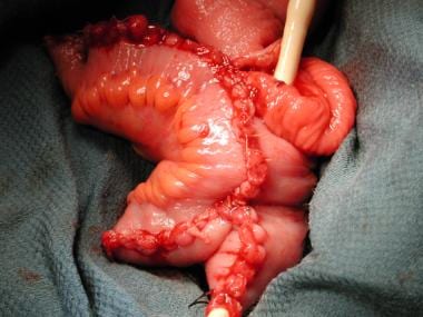
The bowel segment of the Indiana pouch includes the entire ascending colon, a small portion of the transverse colon, and about 7-8 cm of the terminal ileum.
Click to see larger pictureClick to see detailView Full Size Image
Media type:  Photo

Media file 8:  The colon has been opened on its antimesenteric border, and the terminal ileum has been plicated by application of the gastrointestinal anastomosis (GIA) stapler to reduce the lumen to approximately 16F. A 14F catheter is shown traversing the efferent limb. The ileocecal valve also has been bolstered with 2-0 absorbable sutures.
Click to see larger pictureClick to see detailView Full Size Image
Media type:  Photo

Media file 9:  Dissection of the lateral pedicle extending from the internal iliac vessels over the distal ureters. These vessels can be ligated, allowing full mobilization of the ureter down to the bladder.
Click to see larger pictureClick to see detailView Full Size Image
Media type:  Photo

Media file 10:  The posterior peritoneum has been entered, and the rectum is retracted to the right. The posterior plane between the bladder and prostate anteriorly and the rectum posteriorly can then be entered using a combination of sharp and blunt dissection; this defines the posterior pedicles.
Click to see larger pictureClick to see detailView Full Size Image
Media type:  Photo

Media file 11:  The rectum is retracted down and to the left, and the articulating stapler is applied to the posterior pedicle. Three such applications of the stapler ty