Background
With the widespread use of contemporary ultrasonographic techniques in the obstetric period, congenital hydronephrosis has become the most common initial presentation of ureteropelvic junction (UPJ) obstruction. [1] Prior to this, infants often presented with an abdominal mass, and children presented with abdominal pain, nausea, and vomiting. Hematuria and urinary tract infections are also not uncommon presenting symptoms.
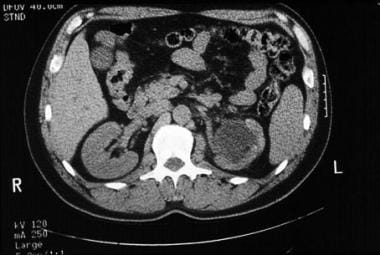
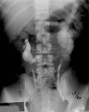
The application of radiographic studies, according to the pediatric urology paradigm, separates cases of congenital hydronephrosis due to UPJ obstruction from cases caused by other etiologies such as posterior urethral valves, megaureter, and vesicoureteral reflux. See the images below.
 CT scan without contrast demonstrating severe left-sided hydronephrosis secondary to ureteropelvic junction obstruction.
History of the Procedure
UPJ obstruction is the most common cause of prenatal hydronephrosis, accounting for 80% of the cases. Dismembered pyeloplasty has been the criterion standard surgical therapy for UPJ obstruction. In the 1980s, with the advent of percutaneous access to the kidney, antegrade endopyelotomy was developed as a less-invasive surgical therapy for UPJ obstruction. More recently, with rapid advancements in optics yielding smaller and more functional ureteroscopes, retrograde endopyelotomy has developed as an even more minimally invasive approach to treat UPJ obstruction. In addition, a retrograde approach using a balloon cutting catheter (Acucise) allows treatment of UPJ obstruction using only fluoroscopic guidance. The most recent advancement in treatment for UPJ obstruction is the development of the laparoscopic dismembered pyeloplasty.
Problem
Urologic texts define UPJ obstruction as an impediment to urinary flow from the renal pelvis into the ureter, which may result in symptoms or renal damage. The problem that clinicians face with UPJ obstruction is determining when poor drainage of the renal collecting system jeopardizes the future function of the affected kidney and the patient's overall health. The answer to this question drives the urological workup of potential UPJ obstruction and results in the application of surgical techniques to alleviate the obstruction and preserve renal function.
Epidemiology
Frequency
Obstruction of the UPJ is one of the most common congenital abnormalities of the urinary tract. Prior to the proliferation of prenatal ultrasonography, most cases of UPJ obstruction were not detected in the first year of life. The frequency of births with unilateral UPJ obstruction is estimated to be 1 case in 5000-8000 live births, and it is bilateral in 6% of these cases. Studies have indicated that UPJ obstruction is more common on the left side than on the right by a left-to-right ratio of 5:2 and that it is has a male-to-female ratio of 5:2. Another study found bilateral UPJ obstruction in 32 of 89 cases. This same study found that the diagnosis was made prenatally in 74 of 89 cases and that most patients diagnosed postnatally presented with an abdominal mass.
Currently, the vast majority of UPJ obstructions (90%) are detected with prenatal ultrasonography. As many as 80% of children identified with prenatal hydronephrosis have no signs or symptoms of their urologic abnormality after birth. An epidemiological study of 11,986 pregnant women who underwent prenatal ultrasound evaluations demonstrated that the overall frequency of congenital abnormalities is 0.5% and that urinary tract abnormalities represent 50% of these cases. [2] Furthermore, others have estimated that the etiology of prenatal hydronephrosis is UPJ obstruction in 50-67% of the cases. Taken together, this suggests that each mother who undergoes prenatal ultrasound examination has a 0.2-0.4% chance of having a neonate with hydronephrosis and these children have a 50% chance of eventually having the diagnosis of UPJ obstruction after birth.
UPJ obstruction is associated with a number of anomalies. A known association exists between UPJ obstruction and horseshoe kidneys. [3] UPJ obstruction has also been found to occur in 17 of 82 patients with ectopic kidneys. A strong relationship also exists between UPJ obstruction and nephrolithiasis. [4] One study found a 20% incidence rate of stones in patients with UPJ obstruction. Finally, no strong evidence exists indicating a hereditary pattern. Only one study has suggested that UPJ obstruction has a genetic etiology, [5] and thus, most cases are believed to be spontaneous in nature.
Etiology
The etiologies of UPJ obstruction are numerous and are classified on an anatomic basis as either extrinsic or intrinsic. Intrinsic causes are inherent to the development and anatomy of the UPJ itself, while extrinsic causes are exterior to the UPJ. Additionally, UPJ obstruction can be classified as primary and secondary. Primary UPJ obstruction is thought to be due to developmental anomalies of the UPJ, while secondary UPJ obstruction is due to other causes, including previous surgery, recurrent stone passage, or infection and vesicoureteral reflux.
Because the pathophysiology is enigmatic, the etiologies of UPJ obstruction are classified on an anatomic basis. Intrinsic etiologies are primarily due to insertional anomalies or functional abnormalities such as an aperistaltic section of smooth muscle. Studies have demonstrated abnormal collagen architecture within the stenotic UPJ.
Another intrinsic etiology that has been described is tissue valves forming mucosal folds and ureteral polyps that may be due to anatomical abnormalities. As first proposed in 1894 by Fenger, these anatomic anomalies may obstruct the ureter at the UPJ in a ball-valve fashion during periods of diuresis.
Insertional anomalies are described as cases in which the ureter inserts into the renal pelvis at a location that is not the most dependent. This prevents efficient drainage of the pelvis during periods of diuresis. However, whether this is the cause or the result of UPJ obstruction is not clear. Because the kidney is not fixed, except by its pedicle, UPJ obstruction can possibly cause dilation of the pelvis, resulting in distortion of the normal renal position and making the ureter appear to insert in an anomalous location. On the other hand, faulty embryogenesis of the renal unit may be the cause of the anomalous ureteral insertion. Some physicians have classified this cause as extrinsic because the pattern of volume-flow relationships across the UPJ is similar to the pattern of other extrinsic etiologies.
Extrinsic etiologies of UPJ obstruction result from lesions that are anatomically exterior to the UPJ itself. These include aberrant or accessory blood vessels, scars from previous surgery, scarring from nephrolithiasis or infection, and other secondary causes such as vesicoureteral reflux.
At the time of surgical repair, accessory vessels to the lower pole of the kidney or early branching of the segmental artery to the lower pole have been found to be associated with cases of UPJ obstruction. Because these vessels pass anterior to the ureter, distension of the pelvis during diuresis has been hypothesized to possibly cause the pelvis to displace anteriorly and hang over these vessels, which produces a kink in the ureter. This kink worsens as the process repeats itself, and the pelvis becomes progressively more hydronephrotic as the result of the developing UPJ obstruction.
Previous surgery, nephrolithiasis, or infections that cause scars and fibrosis around the UPJ and resultant obstruction may also be extrinsic etiologies that trigger UPJ obstruction by the formation of bands of tissue that compress the ureter. Differentiating this from an intrinsic cause may be difficult because a UPJ obstruction may feasibly develop adhesions and scars from the UPJ obstruction, the stone disease, and infections that may follow.
Pathophysiology
The cause of most intrinsic (or primary) UPJ obstruction likely relates to the embryological development of the urinary tract. During the fifth week of gestation, the ureteric bud forms from the wolffian duct and invades the metanephric blastema to begin renal differentiation. The nephrons, in turn, induce the ureteric bud to further divide and branch, leading to the formation of the collecting system (including the UPJ).
The aberrations of this developmental process must cause primary UPJ obstructions. For example, during development, the ureter is believed to become solid and then recanalize later. This is thought to occur mostly at the mid ureter. Incomplete recanalization has been speculated to possibly lead to UPJ obstruction. Additionally, smooth muscle differentiation begins in the bladder at 7 weeks' gestation and reaches the upper ureter by approximately the 16th week. An abnormality in smooth muscle development may lead to a section of ureter that does not appropriately contract and, thus, also to primary UPJ obstruction due to poor peristalsis.
The notion that vesicoureteral reflux can lead to progressive pelvic dilation and, eventually, UPJ obstruction, is logical. Although an association exists between UPJ obstruction and vesicoureteral reflux, no causal relationship has been identified. In a large retrospective review, no increased risk of UPJ obstruction was associated with all grades of vesicoureteral reflux. A 5-fold increased risk of UPJ obstruction existed in patients identified to have grade 5 reflux. The same review demonstrated that of patients with vesicoureteral reflux, only 3.6% also had UPJ obstruction. This small percentage does not support vesicoureteral reflux as a causative etiology of UPJ obstruction. Moreover, the same review demonstrated that when UPJ obstruction is repaired prior to the repair of vesicoureteral reflux, the UPJ obstruction does not recur. If vesicoureteral reflux were a causative etiology of UPJ obstruction, this would not be the case.
Presentation
UPJ obstruction occurs most often in children but may present in persons of any age. The clinical presentation of UPJ obstruction has changed dramatically because of the widespread proliferation of prenatal ultrasonography. Prior to this change, most children presented with an abdominal mass or urosepsis. In this age of ubiquitous prenatal ultrasound, the vast majority of patients with UPJ obstruction present with prenatal hydronephrosis.
Clinical symptoms of UPJ obstruction later in life include urosepsis, failure to thrive, flank pain or mass, and hematuria. As first described by Dietl, the episodes of flank pain, nausea, and vomiting may manifest during periods of rapid diuresis with large volumes of liquid intake (so-called Dietl crisis). [6] This may only manifest after drinking liquids that promote a brisk diuresis such as beer or coffee.
In adults, one study demonstrated that the most common presenting symptoms are flank pain (77%), nephrolithiasis (20%), microscopic hematuria (16%), history of pyelonephritis (14%), gross hematuria (9%), decreased renal function (9%), and gastrointestinal symptoms (5%). Hypertension may also be a rare presenting symptom.
Indications
Once the diagnosis of ureteropelvic junction (UPJ) obstruction has been made, management of depends on the severity of the case. Indications for dismembered pyeloplasty or any other operative therapy are variable. Most clinicians consider the presence of symptoms from the obstruction, such as recurrent flank pain, nausea, and vomiting, to be indications for interventions. Other indications include recurrent urinary tract infections, pyelonephritis, ipsilateral nephrolithiasis, and deterioration in renal function.
Contraindications
While performing an antegrade endopyelotomy, the inability to pass a guidewire through the strictured area is a contraindication to endopyelotomy. Additionally, a stricture longer than 2 cm is generally a contraindication to endopyelotomy.
Some authors believe a crossing vessel is a contraindication to endopyelotomy because success rates are reportedly lower and bleeding complications higher. However, others have challenged these contentions and believe that if the incision is performed in a true lateral position, endopyelotomy is safe and has an acceptable success rate even in the presence of a crossing vessel.
Uncorrected coagulopathy is a contraindication to surgical repair, and thus, referral to an internist or hematologist would be appropriate before undertaking surgical treatment.
-
CT scan without contrast demonstrating severe left-sided hydronephrosis secondary to ureteropelvic junction obstruction.
-
Excretory urogram shows a horseshoe kidney with left hydronephrosis.


