Practice Essentials
In 1858, Wallmann first reported on colloid cysts. In 1921, Dandy accomplished the first successful resection of a colloid cyst through a transcortical-transventricular approach. Less invasive techniques were later developed to reduce the complications associated with the open approach. Freehand aspiration of a colloid cyst was performed first by Gutierrez-Lara et al, [1] and the first stereotactic aspiration of a colloid cyst was performed by Bosch et al. [2] Endoscopic aspiration of a colloid cyst was reported by Powell [3] and has progressively gained consensus as a safe alternative treatment. [4, 5, 6, 7, 8, 9, 10, 11, 12, 13]
Colloid cysts are nonneoplastic epithelium-lined cysts of the central nervous system that almost always arise from the anterior third ventricle roof (immediately posterior to the foramen of Monro). These epithelium-lined, mucin-containing cysts can be found in asymptomatic patients; however, depending on their location, size, and degree of cerebrospinal fluid (CSF) obstruction, patients may present with a variety of neurologic symptoms. The effects can range from headache to, on rare occasions, death when there is acute hydrocephalus. [14, 15]
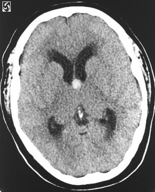
(The image below depicts a colloid cyst at the foramen of Monro causing hydrocephalus.)
Testing
Routine preoperative studies, including a CBC count, chemistry panel, and coagulation studies are performed in addition to various imaging modalities. Lumbar puncture is typically contraindicated in patients with these lesions because of a risk of cerebral herniation in patients who demonstrate noncommunicating hydrocephalus.
The CT scan is an important preoperative study because the viscosity of the cyst contents correlates more closely to the radiodensity visible on a CT scan than to the density visible on MRI. The size of the cysts varies, but most are 5-25 mm. [16] On MRI, the most common appearance is hyperintensity on T1-weighted images and hypointensity on T2-weighted images. The variable MRI signals do not correlate with the fluid density of cyst contents, although MRI is valuable in differentiating a colloid cyst from a basilar tip aneurysm. [17, 18, 19, 16, 19]
Etiology
The etiology of this tumor is still a source of debate. In 1910, Sjovall hypothesized that colloid cysts were remnants of the paraphysis, an embryonic midline structure within the diencephalic roof immediately rostral to the telencephalic border. The cells of the paraphysis are similar to those found in colloid cysts (ie, low columnar epithelial cells without cilia or blepharoplasts). These cysts were called paraphysial cysts for 50 years; however, several reports have been written about colloid cysts found in other locations, including the posterior third ventricle, the fourth ventricle, the septum, and rarely the frontal lobe, cerebellum, and pontomesencephalon. [20]
The origin of colloid cysts continues to be uncertain. Diencephalic ependyma, invagination of neuroepithelium of the ventricle, and the respiratory epithelium of endodermal origin are other etiologic possibilities. One leading theory is that colloid cysts form when ectopic endodermal elements migrate into the velum interpositum during central nervous system embryonic development. The fact that they are rarely seen in children indicates that they must enlarge with time. The cyst wall is lined with a mixed array of epithelial and goblet cells secreting proteinaceous mucinous fluid and may be responsible for the increase in size of the cyst. In addition, cyst cavities may be filled with blood degradation products such as cholesterol crystals.
Treatment
The most common indication for surgery is hydrocephalus associated with a colloid cyst. This usually occurs in the setting of a large cyst that obstructs the foramen of Monro. A more difficult clinical situation occurs when patients present with few or no symptoms and have small colloid cysts and large ventricles. In many cases, these patients may be managed conservatively and observed with serial MRIs, but they should be counseled about the potential symptoms of hydrocephalus. Lastly, patients who have small cysts and normal-sized ventricles are not likely to deteriorate and do not need surgery. If patients are too ill to tolerate surgical resection, then CSF diversion, often requiring bilateral shunts, may be considered. This situation is suboptimal because sudden death has been reported in the absence of acute obstructive hydrocephalus. The surgical approaches of colloid cysts commonly include endoscopy or open microsurgery. Endoscopic techniques have been found to reduce morbidity, including lower rates of infection, infarcts, and seizure, as well as decrease the rate of shunt dependency. The microsurgical approach has been considered the gold standard for treatment because complete resection can be achieved. [21, 22, 23, 24, 25, 26, 27, 28]
Prognosis
Sudden death associated with colloid cysts has been reported, but the risk of sudden death does not seem to correlate with tumor size, degree of ventricular dilatation, or duration of symptoms. Fortunately, the incidence of sudden death appears to be low; therefore, prevention of sudden death should not be used as an indication for surgery in asymptomatic patients with small cysts and no hydrocephalus.
Colloid cysts are usually cured after successful aspiration and complete resection. Hydrocephalus may develop despite removal of the cyst, and periodic CT scans should be performed. Preoperative function partly determines patient outcome; most patients tolerate resection well.
Epidemiology
The estimated incidence of colloid cysts is about one per one million, and colloid cysts account for 0.5-1% of all intracranial tumors. [29]
Eighty percent of the patients with colloid cyst reported in the literature are aged 30-60 years. Approximately 15-20% of all intraventricular masses are colloid cysts. Colloid cysts develop in the rostral aspect of the third ventricle in the foramen of Monro in 99% of cases, and despite their benign histology, they may carry high risks and neurologic complications, with a mortality reported from 3.1% to 10% in symptomatic cases or 1.2% in total. [14, 30]
Although these tumors are considered congenital, their presentation in childhood is rare (the youngest reported case involved a 2-month-old infant). The increased use of CT and MRI has resulted in an increased number of patients being diagnosed. No known genetic relationship has been determined, although familial occurrences of colloid cysts have been reported.
Presentation
Colloid cysts are often found incidentally, but when symptomatic, they present as obstructive hydrocephalus and paroxysmal headaches. These headaches are typically worse in the morning and may be exacerbated by leaning the head forward. Other symptoms are gait disturbances, short-term memory loss, nausea, vomiting, and behavioral changes. [30] Sudden weakness in the lower limbs associated with falls and without loss of consciousness (drop attacks) have been reported. Additionally, symptoms similar to normal-pressure hydrocephalus (eg, dementia, gait disturbance, urinary incontinence) have been associated with the presentation of colloid cysts.
In a study of 155 patients with newly diagnosed colloid cysts, Pollock et al described the following 4 factors associated with cyst-related symptoms [31] :
-
Younger patient age (44 yr vs 57 yr)
-
Cyst size (13 mm vs 8 mm)
-
Ventricular dilation (83% vs 31%)
-
Increased cyst signal on T2-weighted MRI (44% vs 8%)
The most significant variable of these was ventriculomegaly. For patients with enlarged ventricles, patient age (≤50 yr vs >50 yr) was the most important variable because patients aged 50 years or younger with enlarged ventricles were not affected by cyst size.
On rare occasions, a colloid cyst may obstruct the foramen of Monro completely and irreversibly, resulting in sudden loss of consciousness and, if patients are not treated, coma and subsequent death due to herniation. An alternative theory suggests that sudden death in patients with colloid cysts may be related to acute neurogenic cardiac dysfunction (secondary to the acute hydrocephalus) and subsequent cardiac arrest rather than herniation. The risk of sudden death remains difficult to predict. The presence of symptoms may be useful, as one study found that 8% of asymptomatic patients with a colloid cyst of the third ventricle eventually became symptomatic, [31] whereas another study found that 34% of symptomatic patients presented to a hospital with acute deterioration and in some cases sudden death. [32]
Cyst size and extent of ventricular dilatation do not seem to predict for acute deterioration. [33] In a study of 163 patients diagnosed with a colloid cyst of the third ventricle, Beaumont et al demontrated a strong correlation between the diameter of the cyst and the obstructive hydrocephalus. A similar correlation was also demostrated in this study between the hypersignal on FLAIR (fluid-attenuated inversion recovery) images of the cyst and the obstructive hydrocephalus. [14]
-
Axial CT scan that shows a colloid cyst with associated hydrocephalus.
-
Coronal MRI shows a colloid cyst in the roof of the third ventricle. The patient has mild hydrocephalus.
-
Intraoperative photograph through the operating microscope shows a colloid cyst in the Monro foramen. Choroid plexus is observed overlying the cyst, and the thalamostriate vein is along the inferior border.
-
Intraoperative photograph that shows removal of the cyst, leaving a dilated Monro foramen. The third ventricle can be seen through the opening.


