Practice Essentials
Lesions of the spine can be either extradural or intradural, with extradural lesions being located outside of the surrounding dural sac and intradural lesions being within the dural sac. Intradural lesions, furthermore, can be intramedullary or extramedullary, with intramedullary lesions being located within the spinal cord and extramedullary lesions being external to the spinal cord. [1, 2]
Intradural spinal cord tumors are uncommon lesions and fortunately affect only a minority of the population. However, when lesions grow, they result in compression of the spinal cord, which can cause limb dysfunction, cause motor and sensation loss, and, possibly, lead to death. Spinal tumors are classified on the basis of anatomic location as related to the dura mater (lining around the spinal cord) and spinal cord (medullary) as epidural, intradural extramedullary, and intradural intramedullary. Primary spinal tumors are typically intradural in location, whereas extradural spinal tumors are typically due to metastatic disease. [1]
The histopathologic types that account for 95% of intradural intramedullary neoplasms include astrocytomas, ependymomas, and hemangioblastomas. Spinal cord astrocytomas and ependymomas can be further classified as glial cell neoplasms. [3, 4, 5, 6, 7] Intramedullary spinal cord tumors account for approximately 2% of adult and 10% of pediatric central nervous system neoplasms. In adults, 85-90% of intramedullary tumors are the glial subtypes, astrocytoma or ependymoma. Ependymomas account for approximately 60-70% of all spinal cord tumors found in adults, while, in children, 55-65% of intramedullary spinal cord tumors are astrocytomas. Hemangioblastomas account for 5% of tumors, whereas paragangliomas, oligodendrogliomas, and gangliogliomas account for the remaining lesions. (See the image below.)
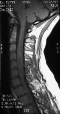 This T1-weighted sagittal MRI is from a 19-year-old man with 4-month history of progressive motor loss and an inability to ambulate. He underwent spinal biopsy that confirmed an intramedullary glioblastoma.
Pathophysiology and pathogenesis
The spinal cord consists of numerous nerve bundles that descend from and ascend to the brain. The spinal cord parenchyma consists of both gray (neurons and supporting glial cells) and white matter (axonal) and tracts that transmit electrical impulses between the brain and the body. These tracts, or circuits, control posture, movement, sensation, and autonomic system function, including bowel, bladder, and sexual function. Neurologic dysfunction develops as the spinal cord tumors enlarge and compress adjacent healthy neural tissue, disrupting these pathways. Upon further compression, patients can lose complete motor function and sensation below the lesion. In addition to weakness and sensory loss, patients may experience pain, particularly at night. This pain is believed to be related to disturbances in venous outflow by the tumor, causing engorgement and swelling of the spinal cord.
The pathogenesis of spinal neoplasms is unknown, but most arise from normal cell types in the region of the spinal cord in which they develop. A genetic predisposition is likely, given the higher incidence in certain familial or syndromic groups (neurofibromatosis). Astrocytomas and ependymomas are more common in patients with neurofibromatosis type 2, which is associated with an abnormality on chromosome 22. In addition, spinal hemangioblastomas can develop in 30% of patients with von Hippel-Lindau syndrome, which is associated with an abnormality on chromosome 3.
Imaging
Observation with serial imaging studies over a variable period is a treatment option for patients who pose a high surgical risk, who are elderly, and/or who only have minimal neurologic signs. MRI is the most accurate and noninvasive technique for imaging the spine and is the imaging modality of choice. Gadolinium (contrast) requires evaluation of kidney function because cases of malignant fibrosis have been reported. General findings include enlargement of the spinal cord and syringomyelia or cystic cavity associated within the lesion. Patients in whom pathology tissue shows a malignant neoplasm may be best treated with radiotherapy because they are expected to have an accelerated deterioration and complete surgical resection is not possible.
Treatment
Pharmacologic treatment of intramedullary spinal cord tumors is of limited benefit. High-dose intravenous corticosteroid therapy may improve neurologic function transiently but is not appropriate for long-term treatment. Optimal treatment options depend on the patient's clinical symptoms and neurologic findings. When and whether to treat these lesions, as well as perform radiosurgery or surgical excision of lesions, remains controversial. However, cures have been reported only after complete surgical resection. Therefore, patients with neurologic symptoms and confirmatory findings from imaging studies may benefit most from surgical excision, with the surgical goal of total gross resection of the lesion. [8, 9, 10, 11]
Intraoperative neuromonitoring has been shown to be of clinical importance during surgical resection. The primary monitoring modalities are somatosensory evoked potentials, transcranial motor evoked potentials via limb muscles or spinal epidural space (D-waves), and dorsal column mapping. [12, 9, 10]
Prognosis
Intramedullary spinal cord neoplasms or tumors are typically histopathologically "benign" or slow growing. However, patients can have more aggressive neoplasms as well as morbidity due to the location of the lesion. Consequently, compared with similar intracranial neoplasms, patients may have a prolonged survival after diagnosis. Primary spinal astrocytomas are frequently high-grade tumors with a poor prognosis and a high rate of postoperative neurologic impairment. [13, 14, 11]
The 5-year survival rate for patients with benign or low-grade spinal cord neoplasms is greater than 90%. In Brotchi's series of 239 patients with low-grade spinal tumors and operative intervention, 5% worsened, 50% stabilized, and 40% improved. [15]
Khalid et al retrospectively assessed survival in histologically confirmed, intramedullary spinal cord astrocytomas in patients 18 years of age and older using the Surveillance, Epidemiology, and End Results (SEER) database, and found that older age, WHO grade IV classification, tumor invasiveness, and subtotal resection were all associated with a worse prognosis. [16]
Presentation
Patients with intramedullary glial spinal cord tumors (ie, ependymomas, astrocytomas) typically present with back pain referred from the level of the lesion, sensory changes, or worsening function. The symptoms can be of a long duration, since these lesions tend to grow slowly and typically have a benign histopathology. Patients with low-grade astrocytomas tend to experience symptoms over a mean duration of 41 months. This is in contrast to patients with malignant astrocytomas, whose symptoms persist for a mean duration of only 4-7 months before diagnosis.
Tumor-specific characteristics
Ependymomas are associated with the following:
-
Mean age at presentation of 43 years
-
Slight female predominance
-
Pain localized to the spine (65%)
-
Pain worse at night or upon awakening
-
Dysesthetic pain (burning pain)
-
Long history of symptoms
-
Myxopapillary variant (mean age of presentation of 21 yr; slight male predominance)
Astrocytomas are associated with the following:
-
Equal male and female prevalence
-
Pain localized to spine
-
Pain worse at night or upon awakening
-
Paresthesias (abnormal sensation)
Hemangioblastomas are associated with the following:
-
Onset of symptoms by the fourth decade of life, 80% symptomatic by age 40 yr
-
Familial disorder (ie, von Hippel-Lindau syndrome) present in a third of patients
-
Decreased posterior column sensation
-
Back pain localized over lesion
Physical examination findings
Sensory findings include the following:
-
Decreased touch, pain, and/or temperature sensation
-
Hyperesthesias
-
Decreased proprioception (inability to localize limbs in space)
-
Abnormal sensation below the level of lesion
-
Abnormal sensation only at level of lesion (suspended level)
Hyperreflexia findings include the following:
-
Hoffman sign for cervical lesions
-
Clonus
-
Extensor plantar response (Babinski sign)
Other findings include the following:
-
Motor weakness (late finding)
-
Spasticity
-
Increased tone
-
Muscle atrophy (late finding)
Relevant Anatomy
Arterial
Understanding the normal spinal cord vascular supply is essential to treating intramedullary spinal cord lesions, specifically because these vessels may have a variable and inconsistent distribution.
The great vessels (aorta, carotids) contribute arterial supply to the spinal cord via segmental arteries, which further branch into medullary and radicular arteries. The radicular artery provides extramedullary blood supply to the nerve root and dura; the medullary artery bifurcates into anterior and posterior divisions to form the spinal arteries. One anterior and 2 posterior spinal arteries then transverse the longitudinal axis of the spinal cord and provide the blood supply to the spinal cord. Neoplasms acquire their blood supply by leaching blood from these vessels.
Venous
The venous plexus of the spinal column, termed the Batson plexus, is unlike other venous systems in the body because the veins do not contain valves. Therefore, blood can have pathologic retrograde flow. This retrograde flow blood can back up and cause venous congestion. This can manifest as venous hypertension. Because oxygenated blood cannot pass through the spinal cord because of the congestion of outflow, patients present with progressive neurologic dysfunction.
Spinal cord
The spinal cord parenchyma consists of a central canal surrounded by an H-shaped gray matter region that contains neurons. Outer myelinated nerve tracts, termed white matter, surround the central gray matter. The central canal represents an embryologic remnant from neurulation of the neural plate and is lined with ependymal cells. Ependymomas arise from these cells and, therefore, are typically located centrally in the spinal cord parenchyma. In contrast, astrocytes support gray matter neurons and white matter axons. Neoplastic transformation of these supporting cells results in the development of astrocytomas and may occur almost anywhere within the cord.
-
This T1-weighted sagittal MRI is from a 19-year-old man with 4-month history of progressive motor loss and an inability to ambulate. He underwent spinal biopsy that confirmed an intramedullary glioblastoma.
-
This T2-weighted MRI is from a 19-year-old man with 4-month history of progressive motor loss and an inability to ambulate. He underwent spinal biopsy that confirmed an intramedullary glioblastoma.
-
This 28-year-old man presented with progressive tetraplegia. T2-weighted sagittal MRI illustrates an intramedullary ependymoma, confirmed with pathologic evaluation. Note the cysts at the cranial (top) and caudal (bottom) of the tumor.
-
Postoperative axial MRI (patient from Image 3) at distal region of resection. A complete or gross total resection was obtained and confirmed with postoperative MRI. Note the cyst cavity has collapsed and the spinal cord is significantly atrophied. Despite the small size of the spinal cord, the patient experienced significant improvement in his neurologic function postoperatively.


