Continually Updated Clinical Reference
 
 
  All Sources     eMedicine     Medscape     Drug Reference     MEDLINE
 
eMedicine - Paraphimosis : Article by

Quick Find
Authors & Editors
Introduction
Indications
RELEVANT ANATOMY
Contraindications
Treatment
Complications
Outcome And Prognosis
Acknowledgments
Multimedia
References


Patient Education
Men's Health Center

Foreskin Problems Overview

Foreskin Problems Causes

Foreskin Problems Symptoms

Foreskin Problems Treatment

Circumcision Overview

Circumcision Treatment


Author: Jeffrey M Donohoe, MD, Assistant Professor of Pediatric Urology, Department of Surgery, Division of Urology, Medical College of Georgia

Jeffrey M Donohoe is a member of the following medical societies: American Academy of Pediatrics and American Urological Association

Coauthor(s): Hye Kim, RPh, Investigational Drug Pharmacist, Pharmacy Services, Johns Hopkins Hospital; James A Brown, MD, FACS, Associate Professor, Department of Surgery, Medical College of Georgia; Consulting Staff, Head of Urologic Oncology, Veterans Affairs Medical Center

Editors: Allen Donald Seftel, MD, Department of Urology, Associate Professor, Case Western Reserve University; Francisco Talavera, PharmD, PhD, Senior Pharmacy Editor, eMedicine; Shlomo Raz, MD, Professor, Department of Surgery, Division of Urology, University of California at Los Angeles School of Medicine; J Stuart Wolf, Jr, MD, FACS, David A Bloom Professor of Urology, Director, Division of Minimally Invasive Urology, Department of Urology, University of Michigan Medical Center; Stephen W Leslie, MD, FACS, Founder and Medical Director of the Lorain Kidney Stone Research Center, Clinical Assistant Professor, Department of Urology, Medical College of Ohio

Author and Editor Disclosure

Synonyms and related keywords: painful swelling of prepuce, swollen foreskin, prepuce swelling, phimosis, penile pain, painful penis, infected penis, foreskin, prepuce, edematous prepuce, manual compression

Paraphimosis is an uncommon condition in which the foreskin, once pulled back behind the glans penis, cannot be brought down to its original position, thus constituting one of the few urologic emergencies encountered in general practice (see Image 1). Like phimosis, paraphimosis occurs only in uncircumcised or partly circumcised males.

Paraphimosis is different from phimosis, a nonemergency condition in which the foreskin cannot be pulled back behind the glans penis.

Patients with mild forms of paraphimosis have excellent outcomes, but severe paraphimosis can lead to dire consequences; therefore, view paraphimosis as a urologic emergency.

Paraphimosis may occur when the foreskin has been pulled back behind the head of the penis for an extended period and is often caused by well-meaning health professionals secondary to penile examination or urethral instrumentation.

When paraphimosis is suspected, immediately obtain a urology consult for proper evaluation and diagnosis. Prompt attention and treatment of this emergency should lead to a favorable outcome.

Frequency

Paraphimosis is a relatively uncommon condition and occurs less often than phimosis.

Paraphimosis is almost always an iatrogenically or inadvertently induced condition. The condition occurs more often in hospitals and nursing homes than in the private community. In the private community, either the affected individual or a parent often retracts the prepuce and then inadvertently leaves it in its retracted position. In most cases, the foreskin reduces on its own and therefore precludes paraphimosis; however, if the slightest resistance to retraction of the prepuce is present, leaving it in this state predisposes it to paraphimosis. As edema accumulates, the condition worsens.

According to the National Hospital Discharge Survey, a trend in the United States over the last 30-40 years has been toward noncircumcision. Circumcision rates, which were at an all-time high of 78-80% in the mid-to-late 1960s, decreased to 55-60% in 2003. With more uncircumcised individuals, paraphimosis has the potential to become more common. Because paraphimosis is a condition that is almost always iatrogenically or inadvertently induced, simple education and clarification of proper prepuce care to parents, the individuals themselves, and health care professionals may be all that is required to prevent this problem.

Etiology

Most frequently, paraphimosis occurs after retraction of the foreskin during detailed penile examination, cleaning, urethral catheterization, or cystoscopy. Being prudent with foreskin manipulation is the most important key in preventing paraphimosis.

Development of paraphimosis after catheterization is not uncommon. Before the insertion of a urethral catheter, a health professional retracts the foreskin to sterilely prepare and drape the glans penis. The retracted foreskin may be left in that manner for several hours to days. The failure to restore the prepuce to its original position sometimes leads to the development of paraphimosis.

A more unusual cause of paraphimosis is self-infliction, such as piercing with a penile ring into the glans. Paraphimosis secondary to erections has also been reported.

Pathophysiology

When the foreskin becomes trapped behind the corona for a prolonged period, a tight band of tissue forms around the penis. This constricting ring impairs blood and lymphatic flow from the glans penis and prepuce. The ensuing tissue ischemia and vascular engorgement cause painful swelling of the glans and prepuce and may eventually cause gangrene or autoamputation of the distal penis.

When diagnosed early, paraphimosis can be remedied easily with simple manual reduction in combination with other conservative measures.

Patients with severe paraphimosis that proves refractory to conservative therapy require a bedside emergency dorsal slit procedure to save the penis. At a later date, a formal circumcision can be performed in the operating room.

Clinical

Adult patients with symptomatic paraphimosis most often report penile pain. Children may report obstructive voiding symptoms.

On examination, the glans penis is enlarged and congested with a collar of edematous foreskin. A constricting band of tissue is noted directly behind the head of the penis (see Image 2). The remainder of the penile shaft is unremarkable. An indwelling urethral catheter is often present. Removing the catheter may help treat paraphimosis.

If paraphimosis is left untreated for too long, necrosis of the glans penis can occur. Partial amputation of the distal penis has been reported (see Image 3).

In the pediatric population, paraphimosis may manifest as acute urinary obstruction.


Indications for an emergency dorsal slit procedure include phimosis and paraphimosis that have proven refractory to more conservative measures.

Emergency dorsal slit procedures are generally reserved for severe or complex paraphimosis.

At a later date, a formal circumcision can be performed as an outpatient procedure. Prepuce-sparing procedures have been described and may be appropriate to perform if the individual must retain intact foreskin; however, the best way to ensure that paraphimosis will not recur is to perform circumcision.


The penis is divided into 3 parts.

  • The root of the penis lies under the pubic bone and provides stability when the penis is erect.
  • The body of the penis constitutes the major portion of the penis and is composed of 2 cavernosal bodies (ie, corpora cavernosa) and a corpus spongiosum (ie, head of the penis). The male urethra traverses through the corpus spongiosum and exits from the meatus. The cavernosal bodies produce an erection when filled with blood.
  • The glans is the distal expansion of the corpus spongiosum usually covered by the loose skin of the prepuce in uncircumcised individuals. A collar of tissue immediately behind the glans penis is known as the coronal sulcus.

The penis is innervated by the left and right dorsal nerves (main sensory nerve supply), which are branches of the pudendal nerve.

The penis is a highly vascular organ supplied by the internal pudendal artery, which arises from the internal iliac artery and then branches into the deep penile artery, the bulbar artery, and the urethral artery.

The deep penile artery becomes the cavernosal arteries, which supply the entire corpus cavernosum. The urethral artery supplies the glans penis and the corpus spongiosum. The bulbar artery nourishes the bulbar urethra and the bulbospongiosus muscle.


Do not consider circumcision in a neonate with hypospadias, a dorsal hood deformity, or a small penis. Refer the neonate to a urologist.


Medical therapy

Medical therapy involves reassuring the patient, reducing the preputial edema, and restoring the prepuce to its original position and condition. Several methods of reducing the penile swelling have been described. Ice packs, penile wraps, and manual compression mechanically disperse the penile and preputial edema, while granulated sugar1 and hyaluronidase2 have been reported as effective agents to reduce swelling. If a Foley catheter is present, remove it temporarily until the paraphimosis is resolved.

The author has never used any management options other than gentle reassurance, local anesthesia (1% lidocaine without epinephrine), intravenous sedation with morphine or midazolam (Versed) (in select cases), and then manual compression with simultaneous manual reduction. The author identifies the location of the cicatricial ring, reduces the edema distal to it, and, as quickly and efficiently as possible, reduces the cicatricial ring with one hand while the second hand has been compressing the glans and forcing out the edema. After 2 or 3 solid attempts, the author resorts to a dorsal slit procedure as described in Surgical therapy.

Several other methods to effectively reduce the glanular and prepucial edema prior to reduction of the foreskin have been described in the literature. Some of these methods are described are as follows:

  • Wrap the penis in plastic and apply ice packs.
  • Use compressive elastic dressings.
  • Apply direct circumferential manual compression. (Application of a topical anesthetic such as 2% lidocaine gel or eutectic mixture of local anesthetics cream [2.5% prilocaine, 2.5% lidocaine] to the penile skin a few minutes to an hour before penile manipulation reduces pain and helps patients, particularly children, tolerate the procedure.)
  • Apply granulated sugar to the surface of the edematous prepuce, and cover it with a condom or a finger of a rubber glove. The hypotonic fluid from the swollen foreskin moves down the osmotic gradient into the hypertonic sugar, which results in a reduction of the preputial edema. This treatment is based on the principle that fluid transfer occurs via an osmotic gradient.
  • Using a tuberculin syringe, inject 1 mL of hyaluronidase (150 U/cc Wydase) directly into several sites of the edematous prepuce. Hyaluronidase breaks down hyaluronic acid in connective tissue and enhances fluid diffusion between tissue planes, decreasing preputial swelling and resulting in almost immediate resolution of the edema. (The use of hyaluronidase in the pediatric population has been well documented.)

Regardless of the method chosen, when the preputial swelling and edema have subsided, correct the paraphimosis by gentle manual reduction (see Image 4).

To reposition the prepuce, place both thumbs on the glans penis and wrap the fingers behind the prepuce. Apply a gentle, steady pressure to the prepuce with counterpressure to the glans penis as the prepuce is pulled down.

When performed properly, the constricting band of tissue should come down to completely cover the glans with the prepuce. If the prepuce comes down but the constricting band remains behind, the paraphimosis has not been reduced properly or sufficiently.

In patients who are determined to retain the appearance of an uncircumcised phallus, the author has the patient apply triamcinolone cream 0.1% to the affected area to possibly reduce the fibrosis of the ring. This has been described in the treatment for phimosis and has proven efficacy in temporarily preventing recurrent phimosis, decreasing the need for circumcision. After 6 weeks of triamcinolone application, if the prepuce can easily be retracted and reduced, the patient may proceed as such, but the risk for recurrent phimosis and paraphimosis remains. More often than not, the author ultimately performs circumcision.

Surgical therapy

The puncture technique,3, 4 a minimally invasive procedure, and blood aspiration are common therapies to decompress the edematous prepuce.

To perform the puncture technique, use an 18- or 21-gauge hypodermic needle to puncture the edematous prepuce at multiple sites and release the trapped fluid (see Image 5). External drainage results in rapid resolution of edema, which is followed by manual reduction of the foreskin.

Alternatively, blood aspiration of the penis may be attempted after a tourniquet has been applied.

If a severely constricting band of tissue precludes all forms of conservative or minimally invasive therapy, an emergency bedside dorsal slit procedure may be performed, followed by a delayed circumcision.

The penis is prepared and draped. The cicatricial ring is identified. An assistant may be needed to retract the edematous glans away from the incision site. The ring is crushed with a hemostat at the 12-o'clock position and then cut with scissors. The entire ring must be incised. It is usually no more than 5-10 mm thick. Once this is done, the prepuce can easily be reduced. Sutures, if necessary, are placed to control bleeding from the cut edges of the ring. Long-term management is discussed with the patient. Options include surgical treatment via circumcision or conservative therapy via application of triamcinolone cream for 6 weeks and watchful waiting.

Circumcision is the definitive therapy for paraphimosis.

Preoperative details

Obtaining a properly informed consent before a circumcision is performed is critical. Inform patients, parents, and/or caregivers of the potential risks of bleeding, infection, suture disruption, urethral injury, and too much or too little skin being removed. Also inform patients that circumcision does not affect the length or girth of the penis.

Instruct patients to abstain from genital stimulation for up to 6 weeks after surgery. Inadvertent erections can strain suture lines and cause incisions to break down.

Patients having circumcisions for recurrent balanitis should be free of infection before the procedure.

Intraoperative details

Place the patient in a supine position. Preparation includes a thorough surgical scrub of the genital area with povidone-iodine solution. Shaving or clipping pubic hair is unnecessary.

Either general or local anesthetics may be used. General anesthesia is recommended for children, while local anesthesia is recommended for adults.

Local anesthesia can be accomplished by a dorsal penile nerve block with a ring block. Equal volumes of 0.5% bupivacaine (Marcaine) and 1-2% lidocaine (Xylocaine) without epinephrine are commonly used. The maximum recommended dose of lidocaine without epinephrine is 4.5 mg/kg or 315 mg for a 70-kg male.

  • Dorsal penile block: Insert a short 25-gauge needle anterior to the pubic arch at the 10-o'clock position until the Buck fascia is encountered. Insert the needle through the Buck fascia, but remain outside of the corporal bodies. Aspirate to make sure the needle is not in a corporal body. Inject 10 mL of 1% lidocaine solution. Repeat the process at the 2-o'clock position.
  • Ring block: Insert a short 25-gauge needle at the base of the penis until the Buck fascia is encountered. Remain outside of the corporal bodies. Inject the anesthetic into the Buck fascia circumferentially around the base of the penis.
  • A combination of dorsal penile and ring blocks should provide adequate local anesthesia. If not, inject additional anesthetic directly into the incision line.

To perform a dorsal slit procedure for emergent reduction of paraphimosis, use the following technique:

  • Identify the constricting band of prepuce.
  • Place 2 straight hemostats at the 12-o'clock position to crush the constricting foreskin (see Image 6).
  • After 1 full minute, sharply incise the crushed tissues between 2 hemostats (see Image 7).
  • In running fashion, close each edge of the incision with 4.0-5.0 absorbable sutures (children) or 3.0-4.0 absorbable sutures (adults) (see Image 8).

When performing a circumcision, either the dorsal slit or sleeve techniques are commonly used. The dorsal slit technique is preferred for patients with severe phimosis and those with paraphimosis in whom intraoperative manipulation of the foreskin is difficult.

To perform the dorsal slit, use the following technique:

  • Sharply incise the foreskin at the 12-o'clock position, perpendicular to the corona.
  • With scissors, excise the foreskin at its base.
  • Ligate superficial veins.
  • Use electrocautery to obtain hemostasis.
  • Circumferentially approximate the proximal and distal edges of the foreskin with 4.0-5.0 absorbable sutures in an interrupted fashion (see Image 9).

The sleeve technique is an alternative method for circumcision and may be used as a primary surgical procedure or as a secondary definitive operation after an emergency dorsal slit has been made at bedside.

  • After administering anesthesia, outline the redundant foreskin using a surgical marker.
  • Incise the proximal and distal skirts of the foreskin so that a sleeve of foreskin is present between the incisions (see Image 10).
  • Remove the sleeve using electrocautery (see Image 11).
  • Use electrocautery and ligatures to achieve adequate hemostasis before circumferentially approximating the edges of the foreskin with 3.0-5.0 absorbable sutures in an interrupted fashion (see Image 12).

Postoperative details

After the circumcision, petroleum jelly and sterile gauze or petrolatum gauze dressings may be applied over the sutures, followed by a sterile white gauze dressing. Prescribe oral narcotics and discharge the patient. Some surgeons prescribe oral antibiotics as well.

Remove the dressing 24-48 hours after surgery. Advise patients to wear loose-fitting clothes, to gently wash the wound daily for the next 5-7 days, and to refrain from any sexual activity for the next 6 weeks to prevent breakdown of the sutures and incision line. Some surgeons additionally recommend keeping the wound completely dry to avoid inadvertent infection of the suture line.

One ampule of amyl nitrate may be used in instances of postoperative erections.

Follow-up

Patients generally have a follow-up examination in 2-3 weeks to check the wound. Assess the wound for signs of infection and inspect the suture line.

For excellent patient education resources, visit eMedicine's Men's Health Center. Also, see eMedicine's patient education articles Foreskin Problems and Circumcision.


Complications of paraphimosis include pain, infection, and swelling of the glans penis. The distal portion of the penis can become ischemic and even necrotic.

Potential complications involved with any circumcision include bleeding, infection, shortening of penile skin, and urethral injury.

Postoperative bleeding is the most common complication. Meticulous hemostasis during the initial surgery is the rule. Bleeding may occur if a scab is pulled off during removal of the dressing. This bleeding can often be controlled with direct pressure. Rarely, electrocautery or ligature may be required.

Infection after circumcision is uncommon. If acquired, it may be treated with oral antibiotics.

Urethral injury is extremely rare.


Paraphimosis does not recur after a proper circumcision.

Outcome after a dorsal slit procedure or a circumcision is excellent. Sometimes, patients with a favorable outcome from dorsal slit procedures decline circumcision.


The authors and editors of eMedicine gratefully acknowledge the contributions of previous author, Jong M. Choe, MD, FACS, to the development and writing of this article.


Media file 1:  
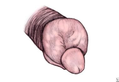
Illustration of paraphimosis. The foreskin is swollen and edematous. A constricting collar or band is present behind the glans penis.
Click to see larger pictureClick to see detailView Full Size Image
Media type:  Image

Media file 2:  Mild-to-moderate form of paraphimosis. The treatment involves manual reduction, puncture technique, or medical therapy.
Click to see larger pictureClick to see detailView Full Size Image
Media type:  Photo

Media file 3:  Severe form of paraphimosis. The distal penis has begun the process of autoamputation.
Click to see larger pictureClick to see detailView Full Size Image
Media type:  Photo

Media file 4:  This demonstrates the technique of manually reducing the paraphimotic foreskin.
Click to see larger pictureClick to see detailView Full Size Image
Media type:  Photo

Media file 5:  The puncture method to relieve preputial edema resulting from paraphimosis. Using a needle, several punctures are made in the foreskin to relieve the trapped fluid.
Click to see larger pictureClick to see detailView Full Size Image
Media type:  Image

Media file 6:  The dorsal slit technique to treat paraphimosis is begun by crushing the foreskin at the 12-o'clock position with 2 straight hemostats. A sharp incision is made between the hemostats.
Click to see larger pictureClick to see detailView Full Size Image
Media type:  Photo

Media file 7:  In this case, a dorsal slit procedure to treat paraphimosis has been performed halfway. Note that the edges of the incision have been oversewn with absorbable sutures.
Click to see larger pictureClick to see detailView Full Size Image
Media type:  Photo

Media file 8:  A finished product of the dorsal slit procedure to treat paraphimosis. Note that the cosmetic appearance of the completed dorsal slit is similar to that of a finished circumcision.
Click to see larger pictureClick to see detailView Full Size Image
Media type:  Photo

Media file 9:  To remove the foreskin completely after a dorsal slit procedure to treat paraphimosis, a circumcision is performed. The circumcision is begun, circumscribing proximal and distal skin incisions to create a sleeve of excess foreskin that is removed. This sleeve of excess foreskin is crushed at the 12-o'clock position for hemostasis.
Click to see larger pictureClick to see detailView Full Size Image
Media type:  Photo

Media file 10:  During circumcision, the foreskin is sharply incised between 2 hemostats.
Click to see larger pictureClick to see detailView Full Size Image
Media type:  Photo

Media file 11:  Excess foreskin is removed during a circumcision. The shaft of the penis is displaced downward with a sponge while the foreskin is peeled off using electrocautery.
Click to see larger pictureClick to see detailView Full Size Image
Media type:  Photo

Media file 12:  After the excess foreskin has been removed by circumcision, the preputial skin edges are reapproximated with absorbable sutures. The operation is completed.
Click to see larger pictureClick to see detailView Full Size Image
Media type:  Photo


  1. Cahill D, Rane A. Reduction of paraphimosis with granulated sugar. BJU Int. Feb 1999;83(3):362. [Medline].
  2. Litzky GM. Reduction of paraphimosis with hyaluronidase. Urology. Jul 1997;50(1):160. [Medline].
  3. Finkelstein JA. "Puncture" technique for treating paraphimosis. Pediatr Emerg Care. Apr 1994;10(2):127. [Medline].
  4. Hamdy FC, Hastie KJ. Treatment for paraphimosis: the 'puncture' technique. Br J Surg. Oct 1990;77(10):1186. [Medline].
  5. Baigrie RJ. Treatment for paraphimosis. Br J Surg. Mar 1991;78(3):378. [Medline].
  6. Fuenfer MM, Najmaldin A. Emergency reduction of paraphimosis. Eur J Pediatr Surg. Dec 1994;4(6):370-1. [Medline].
  7. Gausche M. Genitourinary surgical emergencies. Pediatr Ann. Aug 1996;25(8):458-64; quiz 465-7. [Medline].
  8. Hansen RB, Olsen LH, Langkilde NC. Piercing of the glans penis. Scand J Urol Nephrol. May 1998;32(3):219-20. [Medline].
  9. Higgins SP. Painful swelling of the prepuce occurring during penile erection. Genitourin Med. Dec 1996;72(6):426. [Medline].
  10. Hollowood AD, Sibley GN. Non-painful paraphimosis causing partial amputation. Br J Urol. Dec 1997;80(6):958. [Medline].
  11. Jones SA, Flynn RJ. An unusual (and somewhat piercing) cause of paraphimosis. Br J Urol. Nov 1996;78(5):803-4. [Medline].
  12. Olson C. Emergency treatment of paraphimosis. Can Fam Physician. Jun 1998;44:1253-4, 1257. [Medline].
  13. Raveenthiran V. Reduction of paraphimosis: a technique based on pathophysiology. Br J Surg. Sep 1996;83(9):1247. [Medline].
  14. Samm BJ, Dmochowski RR. Urologic emergencies. Trauma injuries and conditions affecting the penis, scrotum, and testicles. Postgrad Med. Oct 1996;100(4):187-90, 193-4, 199-200. [Medline].

Paraphimosis excerpt

Article Last Updated: Aug 25, 2006