Continually Updated Clinical Reference
 
 
  All Sources     eMedicine     Medscape     Drug Reference     MEDLINE
 
eMedicine - Phimosis, Adult Circumcision, and Buried Penis : Article by

Quick Find
Authors & Editors
Introduction
Indications
Relevant Anatomy
Contraindications
Treatment
Complications
Outcome And Prognosis
Acknowledgments
Multimedia
References


Patient Education
Men's Health Center

Foreskin Problems Overview

Foreskin Problems Causes

Foreskin Problems Symptoms

Foreskin Problems Treatment

Circumcision Overview

Circumcision Treatment


Author: Richard A Santucci, MD, FACS, Chief of Urology, Detroit Receiving Hospital; Specialist-in-Chief of Urology, Detroit Medical Center; Chief of Urologic Trauma Surgery, Sinai Grace Hospital; Director, The Center for Urologic Reconstruction

Richard A Santucci is a member of the following medical societies: American College of Surgeons, American Urological Association, and Société Internationale d'Urologie (International Society of Urology)

Coauthor(s): Hye Kim, RPh, Investigational Drug Pharmacist, Pharmacy Services, Johns Hopkins Hospital; Ryan P Terlecki, MD, Staff Physician, Department of Urology, Wayne State University Health Center

Editors: Leonard Gabriel Gomella, MD, FACS, The Bernard W Godwin Professor of Prostate Cancer Chairman, Department of Urology, Associate Director of Clinical Affairs, Kimmel Cancer Center, Thomas Jefferson University; Francisco Talavera, PharmD, PhD, Senior Pharmacy Editor, eMedicine; J Stuart Wolf, Jr, MD, FACS, David A Bloom Professor of Urology, Director, Division of Minimally Invasive Urology, Department of Urology, University of Michigan Medical Center; Stephen W Leslie, MD, FACS, Founder and Medical Director of the Lorain Kidney Stone Research Center, Clinical Assistant Professor, Department of Urology, Medical College of Ohio

Author and Editor Disclosure

Synonyms and related keywords: phimosis, elective circumcision, foreskin, buried penis, concealed penis, inability to retract the foreskin, physiologic phimosis, pathologic phimosis, congenital phimosis, adherent prepuce, adherent foreskin, paraphimosis, inability to pull down the foreskin, infection of the foreskin, balanitis, infection of the head of the penis, balanoposthitis, penile carcinoma, diabetes mellitus, urinary tract infection, UTI, cystitis

Phimosis

Phimosis is defined as the inability of the prepuce (foreskin) to be retracted behind the glans penis in uncircumcised males.

Nearly all males are born with congenital phimosis, a benign condition that resolves in the overwhelming majority of infants as they transition into childhood. In 1949, Douglas Gairdner showed that only 4% of infants had a fully retractable foreskin at birth but that 90% did by age 3 years. Contemporary work demonstrates that only 1% of males aged 17 years still have an unretractable foreskin. Adult phimosis (ie, pathologic or true phimosis) may occur secondary to poor hygiene or an underlying medical condition (eg, diabetes mellitus).

Uncomplicated pathologic phimosis is usually amenable to conservative medical treatment. Failure of medical treatment warrants surgical intervention, usually in the traditional form of a circumcision or preputioplasty.

Although phimosis is the most common indication for adult circumcision, other reported indications include paraphimosis, balanitis without phimosis, condyloma, redundant foreskin, and patient choice.

Buried penis

Buried penis was described in the early 20th century as a penis of normal size that lacks an appropriate sheath of skin and is located beneath the integument of the abdomen, thigh, or scrotum. This condition is more common in children, usually presenting in neonates or obese prepubertal boys; however, it can also be seen in adults and has been observed in both circumcised and uncircumcised individuals. Marginal cases may not be diagnosed until adulthood, when increased fat deposition accentuates the problem.

Several classification systems of buried penis have been proposed, although none has been universally adopted in the literature. Maizels et al (1986) differentiated between the terms concealed (before circumcision), trapped (cicatricial [scarred] after circumcision), and buried (associated with adolescence and obesity).1

In most congenital pediatric cases, the buried penis is self-limited. In untreated adults, however, the condition tends to worsen as the abdominal pannus continues to grow.

History of the Procedure

Circumcision and adult circumcision

Circumcision is one of the earliest elective operations known to man. Historically, this procedure has been performed for a variety of religious reasons, social reasons, or both. The practice is considered a commandment in Jewish law and is considered a rule of cleanliness in Islam, although it is not mentioned in the Quran. In biblical times, mothers were responsible for circumcising their newborn sons, with mohels gradually taking over. Priests in ancient Egypt would perform the procedure with their gold-impregnated thumbnail. Female circumcision, which is likely better termed "genital mutilation," has been practiced for centuries by some cultures but is an unacceptable practice and without medical benefit.

Adult circumcision for phimosis is described in textbooks dating from the early 19th century. Alternative procedures for adult phimosis were described as early as 1900 by Cloquet. Surgical attempts to restore the prepuce are also well documented, going as far back as the Old Testament. However, potential psychological and surgical complications have led to closer scrutiny of routine neonatal circumcision. Currently, the American Academy of Pediatrics (AAP) neither recommends nor condemns routine neonatal circumcision.

Studies suggest that circumcised boys have a lower risk of urinary tract infections (UTIs). To put this in perspective, the approximate chance of a UTI occurring in the first year of life is 1 in 100 in uncircumcised boys and 1 in 1000 in circumcised boys. A lower risk of malignancy is also reported in studies of circumcised men, although the incidence is also rare in uncircumcised men. Of note, this decreased risk seems to be associated only with infant circumcision and not with adult procedures.

The theory that circumcision contributes to prevention of sexually transmitted diseases (STDs) was encouraged by a 19th-century report of lower rates of syphilis in Jewish men. Van Howe et al (1999), in a literature review, found no clear evidence that circumcision prevents STDs.2 Some researchers contend that the risk of HIV infection is higher in uncircumcised men. Other studies, however, show a higher risk in circumcised men, and still others show no difference between the two.

Surgical correction of buried penis

The first description of the buried penis was in 1919 by Keyes. The first attempted surgical correction of this problem was by Schloss in 1959, and in 1968, successful correction was performed in an adult by Glanz. Since that time, numerous techniques have been developed.

Problem

Phimosis

Phimosis is defined as a condition in which the foreskin cannot be retracted behind the head of the penis. Depending on the situation, this condition may be considered either physiologic or pathologic.

Physiologic, or congenital, phimosis is the normal condition of the newborn male. In 90% of cases, natural separation allows the foreskin to retract by age 3 years. However, phimosis persisting into late adolescence or early adulthood need not be considered abnormal.

The entity of pathologic, or true, phimosis is far less common and can be present in children or adults. This is associated with cicatricial scarring of the prepuce that is often white in appearance. Phimosis may occur after circumcision if redundant inner prepuce slides back over the glans, with subsequent cicatricial scarring and contraction.

Buried penis

Buried penis is a true congenital disorder in which a penis of normal size lacks the proper sheath of skin and lies hidden beneath the integument of the abdomen, thigh, or scrotum. The literature, on occasion, also refers to this condition as a hidden or concealed penis. Trapped penis is a condition in which the penis becomes inconspicuous secondary to a cicatricial scar, usually after overzealous circumcision. Webbed penis occurs when the penile shaft is obscured secondary to scrotal skin webs at the penoscrotal junction. Micropenis represents a penis less than 2 standard deviations below the mean in length when measured in the stretched state. Diminutive penis is seen when the penis is small, malformed, or both secondary to epispadias, exstrophy, severe hypospadias, chromosomal abnormalities, or intersex conditions.

Frequency

Phimosis

Nearly all males are born with physiologic phimosis. Data have shown that the foreskin is retractable in 90% of boys by age 3 years. Only 1% of boys have physiologic phimosis persisting until age 17 years. Thus, most healthy adult men should not have phimosis; presence of the disorder in an adult male should raise the suspicion of balanitis (infection of the foreskin), balanoposthitis (infection of glans and foreskin), diabetes, or malignancy.

Approximately 1 in 6 men in the world are circumcised. In the United States, circumcision is the fifth most common procedure, and in 1992, 62% of newborn males in the United States had their foreskins removed.

Buried penis

Congenital buried penis is an uncommon phenomenon. The incidence of buried penis in adulthood is unknown, but it is highly likely that many cases go unreported.

Etiology

Phimosis

Physiologic phimosis is the rule in newborn males. Formation of the prepuce is complete by 16 weeks' gestation. The inner prepuce and glans penis share a common, fused mucosal epithelium at birth. This epithelium separates by desquamation over time as the proper hormonal and growth factors are produced. Thus, neonatal circumcision is a surgical treatment of normal anatomy.

Pathologic, or true, phimosis has several different etiologies. The most common cause is infection, such as posthitis, balanitis, or a combination of the two (balanoposthitis). Diabetes mellitus may predispose to such infections.

Adult circumcision is most commonly done for phimosis. When circumcision is performed for phimosis, 25-46% of removed foreskins are histologically normal. Other indications for adult circumcision include balanitis xerotica obliterans (BXO), infection without phimosis; paraphimosis; Bowen disease; carcinoma; condylomas (warts); trauma; religious or social reasons; and personal preference.

Buried penis

Various etiologic factors have been proposed to explain congenital buried penis. Recent literature favors dysgenetic dartos tissue with abnormal attachments proximally and to the dorsal cavernosum. A prominent prepubic fat pad is also a common primary factor in addition to dysgenetic dartos fascia. Secondary buried penis may be the result of an overzealous circumcision with subsequent cicatricial scar (trapped penis), a large hernia, or a hydrocele.

Affected adults are commonly obese and often have a history of trauma or surgery. Adults with this condition may have undergone abdominoplasty with overzealous release of attachments between the Scarpa and dartos fasciae, penile-lengthening procedures, or other genitoinguinal surgery.

Pathophysiology

Phimosis

The foreskin of an uncircumcised child should not be forcefully retracted. This may result in significant bleeding, as well as glanular excoriation and injury. Consequently, dense fibrous adhesions may form during the healing process, leading to true pathologic phimosis.

Circumcision has been promoted as a means of reducing the risk of UTIs, which are more common in uncircumcised males younger than 6 months. The risk in circumcised infants is approximately 1 in 1000, whereas the risk in uncircumcised infants is about 1 in 100. Some researchers contend that the risk of UTI in these children is not high enough to warrant mandatory circumcision. Interestingly, nongonococcal urethritis (NGU) may be more common in circumcised men. A multicenter study suggests, however, that if a child has associated vesicoureteral reflux (VUR), the benefit of reduced risk of infection may be a valid reason for surgery.

Another cited indication for circumcision is prevention of STDs. Numerous case-control studies concerning the relationship between the foreskin and HIV infection have been published with inconsistent results and no definite link. Therefore, it seems that STD prevention is not a justification for routine circumcision. Of note, of the developed nations, the United States has one of the highest rates of STDs, HIV infection, and male circumcision.

Infant circumcision seems to decrease the risk of penile cancer, whereas later circumcision does not. Penile cancer is a rare disease in the United States, with an incidence of 1.5 per 100,000 people. In developing countries, the incidence is higher and accounts for up to 10% of malignancies in some African and South American nations. Although primarily a disease of older men, penile cancer has been reported in children. The lowest incidence has been reported in Jews, with a similar incidence in Muslims; both groups have high rates of neonatal circumcision.

Daling et al performed a population-based case-control study in 2005 that looked at the importance of circumcision in patients with penile cancer. Preputial status was not found to be a statistically significant factor in penile cancer. The investigators concluded that the role of circumcision in penile cancer prevention is unclear.3 Several studies suggest that poor hygiene may be a stronger risk factor than circumcision status. Although smegma has been implicated as a carcinogenic agent, definitive evidence is lacking.

Adult phimosis may be secondary to repeated episodes of balanitis or balanoposthitis. Such infections are commonly due to poor personal hygiene (failure to regularly clean under the foreskin).

Phimosis may be a presenting symptom of early diabetes mellitus. When the residual urine of a patient with diabetes mellitus becomes trapped under the foreskin, the combination of a moist environment and glucose in the urine may lead to a proliferation of bacteria, with subsequent infection, scarring, and eventual phimosis.

Buried penis

A properly formed penis is normally present by 16 weeks' gestation. Congenital buried penis is caused by a developmental anomaly in which the dartos fascia has not developed into the normal elastic configuration to allow the penile skin to move freely over the deeper tissues of the penile shaft. Instead, the dartos layer is inelastic, which prevents the forward extension of the penis and holds it buried under the pubis.

Other possible contributing factors to congenital buried penis include excess prepubic fat, scrotal webbing, deficient penile skin, loose skin, an abnormally low position at which the crura separate, abnormal attachments between the Buck fascia and the tunica albuginea, and insufficient attachment of dartos fascia and skin to the Buck fascia.

The pathophysiology of buried penis in adults differs from that in children and includes iatrogenically induced scar contracture with concurrent descent of the abdominal fat pad. Because the penis is suspended from the pubis by the suspensory ligament, it remains fixed, unlike the prepubic fat. As fat descends over the penis, excessive moisture and bacterial overgrowth may occur. Chronic infection may lead to skin maceration and more scar contracture, further aggravating the problem. In many children, this condition is self-limited. However, in adults, total body fat content typically increases with age, causing the buried penis to worsen with time.

Clinical

Phimosis

Congenital or physiologic phimosis is clinically asymptomatic and not a cause for concern. It is often associated with "ballooning" of the foreskin during voiding. This is a self-limited phenomenon that, in the absence of pathologic phimosis, does not indicate urinary obstruction. Pathologic, or true, phimosis is far less common. Symptoms include skin irritation, dysuria, bleeding, and occasionally enuresis or urinary retention. Physical examination usually reveals white cicatricial scarring at the preputial ring. Meuli et al (1994) devised the following scoring system to rate the severity of phimosis:4

  • Grade I - Fully retractable prepuce with stenotic ring in the shaft
  • Grade II - Partial retractability with partial exposure of the glans
  • Grade III - Partial retractability with exposure of the meatus only
  • Grade IV - No retractability

Pathologic phimosis may be due to BXO, a genital form of lichen sclerosus et atrophicus. This condition affects both men and boys and represents an absolute indication for circumcision, which may be curative. The etiology of BXO is unknown, and it may represent a premalignant state. Clinically, it presents as severe phimosis and possibly meatal stenosis, glanular lesions, or both.

In older men, when the phimosis is severe, the distal foreskin often appears swollen and erythematous with cracked fissures (see Image 1). Men who are affected report pain and discomfort during sexual activity or when they attempt to retract the foreskin. Unlike in the pediatric population, lower urinary tract voiding symptoms are absent. In older men, acquired phimosis is often associated with poor hygiene but may be a product of diabetes mellitus.

All uncircumcised adult men should have the foreskin retracted to exclude occult carcinoma as a part of a complete urologic examination. Squamous cell carcinoma of the penis may manifest itself as an ulcerated fungating mass of the glans or the prepuce. Alternatively, carcinoma in situ or penile carcinoma may appear as a velvety macular lesion of the glans (erythroplasia of Queyrat) or the penile shaft (Bowen disease).

Buried penis

Most pediatric cases of buried penis present in neonates or prepubertal boys. The most common age range of patients at presentation is 6 months to 1 year. Adolescents presenting with buried penis are usually obese, and weight loss should be advised. Patients may be uncircumcised or circumcised; the latter complicates repair. One series found that 77% of children presenting with buried penis had been previously circumcised, emphasizing a need for pediatric urologists to educate primary care physicians.

The reasons for presentation are varied. Often, parents are concerned because they are unable to see the penis, which may also make proper hygiene difficult. Occasionally, they may witness ballooning of the foreskin with voiding, and children may be persistently wet if they are voiding into the preputial sac.

Adolescents may report dysuria, dribbling between voids, trouble directing their urinary stream because of difficulty holding the penis, or embarrassment in the locker room. Some patients have a history of balanitis and balanoposthitis, and some have undergone a radical circumcision or even multiple circumcisions.

In addition to some of the symptoms seen in children, adults may present with sexual complaints. These include painful erection, sexual embarrassment, and difficulty with vaginal penetration, especially if the tip of the glans does not project past the male escutcheon. This condition may lead to the inability to void in a standing position and may also cause the patient to soil himself while urinating in the seated position. Obesity and diabetes mellitus are commonly associated comorbidities.

On physical examination, the penis may be concealed because it is buried in prepubic tissues; buried and enclosed in scrotal tissue (penis palmatus); trapped by phimosis, traumatic scar tissue, or postcircumcision cicatrix; or hidden secondary to a large hernia or hydrocele. A smooth transition from prepubic to penile skin indicates a buried penis. Trapped penis demonstrates a circumferential groove at the base of the penis. Only Maizels (1986)1 and Burkholder (1983)5 have noted an association with renal anomalies. Other genitourinary anomalies have not been associated with this condition.


Phimosis

The main medical indication for circumcision in children is pathologic phimosis. In a prospective long-term study, 40% of boys treated for phimosis were found to have BXO, which has been linked to the development of penile squamous cell carcinoma (SCC). Although potent topical steroids may allow improvement and slow progression, total circumcision is the treatment of choice for BXO and may be curative.

Recurrent balanoposthitis, which affects 1% of boys, is also considered a relative indication for circumcision. However, this condition tends to be self-limited, and even if balanoposthitis is recurrent, preputioplasty and topical steroids represent alternatives to circumcision. In patients with balanoposthitis who are sufficiently troubled to warrant surgical intervention, circumcision always solves the problem.

Paraphimosis results from abuse or accident, not disease, of the foreskin and can be seen at any age. It represents the second most common indication for adult circumcision. Infants may present with paraphimosis if their parents have retracted the prepuce and failed to pull it forward thereafter. Reduction of the foreskin under sedation is almost always possible. However, in some situations, a dorsal slit or circumcision is required (see Paraphimosis). Unrecognized chronic paraphimosis or delay in diagnosis may result in urinary retention or even penile autoamputation.

Other indications for circumcision that are less common include small preputial tumors, multiple preputial cysts or condylomas, and penile lymphedema. A reasonable case may be made for circumcising boys with VUR who suffer from UTIs. In addition, the foreskin may be removed to perform a biopsy of lesions hidden under the prepuce or for definitive radiation therapy for penile cancer.

Occasions arise in which urethral instrumentation—in the form of a cystoscopy or Foley catheterization—is necessary. This may be quite problematic in an adult man affected by severe phimosis. In such instances, an emergency bedside dorsal slit can be performed safely and expeditiously. After being discharged, the patient may proceed to have a formal circumcision.

Many circumcisions are done for social or religious reasons. Interestingly, only 1 out of 6 schools of Islamic law consider circumcision obligatory, whereas others feel it is to be recommended. Among religious Jews, circumcision is felt to be a commandment from their creator.

In summary, common indications for circumcision include the following:

  • Phimosis
  • Paraphimosis
  • Recurrent balanitis or balanoposthitis
  • Social or religious reasons

Buried penis

The primary reason that children are referred for correction of the buried penis is cosmesis. In the neonate, observation seems to be a viable option. Children younger than 3 years have a 58% chance of spontaneous resolution. Some pediatric urologists insist that this condition is a developmental stage that will resolve by the time of puberty and feel that correction should therefore be deferred. Evidence exists, however, that spontaneous resolution does not always occur. Also, in men and adolescents, measures such as diet and exercise are unlikely to be effective.

Other authors feel that after age 3 years, buried penis requires correction. The primary reason cited is the importance of being able to void while standing during the period of toilet training. Other indications for repair are numerous. For example, a concealed penis can hamper proper hygiene, trap urine, and complicate voiding. This can lead to repeated infections, secondary phimosis, or even urinary retention. In addition, numerous investigators feel that children with buried penis are at risk for psychological and social trauma, even from an early age. Obese boys with a buried penis may be ostracized by their peers and withdraw socially. Surgery often relieves anxiety and may improve self-image.

In adults, the condition of a buried penis tends to get worse with time as men accumulate more fat. The cicatricial scar will not loosen on its own over time. Urinary and sexual complications can have a significantly negative impact on daily life. Therefore, surgery is likely necessary in these patients.


The penis is composed of paired corpora cavernosa, the crura of which are attached to the pubic arch, and the corpus spongiosum (see Image 16). The proximal portion of the corpus spongiosum is referred to as the bulb of the penis, and the glans represents the distal expansion. The urethra traverses the corpus spongiosum to exit at the meatus. The cavernosal bodies produce the male erection when they are engorged with blood.

The fascial layers of the penis are continuous with the fascial layers of the perineum and lower abdomen. Dartos fascia represents the superficial penile fascia. Deep to this lies the Buck fascia, which covers the tunica albuginea of the penile bodies. Proximally, the Buck fascia is in continuity with the suspensory ligament of the penis, which attaches to the symphysis pubis.

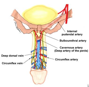
The penis is supplied by a superficial system of arteries arising from the external pudendal arteries and a deep system of arteries stemming from the internal pudendal arteries. The superficial blood supply lies in the superficial penile fascia and supplies the penile skin and prepuce. The internal pudendal artery, which arises from the hypogastric artery, gives rise to the penile artery. The penile artery then gives rise to the bulbourethral artery, the urethral artery, and the cavernous artery (deep artery of the penis) before terminating as the dorsal artery of the penis (see Images 14-15).

Somatic nerve supply to the penis comes by way of the pudendal nerves, which eventually produce the dorsal nerves of the penis on each side. Although cutaneous innervation to the penis is primarily from branches of the pudendal nerve, the proximal portion is supplied by the ilioinguinal nerve after it leaves the superficial inguinal ring. The prepuce has somatosensory innervation by the dorsal nerve of the penis and branches of the perineal nerve. The glans is primarily innervated by free nerve endings and has poor fine-touch discrimination.


Circumcision is generally not performed in children born prematurely or those with blood dyscrasias. It should not be performed in children with congenital penile anomalies such as the following:

  • Hypospadias
  • Epispadias
  • Chordee
  • Penile webbing
  • Buried penis


Medical therapy

Phimosis

Medical management of phimosis has been achieved by the application of steroid creams (0.05% betamethasone). The usual regimen is application of the steroid cream once or twice daily for 4-6 weeks. Studies have shown a success rate of 87% with this treatment. Higher rates of success have been reported with concomitant preputial stretching exercises.

If a patient has concomitant balanitis or balanoposthitis, depending on the etiology, he may be treated with topical antibiotics or antifungals. Patients with diabetes mellitus should be advised on proper serum glucose control.

Buried penis

Buried penis cannot be treated medically. The separate entity of trapped penis following neonatal circumcision has been successfully treated with topical betamethasone and manual retraction.

Surgical therapy

Phimosis

In the United States, circumcision is the surgical treatment of choice for correction of phimosis. In European countries, however, preputioplasty is often used. Although these techniques are outside the scope of this discussion, patients should be aware that these prepuce-preserving methods exist.

Adult circumcision can be performed under local or regional anesthesia and commonly involves either the dorsal slit or the sleeve technique. The dorsal slit is often useful in patients with phimosis, and the sleeve technique may allow for better hemostasis in patients with large subcutaneous veins.

A properly performed circumcision eliminates phimosis, as well as the risks of paraphimosis and frenular tears or bleeding associated with sexual intercourse.

Buried penis

Numerous techniques have been described for repairing the buried penis. Variations have been proposed for different presumed etiologies and to simplify the procedure. Recurrence and the need for subsequent procedures are possibilities. High rates of success have been reported for both pediatric and adult patients.

Preoperative details

Obtaining proper informed consent before the procedure is critical. In particular, inform patients of potential risks, which include bleeding, hematoma formation, infection, suture disruption, inadvertent injury to the urethra or glans, removal of too much or too little skin, and a change of sensation during intercourse. With regard to surgery for the buried penis, patients should be aware of a 1-15% chance of recurrence (secondary concealment) that may necessitate reoperation.

Patients undergoing circumcision for recurrent balanitis should be free from infection before the procedure.

Circumcision may be performed in the office under a local anesthetic or in the operating room under a regional or general anesthetic. For the anxious patient undergoing an office-based procedure, diazepam (Valium), 2-5 mg administered orally 1 hour before the procedure, is often effective.

Drug Name - Diazepam (Valium) - Benzodiazepine derivative anxiolytic used for treatment of anxiety disorders or for short-term relief of anxiety (ie, circumcision, vasectomy).
Adult Dose - 2-10 mg PO bid/qid
Pediatric Dose - <6 months: Not recommended
> 6 months: 1-2.5 mg tid/qid
Contraindications - Documented hypersensitivity; narrow-angle glaucoma
Interactions - Phenothiazines, barbiturates, alcohol, and monoamine oxidase inhibitors (MAOIs) increase CNS toxicity when administered concurrently
Pregnancy D - Fetal risk shown in humans; use only if benefits outweigh risk to fetus
Precautions - Fatigue, drowsiness, and ataxia are most common adverse effects; less common adverse effects include urinary retention, confusion, depression, and slurred speech; caution with other CNS depressants, low albumin levels, or hepatic disease (may increase toxicity)

Intraoperative details

Surgical Repair for Buried Penis

With the patient in a supine position, the genital area is prepared with povidone-iodine solution. Shaving or clipping of the pubic hair is usually unnecessary.

A general anesthetic is recommended for children. A local anesthetic may be used for adults. Local anesthesia is accomplished by administering a dorsal penile nerve block followed by a ring block.

A mixture of equal volumes of 0.5% bupivacaine (Marcaine) and 1-2% lidocaine (Xylocaine) without epinephrine is common. A potential complication of epinephrine use is local tissue ischemia. The maximum recommended dose of lidocaine without epinephrine is 4.5 mg/kg, or 315 mg for a 70-kg male.

Dorsal slit circumcision

To perform the dorsal slit circumcision, clamp the foreskin at the 12-o'clock position with 2 straight hemostats to limit bleeding (see Image 2).

Sharply incise the tissues between the 2 clamps perpendicular to the corona (see Image 3). Make the proximal circumscribing incision (see Image 4).

Excise the foreskin at its base (approximately 1 cm proximal to the coronal sulcus) with scissors to produce a cosmetically attractive circumcised penis (see Image 5). The amount of skin left below the coronal sulcus should be no more than 1 cm long to prevent edema, adhesions, and, occasionally, paraphimosis.

Ligate superficial veins and obtain hemostasis with electrocautery.

Circumferentially approximate the proximal and distal edges of the foreskin with 4.0-5.0 absorbable sutures (children) or 3.0-4.0 absorbable sutures (adults) in an interrupted fashion (see Image 6).

During the circumcision, the use of a thin tapered (noncutting) needle is recommended because it is less traumatic to the tissues and causes less bleeding.

Bacitracin ointment is used to lubricate the suture material (eg, chromic) to facilitate passage of the suture through the delicate skin tissues. This is especially helpful in the pediatric population. Alternatively, mineral oil may be used as a lubricant.

Sleeve technique

The sleeve technique is an attractive alternative for circumcision.

After proper anesthesia has been achieved, mark the redundant foreskin with a marker.

Before making the initial incision, take care to measure out the correct amount of foreskin to be removed. This is accomplished by gently stretching out the penis with an index finger applied to the penoscrotal junction and noting the indentation of the coronal sulcus through the penile skin.

Outline the coronal sulcus with a marker. This is the proximal skirt of the circumscribing incision (see Image 7). The distal skirt of the circumscribing incision is approximately 1 cm proximal to the corona (see Image 8). When this is properly performed, a sleeve of foreskin is present between the 2 incisions, and the remaining penile skin does not remain too short or too long.

Clamp the redundant foreskin between the circumcision incisions at the 12-o'clock position and incise it (see Image 9).

Remove the sleeve by using electrocautery (see Image 10).

Obtain hemostasis with cautery and ligatures. If circumcision has been performed properly, the proximal and distal skirts should approximate well without tension (see Image 11).

Once hemostasis has been achieved, circumferentially approximate the edges of the foreskin with 3.0-5.0 absorbable sutures in either a running 4-quadrant closure or an interrupted fashion (see Image 12). Correct technique allows for proper cosmesis (see Image 13).

Surgical Techniques for Buried Penis

In pediatric cases, sources have described the essential nature of dividing dysgenetic dartos bands and fixation of the dartos fascia to the Buck fascia dorsally in the midline, ventrally over the corpus spongiosum, and proximally along the penile shaft. Care must be taken to avoid injury to either the urethra or the neurovascular bundles.

Surgery is usually necessary for the suprapubic fat pad in adults, but opinions vary as to the value of its removal in children. The same controversy exists regarding whether or not to take down the suspensory ligament.

In 2004, Frenkl et al described a simple technique for the repair of buried penis in children, as follows:6

  • First, a traction suture is placed in the glans, and local anesthetic is instilled along the subcoronal circumference.
  • A circumcision incision is then used and, using the Buck fascia as the dissection plane, the penis is degloved to the penopubic junction.
  • As the next step in the operation, numerous authors describe sharp dissection of dysgenetic dartos fibers.
  • Fixation is then performed between the Buck fascia and the dermis at the base of the penile shaft with 5-0 sutures at the 3- and 9-o'clock positions.
  • The circumferential incision is then reapproximated at its origin with interrupted absorbable suture.

Cromie et al (1998) applied a similar technique to adults for correction of buried penis that involved making a circumferential incision of the inner preputial skin layer proximal to the corona, unfurling the incised skin from the penile shaft, and leaving a coronal collar of approximately 1 cm.7

Brisson et al (2001) add a longitudinal incision on the penile shaft ventrally, along the median raphe. They utilize this exposure to allow for additional points of fixation between the skin and the tunica albuginea.8

In 1998, Donatucci and Ritter reported their technique for correction of buried penis in adults as follows:9

  • If a pannus is to be resected, the amount to be removed is estimated with consideration of avoiding undue tension upon closure. Both liposuction and panniculectomy have been used in these operations. Generally, if excess skin with poor tone is present, panniculectomy should be performed instead of liposuction.
  • Resection is done in an elliptical fashion down to the mons pubis, which is mobilized to the base of the penis. The mons pubis is then resuspended with sutures between the superficial fascia at the penile base and the deep fascia of the abdominal wall. The wound is closed over a drain with gentle pressure over the pubic area.
  • A traction suture is placed in the glans to facilitate dissection, and a Foley catheter is used to protect the urethra. The scar contracture is released, and the penis is degloved to the base along the Buck fascia, with preservation of neurovascular bundles. Adhesions are separated as they are encountered, and chordee is corrected.
  • Artificial erection is utilized to confirm chordee correction and to determine tissue needs for optimal construction. This maneuver is performed using a Penrose drain as a tourniquet at the penile base and injecting 50-100 mL of sterile saline into the corpora cavernosa with a 21-gauge butterfly catheter. If adequate length is not obtained, the suspensory ligament is divided and resuspended from the symphysis pubis with 1-0 silk sutures with the penis in traction.
  • If a patient has a minimal deficiency of longitudinal penile skin, a plasty rearrangement of this skin can often be accomplished. If a severe deficiency of skin is appreciated, a split-thickness skin graft may be used, either from a panniculectomy specimen (if available) or from the lateral thigh. (Other authors, such as Chopra et al [2002], feel that a full-thickness skin graft is more appropriate.10) Once fixation between the skin, dartos, and tunica albuginea at the base of the penis has been performed, as described previously, skin grafts are applied in spiral fashion. If the wound bed is inadequate for grafting because of scarring, the use of flaps may be considered.
  • A penile block at the conclusion of the procedure may provide better postoperative pain control.

Postoperative details

Adult circumcision

Following adult circumcision, many urologists choose not to use a dressing. However, either petroleum jelly and sterile gauze or Xeroform petrolatum gauze may be wrapped around the penis circumferentially, followed by application of sterile gauze and a nonadhesive elastic wrap.

For patients who have undergone adult circumcision, discharge medications should include oral narcotics. The dressing is removed 24-48 hours after surgery. The patient should wear loose-fitting briefs after surgery, and he may shower but should wash gently around the incision site

Full recovery may require 4-6 weeks of complete sexual abstinence.

Buried-penis surgery

Following surgery for the buried penis, a Foley catheter is usually left in place, and the penile shaft is covered with a foam dressing, both of which are removed in 48 hours. For patients who have undergone flap resurfacing, an antibiotic ointment dressing is used. In patients who have undergone a panniculectomy, a pressure garment may be worn over the surgical site for 4-6 weeks. In patients who have undergone liposuction, a pressure dressing is left in place for 7 days.

Children have few restrictions after surgical correction of the buried penis. Adults, however, must refrain from strenuous activity, heavy lifting, and sexual activity for 6 weeks after operation.

Follow-up

Routine follow-up after adult circumcision is not mandatory, but patients may be seen 2-3 weeks after the operation so that the incision can be examined to see if it has appropriately healed. Following buried-penis repair, both pediatric and adult patients may be seen at 2 and 6 weeks after surgery.

For excellent patient education resources, visit eMedicine's Men's Health Center. Also, see eMedicine's patient education articles Foreskin Problems and Circumcision.


Circumcision

Circumcision is a safe surgical procedure that is well tolerated in nearly all patients. However, as with any surgical procedure, complications are possible. Examples include bleeding, infection, hematoma, swelling, pain, and poor cosmesis.

Postoperative bleeding is the most common complication. It usually occurs when a scab is pulled off as the dressing is being removed. However, sutures can also be torn by erections before healing is completed. Bleeding can often be controlled with direct pressure. Rarely, electrocautery or ligature may be required.

Infection after circumcision is uncommon and, if encountered, may be treated with local or oral antibiotics.

Urethral injury is extremely rare. A urethral injury that can result from circumcision is a subcoronal urethrocutaneous fistula. This condition necessitates surgical correction.

Suture sinus tracts can occur if a simple suture becomes epithelialized before suture absorption. These frequently go unnoticed and are likely inconsequential.

Postoperative adhesions may lead to formation of skin bridges between the circumcision incision and the glans. These adhesions are typically on the dorsal surface and therefore may present an obstacle to proper hygiene.

A study by Fink et al (2002) examined the sexual effects of adult circumcision.11 They found that circumcision in the adult appears to result in worsened erectile function and decreased penile sensitivity. However, sexual activity did not change, and satisfaction was actually improved. Given that 93% of the patients in that study had surgery for pathology, circumcision may have alleviated enough sexual apprehension to allow unmasking of previously unidentified erectile dysfunction. Although many investigators feel that circumcision reduces penile sensitivity, an increase in ejaculatory latency time may, in some cases, be considered an advantage.

In evaluating the effect of circumcision on a woman's sexual enjoyment, O'Hara et al (1999) reported that the women surveyed preferred intercourse with uncircumcised men.12 The authors suggest that coitus with a circumcised partner reduces vaginal secretions and decreases continual clitoral stimulation. However, a number of the women surveyed were recruited via the newsletter of an anticircumcision organization.

Buried penis

Correction of the buried penis is a simple and effective procedure with few recurrences or complications. Two series that reported on the long-term outcome of pediatric cases found that the correction resulted in excellent long-term cosmetic results. Higher success rates were found in patients who had the correction as toddlers than in those who had the correction as adolescents. Herndon et al (2003) found that most parents felt that the surgery eliminated associated negative feelings and that it enhanced penile appearance and facilitated better hygiene.13

Most of the complications that are reported after surgery for buried-penis correction are temporary. Reported rates of recurrent retraction, however, range from 1-15% and often require additional surgery. Observation is a viable option for mild cases. Ventral edema has been reported in 1-11% of cases and usually resolves spontaneously. Dissatisfaction with cosmesis is possible, but the physician should preoperatively assess for unrealistic expectations. Postoperative sexual dysfunction has been reported, but conditions predisposing to buried penis often carry a higher likelihood of erectile dysfunction. Pain with erection may be seen with suturing of the Buck fascia to the pubic periosteum.

Patients who have undergone penile reconstruction may present with poor graft healing, flap necrosis, or complaints of decreased sensitivity in the grafted area. Patients may also present with persistent redundancy of the penile skin or reaccumulation of the abdominal fat pad.


Phimosis

Phimosis does not recur after proper circumcision. If too much penile skin is left, a repeat circumcision may be necessary for medical or cosmetic reasons. In adults, some permanent skin-color discrepancy along the suture line of the circumcision may occur. Overall, careful attention to proper surgical technique will allow for a pleasing cosmetic result.

Reported results of buried-penis repair in pediatric and adult cases have generally been good. Surgery often provides immediate excellent cosmetic results with low rates of complications. Brisson et al (2001) contend that both patients and their parents derive psychological benefits from the procedure.8 This assertion seems to be confirmed by postoperative surveys. In addition to improved confidence, improvement in personal hygiene and voiding result from buried-penis repair in both pediatric and adult patients. Adult patients often also report improved sexual performance.


The authors and editors of eMedicine gratefully acknowledge the contributions of previous author, Jong M. Choe, MD, FACS, to the development and writing of this article.


Media file 1:  Phimotic foreskin. The distal foreskin is edematous, with cracked fissures. The patient was unable to retract the foreskin.
Click to see larger pictureClick to see detailView Full Size Image
Media type:  Photo

Media file 2:  Dorsal-slit technique. The redundant foreskin is clamped at the 12-o'clock position for 2 minutes for hemostasis.
Click to see larger pictureClick to see detailView Full Size Image
Media type:  Photo

Media file 3:  In the dorsal-slit technique, the clamped foreskin is incised sharply between the 2 hemostats.
Click to see larger pictureClick to see detailView Full Size Image
Media type:  Photo

Media file 4:  The dorsal slit is being completed, and the circumscribing incision (proximal skirt) has been made.
Click to see larger pictureClick to see detailView Full Size Image
Media type:  Photo

Media file 5:  Dorsal-slit technique. Redundant foreskin has been excised. The distal circumcision incision is 1 cm from the coronal sulcus.
Click to see larger pictureClick to see detailView Full Size Image
Media type:  Photo

Media file 6:  Dorsal-slit technique. Proximal and distal skirts are approximated circumferentially with absorbable sutures in an interrupted fashion.
Click to see larger pictureClick to see detailView Full Size Image
Media type:  Photo

Media file 7:  In the sleeve technique, the circumcision is started by making a circumscribing proximal incision. The incision is carried down to the Buck fascia.
Click to see larger pictureClick to see detailView Full Size Image
Media type:  Photo

Media file 8:  In the sleeve technique, a distal incision is made 1 cm proximal to the coronal sulcus.
Click to see larger pictureClick to see detailView Full Size Image
Media type:  Photo

Media file 9:  Sleeve technique. Redundant foreskin is clamped at the 12-o'clock position with 2 straight hemostats. Next, the foreskin is incised between the 2 hemostats.
Click to see larger pictureClick to see detailView Full Size Image
Media type:  Photo

Media file 10:  Sleeve technique. The excess foreskin is peeled off. The shaft of the penis is displaced downward using a stack of sponges as the redundant foreskin is removed.
Click to see larger pictureClick to see detailView Full Size Image
Media type:  Photo

Media file 11:  Sleeve technique. Excess foreskin has been removed completely.
Click to see larger pictureClick to see detailView Full Size Image
Media type:  Photo

Media file 12:  Sleeve technique. The edges of the penile skin are approximated with absorbable sutures.
Click to see larger pictureClick to see detailView Full Size Image
Media type:  Photo

Media file 13:  Sleeve technique. The circumcision is completed with excellent cosmetic result.
Click to see larger pictureClick to see detailView Full Size Image
Media type:  Photo

Media file 14:  The arterial blood supply of the penis arises from the internal pudendal artery. The internal pudendal artery gives off branches to the bulbar artery, cavernosal artery, and dorsal penile artery. The bulbar artery continues on as the bulbourethral artery to supply the urethra. The cavernosal artery gives rise to the helicine arteries that are end arteries. The dorsal artery of the penis gives branches off to the circumflex arteries.
Click to see larger pictureClick to see detailView Full Size Image
Media type:  Image

Media file 15:  Dorsal view of the arterial and venous blood supply of the penis.
Click to see larger pictureClick to see detailView Full Size Image
Media type:  Image

Media file 16:  Cross-section through the body of the penis.
Click to see larger pictureClick to see detailView Full Size Image
Media type:  Image

Media file 17:  Preoperative photo of a buried penis in an adult.
Click to see larger pictureClick to see detailView Full Size Image
Media type:  Photo

Media file 18:  Same patient after penile reconstruction and removal of the pannus. Note the elliptical incision and marked improvement in perceived penile length.
Click to see larger pictureClick to see detailView Full Size Image
Media type:  Photo

Media file 19:  Same patient at the conclusion of the procedure. Although not seen in this picture, a Foley catheter may be left in place after the operation.
Click to see larger pictureClick to see detailView Full Size Image
Media type:  Photo

Media file 20:  Concealed penis secondary to a scrotal web.
Click to see larger pictureClick to see detailView Full Size Image
Media type:  Photo

Media file 21:  Repair in this patient involved releasing the scrotal web and degloving the penis. This patient was found to have deficient penile skin for reconstruction.
Click to see larger pictureClick to see detailView Full Size Image
Media type:  Photo

Media file 22:  Same patient after application of a split-thickness skin graft that was harvested from the left thigh.
Click to see larger pictureClick to see detailView Full Size Image
Media type:  Photo


  1. Maizels M, Zaontz M, Donovan J. Surgical correction of the buried penis: description of a classification system and a technique to correct the disorder. J Urol. Jul 1986;136(1 Pt 2):268-71. [Medline].
  2. Van Howe RS. Does circumcision influence sexually transmitted diseases?: a literature review. BJU Int. Jan 1999;83 Suppl 1:52-62. [Medline].
  3. Daling JR, Madeleine MM, Johnson LG, Schwartz SM, Shera KA, Wurscher MA. Penile cancer: importance of circumcision, human papillomavirus and smoking in in situ and invasive disease. Int J Cancer. Sep 10 2005;116(4):606-16. [Medline].
  4. Meuli M, Briner J, Hanimann B, Sacher P. Lichen sclerosus et atrophicus causing phimosis in boys: a prospective study with 5-year followup after complete circumcision. J Urol. Sep 1994;152(3):987-9. [Medline].
  5. Burkholder GV, Newell ME. New surgical treatment for micropenis. J Urol. Apr 1983;129(4):832-4. [Medline].
  6. Frenkl TL, Agarwal S, Caldamone AA. Results of a simplified technique for buried penis repair. J Urol. Feb 2004;171(2 Pt 1):826-8. [Medline].
  7. Cromie WJ, Ritchey ML, Smith RC, Zagaja GP. Anatomical alignment for the correction of buried penis. J Urol. Oct 1998;160(4):1482-4. [Medline].
  8. Brisson P, Patel H, Chan M, Feins N. Penoplasty for buried penis in children: report of 50 cases. J Pediatr Surg. Mar 2001;36(3):421-5. [Medline].
  9. Donatucci CF, Ritter EF. Management of the buried penis in adults. J Urol. Feb 1998;159(2):420-4. [Medline].
  10. Chopra CW, Ayoub NT, Bromfield C, Witt PD. Surgical management of acquired (cicatricial) buried penis in an adult patient. Ann Plast Surg. Nov 2002;49(5):545-9. [Medline].
  11. Fink KS, Carson CC, DeVellis RF. Adult circumcision outcomes study: effect on erectile function, penile sensitivity, sexual activity and satisfaction. J Urol. May 2002;167(5):2113-6. [Medline].
  12. O'Hara K, O'Hara J. The effect of male circumcision on the sexual enjoyment of the female partner. BJU Int. Jan 1999;83 Suppl 1:79-84. [Medline].
  13. Herndon CD, Casale AJ, Cain MP, Rink RC. Long-term outcome of the surgical treatment of concealed penis. J Urol. Oct 2003;170(4 Pt 2):1695-7; discussion 1697. [Medline].
  14. Aaronson IA. Micropenis: medical and surgical implications. J Urol. Jul 1994;152(1):4-14. [Medline].
  15. Adham MN, Teimourian B, Mosca P. Buried penis release in adults with suction lipectomy and abdominoplasty. Plast Reconstr Surg. Sep 2000;106(4):840-4. [Medline].
  16. Ahmed A, Mbibi NH, Dawam D, Kalayi GD. Complications of traditional male circumcision. Ann Trop Paediatr. Mar 1999;19(1):113-7. [Medline].
  17. Alter GJ, Ehrlich RM. A new technique for correction of the hidden penis in children and adults. J Urol. Feb 1999;161(2):455-9. [Medline].
  18. American Academy of Pediatrics. American Academy of Pediatrics: Report of the Task Force on Circumcision. Pediatrics. Aug 1989;84(2):388-91. [Medline].
  19. Babu R, Harrison SK, Hutton KA. Ballooning of the foreskin and physiological phimosis: is there any objective evidence of obstructed voiding?. BJU Int. Aug 2004;94(3):384-7. [Medline].
  20. Baskin LS, Canning DA, Snyder HM 3rd, Duckett JW Jr. Surgical repair of urethral circumcision injuries. J Urol. Dec 1997;158(6):2269-71. [Medline].
  21. Burnakis TG. Amyl nitrite for the treatment of penile tumescence. Hosp Pharm. 1991;26:343.
  22. Chu CC, Chen KC, Diau GY. Topical steroid treatment of phimosis in boys. J Urol. Sep 1999;162(3 Pt 1):861-3. [Medline].
  23. Chuang JH, Chen LY, Shieh CS, Lee SY. Surgical correction of buried penis: a review of 60 cases. J Pediatr Surg. Mar 2001;36(3):426-9. [Medline].
  24. Cold CJ, Taylor JR. The prepuce. BJU Int. Jan 1999;83 Suppl 1:34-44. [Medline].
  25. Craig JC, Knight JF, Sureshkumar P, et al. Effect of circumcision on incidence of urinary tract infection in preschool boys. J Pediatr. Jan 1996;128(1):23-7. [Medline].
  26. Dessanti A, Ginesu G, Iannuccelli M, Balata A. Phimosis. Preputial plasty using transversal widening on the dorsal side with EMLA local anesthetic cream. J Pediatr Surg. Apr 2005;40(4):713-5. [Medline].
  27. Devine. Concealed and webbed penis. In: Hinman F, ed. Atlas of Pediatric Urologic Surgery. Philadelphia, Pa: WB Saunders and Co; 1998:606-9.
  28. Dunsmuir WD, Gordon EM. The history of circumcision. BJU Int. Jan 1999;83 Suppl 1:1-12. [Medline].
  29. Fussell EN, Kaack MB, Cherry R, Roberts JA. Adherence of bacteria to human foreskins. J Urol. Nov 1988;140(5):997-1001. [Medline].
  30. Gillett MD, Rathbun SR, Husmann DA, Clay RP, Kramer SA. Split-thickness skin graft for the management of concealed penis. J Urol. Feb 2005;173(2):579-82. [Medline].
  31. Ginsburg CM, McCracken GH Jr. Urinary tract infections in young infants. Pediatrics. Apr 1982;69(4):409-12. [Medline].
  32. Glass JM. Religious circumcision: a Jewish view. BJU Int. Jan 1999;83 Suppl 1:17-21. [Medline].
  33. Gursel EO, Georgountzos C, Uson AC, et al. Penile cancer. Urology. Jun 1973;1(6):569-78. [Medline].
  34. Hollowood AD, Sibley GN. Non-painful paraphimosis causing partial amputation. Br J Urol. Dec 1997;80(6):958. [Medline].
  35. Holman JR, Stuessi KA. Adult circumcision. Am Fam Physician. Mar 15 1999;59(6):1514-8. [Medline].
  36. Hunter RH. Notes on the Development of the Prepuce. J Anat. Oct 1935;70(Pt 1):68-75. [Medline].
  37. Hutcheson JC. Male neonatal circumcision: indications, controversies and complications. Urol Clin North Am. Aug 2004;31(3):461-7, viii. [Medline].
  38. Johnson DE, Fuerst DE, Ayala AG. Carcinoma of the penis. Experience with 153 cases. Urology. May 1973;1(5):404-8. [Medline].
  39. Kiss A, Király L, Kutasy B, Merksz M. High incidence of balan