Practice Essentials
The horseshoe kidney is the most common type of renal fusion anomaly. It consists of two distinct functioning kidneys on each side of the midline, connected at the lower poles (or rarely at the upper poles) by an isthmus of functioning renal parenchyma or fibrous tissue that crosses the midline of the body. [1]
A urogram protocol computed tomography (CT scanning of the abdomen and pelvis, with and without intravenous contrast and with delayed images) is the best initial radiologic study to determine anatomy and relative kidney function (see Workup). Horseshoe kidneys are susceptible to medical renal disease. These diseases, if present, are treated as indicated.
For patient education information, see What Is Horseshoe Kidney?
Relevant Anatomy
Horseshoe kidneys may be found at any location along the path of normal renal ascent from the pelvis to the mid abdomen. The kidneys may be lower than normal because the isthmus is tethered by the inferior mesenteric artery during renal ascent. The isthmus usually lies anterior to the great vessels, at the level of the third to fifth lumbar vertebra. Rarely, it is posterior to these vessels or runs between them.
The vascular supply is variable and originates from the aorta, the iliac arteries, and the inferior mesenteric artery. Bilateral single renal hilar arteries occur in 30% of cases, and various combinations of single and multiple renal hilar and isthmus vessels are seen in 70% of cases. The isthmus of the kidney may not have a separate blood supply or, in 65% of cases, is supplied by a single vessel from the aorta. The blood supply to the isthmus may arise from the common iliac or inferior mesenteric arteries.
Majos and colleagues compared the number of renal arteries and veins in 94 patients with horseshoe kidneys and 248 patients with normal kidneys and found that venous supply of horseshoe kidneys varies substantially and does not follow any pattern currently used in common classification systems. Horseshoe kidneys have more renal arteries and veins than normal kidneys (4.5 vs 2.41 arteries and 3.78 vs 2.29 veins, respectively) and the number of renal veins in horseshoe kidneys shows less correlation with the number of renal arteries. In addition, the researchers reported a weak correlation between the level of connection of the renal veins and renal arteries to their parental vessels. [2]
The collecting system has a characteristic appearance on intravenous urography or CT delayed phase because of an incomplete inward rotation of the renal pelvis, which cauces it to face anteriorly. The axis of the collecting system is deviated inward at the lower poles because of the lower pole's connection with the isthmus. The ureter may have a high insertion point into the renal pelvis and may cross anteriorly over the isthmus as it descends to the bladder, which can cause drainage problems. Rarely does the collecting system cross the isthmus to the contralateral kidney.
Pathophysiology
By itself, the horseshoe kidney does not produce symptoms. However, by virtue of its embryogenesis and abnormal anatomy, it is predisposed to a higher incidence of disease than the normal kidney. The variable blood supply, presence of the isthmus, high insertion point of the ureter, and abnormal course of the ureter all contribute to these problems. Because of these embryogenic and anatomical factors, the rates of hydronephrosis, stone formation, infection, and certain cancers are higher, resulting in a diseased horseshoe kidney (see image below).
The most common associated finding in horseshoe kidney is ureteropelvic junction (UPJ) obstruction, which occurs in up to 35% of patients and is the cause of many of the problems associated with a horseshoe kidney. Obstruction is classically due to the high insertion of the ureter into the renal pelvis. The crossing of the ureter over the isthmus may also contribute to obstruction. Nonobstructive dilatation can be distinguished from obstructive dilatation using diuresis radioisotope renal scans.
The prevalence of stones in the horseshoe kidney ranges from 20-60% [3] with a recent meta-analysis putting the prevalence toward the middle of that range, at 36%. [4] Stone disease is thought to be due to the associated hydronephrosis or UPJ obstruction that causes urinary stasis, which hinders stone passage. Metabolic factors, as in the normal population, have also been suggested as contributing to stone formation in these patients. The orientation of the calyces also impairs drainage, resulting in stasis. These kidneys appear dilated or abnormal on most imaging studies, although the radionuclide scans are generally accepted as being diagnostic.
Urinary stasis and stone disease also predispose the horseshoe kidney to infection, which occurs in 27-41% of patients. [5] Ascending infection from vesicoureteral reflux is another cause of infection in the horseshoe kidney.
Certain cancers are more common in the horseshoe kidney. [6] This is thought to be due to teratogenic factors present at birth and the susceptibility of the diseased horseshoe kidney to certain cancers. Renal cell carcinoma is the most common renal cancer in a horseshoe kidney, accounting for 45% of all tumors. [7, 8] The incidence of renal cell cancer in the horseshoe kidney is no different from that of the normal kidney.
Transitional cell cancer and sarcoma account for 20% and 7% of tumors, respectively. The relative risk of transitional cell carcinoma in the horseshoe kidney is 3- to 4-fold higher than in a normal kidney. This is thought to be due to chronic obstruction, stones, and/or infection in the affected kidneys.
The incidence of both Wilms and carcinoid tumors is also higher in the horseshoe kidney. Examination of these tumors may provide an insight into the development and embryogenesis of the horseshoe kidney and the predilection of these two tumors to form in the horseshoe kidney.
Wilms tumor accounts for 28% of malignant lesions. The relative risk of Wilms tumor is increased 2-fold. Interestingly, half of these arise from the isthmus.
Renal carcinoids are rare, with only 32 reported cases. Of the 32 cases, 5 of these renal carcinoids arose in a horseshoe kidney. [6, 9] The relative risk of a carcinoid tumor in a patient with a horseshoe kidney is 62 times that found in the normal population. Of the 5 reported carcinoid tumors reported in patients with horseshoe kidneys, 3 have originated in or have involved the isthmus. The location of these tumors in the isthmus may be explained by the embryogenesis involving abnormal migration of posterior nephrogenic cells, leading to the formation of the isthmus. This is a teratogenic event, which may explain this increased incidence of tumor within the isthmus. This theory may also explain the greater incidence of Wilms tumor in the isthmus, as well. When compared with carcinoid tumor arising in a normal kidney, those that arise in a horseshoe kidney follow a more benign course.
Etiology
Two theories regarding the embryogenesis of the horseshoe kidney have been proposed. The classic teaching of mechanical fusion holds that the horseshoe kidney is formed during organogenesis, when the inferior poles of these early kidneys touch, fusing in the lower midline. The theory of mechanical fusion is valid for horseshoe kidneys with a fibrous isthmus. [10]
Alternatively, more recent studies postulate that the abnormal fusion of tissue associated with the parenchymatous isthmus of some horseshoe kidneys is the result of a teratogenic event involving the abnormal migration of posterior nephrogenic cells, which then coalesce to form the isthmus. [11, 10] This teratogenic event may also be responsible for the increased incidence of related congenital anomalies and of certain neoplasias, such as Wilms tumor and carcinoid tumor associated with the isthmus of the horseshoe kidney.
Epidemiology
Horseshoe kidney has been reported in 1 per 400-800 live births. The true incidence probably lies somewhere between these two extremes. Horseshoe kidney is twice as common in males as in females. No genetic determinant is currently known, although horseshoe kidneys have been reported in identical twins and in siblings within the same family.
In India, horseshoe kidneys occur in 1 per 600-700 individuals. [12]
Prognosis
The horseshoe kidney does not complicate pregnancy or delivery. Importantly, it should be noted that the presence of a horseshoe kidney alone does not affect survival. However, because the horseshoe kidney does have a higher propensity to become diseased, longevity depends on the disease process that the affected horseshoe kidney may harbor or develop.
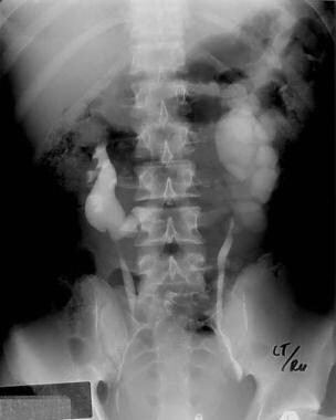
-
Excretory urogram shows a horseshoe kidney with left hydronephrosis.
-
This CT scan demonstrates the isthmus of a horseshoe kidney. Note the uptake of contrast in the isthmus.


