Practice Essentials
Bladder injuries can result from blunt, penetrating, or iatrogenic trauma. [1, 2] The probability of bladder injury varies according to the degree of bladder distention; a full bladder is more susceptible to injury than is an empty one.
Although historically, bladder trauma was uniformly fatal, timely diagnosis and appropriate management now provide excellent outcomes. Early clinical suspicion, coupled with appropriate and reliable radiologic studies, facilitate prompt intervention and successful management. [3] Management varies from conservative approaches that center on maximizing bladder drainage to major surgical procedures aimed at directly repairing the injury.
The most recent American Urological Association Guidelines on Urotrauma, updated in 2020, recommend that "clinicians should perform catheter drainage as treatment for patients with uncomplicated extraperitoneal bladder injuries" whereas "surgeons must perform surgical repair of intraperitoneal bladder rupture in the setting of blunt or penetrating external trauma." [4]
Nevertheless, the literature contains a handful of case reports describing intraperitoneal bladder rupture managed conservatively. Two such reports describe successful treatment of small ruptures in patients with a benign abdomen, using prolonged large-diameter urethral catheter drainage and antibiotic prophylaxis. The authors warn that communication with the peritoneal cavity may persist, and advise open surgical management if clinical deterioration occurs (eg, uremia, infection) or follow-up cystography demonstrates a persistent leak. [5] Two other studies found that patients who undergo open repair of extraperitoneal injuries have lower rates of persistent urine leak than patients treated with urethral catheter drainage. [6, 7]
A retrospective study by Johnson and colleagues concluded that in most patients with extraperitoneal bladder rupture from blunt trauma, conservative management with catheter drainage alone results in outcomes equivalent to those with operative repair. However, these authors authors advocate performing open bladder repair in patients who will be undergoing surgery for other indications. In their analysis of the outcomes in 56 patients treated with catheter drainage and 24 who underwent early cystorrhaphy as a secondary procedure during nonurological interventions, patients treated with catheter dainage alone experienced higher rates of urological complications (P < 0.05), increased length of stay in the intensive care unit (9.0 vs 4.0 days, P=0.02) and hospital (18.9 vs 10.6 days, P=0.02), as well as longer time to negative cystography (25.5 vs 20.0 days, P=0.03). [8]
Collins et al reported on the outcomes of bladder trauma in 192 patients who underwent open surgical repair compared with 192 patients who underwent laparoscopic treatment. The mechanism of injury was blunt trauma in 56% and penetrating trauma in 44%. While there was no significant difference in complications between the groups, the overall mortality rate was lower (4.2%) after open surgical repair than in patients who received laparoscopic treatment (10.4%). [9]
Relevant Anatomy
In adults, the bladder is located in the anterior pelvis and is enveloped by extraperitoneal fat and connective tissue. It is separated from the pubic symphysis by an anterior prevesical space known as the space of Retzius. The dome of the bladder is covered by peritoneum and the bladder neck is fixed to neighboring structures by reflections of the pelvic fascia as well as by true ligaments of the pelvis.
In males, the bladder neck is contiguous with the prostate, which is attached to the pubis by puboprostatic ligaments. In females, pubourethral ligaments support the bladder neck and urethra.
The body of the bladder receives support from the urogenital diaphragm inferiorly and the obturator internus muscles laterally. The superior fascia of the urogenital diaphragm is continuous and includes the pelvic, obturator, and endopelvic fasciae. The inferior fascia of the urogenital diaphragm fuses with Colle's fascia and continues as Scarpa's fascia anteriorly. The dartos muscle and fascia in the scrotum as well as the fascia lata of the thigh are further continuations of this layer.
The type of extravasation (intraperitoneal or extraperitoneal) from a bladder injury depends upon the location of the laceration and its relationship with the peritoneal reflection, as follows:
-
If the perforation is above the peritoneal reflection, on the dome of the bladder, the extravasation is intraperitoneal
-
If the injury is below the peritoneal reflection, and not on the dome of the bladder, the extravasation is extraperitoneal
With an anterosuperior perforation, urinary extravasation may be intraperitoneal, extraperitoneal (space of Retzius), or both. If the tear is posterosuperior, fluid can spread intraperitoneally and retroperitoneally, as well. With bladder rupture, the superior fascia of the urogenital diaphragm, when intact, prohibits extravasated urine from escaping the pelvis, while the inferior fascia of the urogenital diaphragm, when intact, prevents urinary extravasate from flowing into the perineum.
Pathophysiology
Bladder Contusion
Bladder contusion is an incomplete or partial-thickness tear of the bladder. This produces a hematoma within the bladder at the location of injury. Bladder contusion commonly results from blunt trama or extreme physical activity (eg, long-distance running). Patients typically present with gross hematuria. On cystography, the bladder usually appears normal, or it may have a teardrop shape secondary to compression by the hematoma.
Bladder contusion is relatively benign. It is self-limiting and requires no specific therapy, except for rest until hematuria resolves. Nevertheless, it should remain a diagnosis of exclusion. Persistent hematuria or unexplained lower abdominal pain requires further investigation.
Extraperitoneal Bladder Rupture
Traumatic extraperitoneal rupture is usually (89%-100%) associated with pelvic fracture. Previously, the mechanism of injury was believed to be direct perforation by bony fragment or disruption of the pelvic girdle. It is now thought that pelvic fracture is likely coincidental and that bladder rupture most often is a direct result of deceleration injury and fluid inertia coupled with the shearing force created by pelvic ring deformation.
Extraperitoneal rupture is usually associated with fracture of the anterior pubic arch. When this occurs, the anterolateral aspect of the bladder is typically perforated by bony spicules. Forceful disruption of the bony pelvis or the puboprostatic ligaments also tears the bladder wall. In such instances, the degree of bladder injury is directly related to the severity of the fracture.
A mechanism similar to intraperitoneal bladder rupture is thought to underly some extraperitoneal bladder injuries. Specifically, this is the combination of trauma with bladder overdistention, leading to a burst injury.
The classic cystographic finding is contrast extravasation around the base of the bladder, confined to the perivesical space. Often, areas of contrast extravasation shaped like flames, feathers, or starbursts are noted adjacent to the bladder. Additionally, the bladder may assume a teardrop shape secondary to compression from a pelvic hematoma.
With a more complex injury, contrast material can extend to the thigh, penis, perineum, or into the anterior abdominal wall. Extravasation will reach the scrotum when the superior fascia of the urogenital diaphragm, or the urogenital diaphragm itself, becomes disrupted.
If the inferior fascia of the urogenital diaphragm is violated, contrast material will reach the thigh and penis within the confines of the Colles fascia. Rarely, contrast may extravasate into the thigh through the obturator foramen or into the anterior abdominal wall through contiguous tissue planes. Sometimes, extravasation of contrast through the inguinal canal and into the scrotum or labia majora can occur. See the image below.
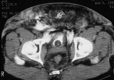 CT scan of extraperitoneal bladder rupture. The contrast extravasates from the bladder into the prevesical space.
Intraperitoneal Bladder Rupture
Classic intraperitoneal rupture is described as large horizontal tears in the bladder dome. This is the least supported area of the bladder and only portion of the organ covered by peritoneum. In such cases, the mechanism of injury is a sudden large increase in intravesical fluid pressure that overcomes the mechanical strength of the bladder wall. This is more likey to occur at greater bladder volumes, as the detrusor muscle fibers are more widely separated along the thinned and stretched bladder wall, offering a lower resistance to spikes in intravesical fluid pressure.
Intraperitoneal bladder rupture generally occurs as the result of a direct blow to a distended urinary bladder. Deceleration injuries can also cause such phenomena. This type of injury is most common in alcoholics and victims of seatbelt or steering wheel trauma. Otherwise, it is more common in children due to the relative intraabdominal bladder position that persists until approximately 20 years of age.
Since urine will generally continue to drain into the abdomen through the open bladder wall defect, intraperitoneal ruptures may go undiagnosed for variable lengths of time. Metabolic and electrolyte abnormalities (eg, hyperkalemia, hypernatremia, uremia, acidosis) may occur as urine is reabsorbed through the peritoneal cavity. Additionally, such patients may appear anuric.
The diagnosis is established when urinary ascites are recovered during paracentesis or the leak is confirmed on imaging. Intraperitoneal rupture demonstrates contrast extravasation into the peritoneal cavity. The contrast media will often outline loops of bowel, fill the paracolic gutters, and pool under the diaphragm. See the image below.
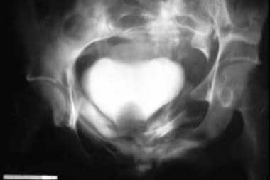
 Cystogram of intraperitoneal bladder rupture. The contrast enters the intraperitoneal cavity and outlines loops of bowel.
Combination of Intraperitoneal and Extraperitoneal Ruptures
Diagnostic imaging with cystogram will reveal contrast outlining the abdominal viscera and perivesical space. Oftentimes this may be observed in penetrating trauma, where the bladder is traversed by a high-velocity bullet, impaled by a knife, or penetrated by another foreign body. This through-and-through injury creates a combined intraperitoneal and extraperitoneal bladder rupture. See the image below.
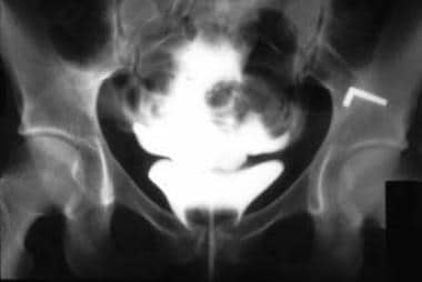
 Cystogram of extraperitoneal bladder rupture. Note the fractured pelvis and contrast extravasation into the space of Retzius.
The high incidence of associated abdominal visceral and vascular injury mandates surgical exploration in virtually every case of combined intraperitoneal and extraperitoneal rupture. Cystography can be falsely negative in penetrating bladder injuries secondary to small-caliber wounds, although the capabilities of cross-sectional imaging with computed tomographic cystography have improved recently. For full discussion, see Bladder Trauma Imaging. However, it is often not the suspected bladder injury alone that drives the consideration for operative intervention. As a result, the diagnosis of such injuries is commonly made during exploratory laparotomy.
Etiology
Blunt Trauma
Deceleration injuries usually produce both bladder trauma (rupture) and pelvic fractures (which can cause bladder perforation). Accordingly, approximately 10% of patients with pelvic fracture also have significant bladder injury. The propensity of the bladder to sustain injury is positively associated with its degree of distention at the time of trauma. When the bladder is full, a blunt blow to the abdomen, as with a punch or kick, can rupture the bladder. Bladder rupture has been documented in children struck in the abdomen by a soccer ball while playing the sport. [10, 11, 12]
Penetrating Trauma
Both gunshot and stabbing are examples of penetrating trauma. Often, these patients incur concomitant injury to other abdominal and/or pelvic organs.
Obstetric Trauma
During prolonged labor or a difficult forceps delivery, persistent pressure from the fetal head against the mother's pubis can lead to bladder necrosis. Direct laceration of the urinary bladder is reported in 0.3% of women undergoing a cesarean delivery. Previous cesarean deliveries with resultant adhesions are a risk factor for such, as undue scarring may obliterate normal tissue planes. Unrecognized bladder injuries may lead to vesicouterine fistulas and other problems.
Gynecologic Trauma
Bladder injury may occur during vaginal or abdominal hysterectomy. Blind dissection in the incorrect tissue plane between the base of the bladder and the cervical fascia is generally the maneuver implicated in such cases.
Urologic Trauma
Perforations of the bladder during bladder biopsy, cystolitholapaxy, transurethral resection of the prostate (TURP), or transurethral resection of bladder tumor (TURBT) are not uncommon. The incidence of bladder perforation with bladder biopsy is reportedly as high as 36%.
Orthopedic Trauma
Orthopedic hardware can easily perforate the urinary bladder, particularly during internal fixation of pelvic fractures. Additionally, thermal injuries to the bladder may occur during the setting of cement substances used to seat arthroplasty prosthetics.
Idiopathic Bladder Trauma
Patients diagnosed with alcoholism and individuals who chronically imbibe a large quantity of fluids are susceptible to idiopathic bladder injury. [13] Previous bladder surgery is a risk factor for such, as areas of scarring are weakened and prone to rupture. In reported cases, all bladder ruptures were intraperitoneal. This type of injury may result from a combination of bladder overdistention and minor external trauma, such as that from a minor stumble or fall.
Epidemiology
Bladder injuries occur in about 1.6% of patients with blunt abdominal trauma. Approximately 60% of bladder injuries are extraperitoneal, 30% are intraperitoneal, and the remaining 10% are both extra- and intraperitoneal. [14]
Frequency of bladder rupture varies according to the mechanism of injury, as follows:
-
External trauma (82%)
-
Iatrogenic (14%)
-
Intoxication (2.9%)
-
Spontaneous (< 1%)
Approximately 60%-85% of bladder injuries result from blunt trauma, while 15%-40% are from penetrating injury. [15] The most common mechanisms of blunt trauma are motor vehicle collision (87%), fall (7%), and assault (6%). In penetrating trauma, the most frequent culprit is gunshot wound (85%), followed by stabbing (15%).
Approximately 10%-25% of patients with pelvic fracture also have urethral trauma. Conversely, 10%-29% of patients with posterior urethral disruption have an associated bladder rupture.
Traumatic Bladder Rupture
Extraperitoneal bladder perforation accounts for 50%-71% of bladder rupture, while 25%-43% are intraperitoneal, and 7%-14% are combined. [16, 17] The incidence of intraperitoneal bladder rupture is significantly higher in children because of the predominantly intraabdominal location of the bladder before puberty.
Combined intraperitoneal and extraperitoneal rupture accounts for approximately 10% of all perforating traumatic bladder injuries. Mortality rates in these patients approach 60% while only 17%-22% of overall bladder injury results in death. This emphasizes the severity of the concomitant injuries associated with combined bladder rupture.
Associated bowel injuries
Among patients with bladder trauma from gunshot, an 83% incidence of associated bowel injury is reported. Colon injuries are noted in 33% of patients with stab wounds, while vascular injuries occur in nearly 82% of patients with a penetrating trauma and carry a 63% mortality rate.
Prognosis
Traumatic bladder rupture, once uniformly fatal, is currently managed successfully with or without surgery, depending upon the type of injury. It is difficult to cite a single specific rate of successful bladder repair due to the wide variety of concurrent trauma these patients often present with. Regardless, critical to the successful management of traumatic bladder rupture are a timely evaluation, accurate diagnosis, and proper management based on the location and severity of the bladder leak.
Recovery after bladder trauma, with or without surgical repair, involves decompression and allowing time for the bladder to heal. In certain situations, surgical repair may help speed healing and overall recovery times. In particular, one study showed that extraperitoneal bladder ruptures repaired during surgery for other injuries were associated with better outcomes—specifically, lower urologic complication rates, shorter intensive care unit stays, decreased length of hospitalization, and faster time to confirmatory x-ray showing no further bladder leakage.ref8}
In general, the bladder heals well and most patients recover normal bladder function. However, in the setting of severe trauma, recovery may be challenged by other concomitant injuries. In particular, with bladder trauma that disrupts the bladder neck, or severe trauma that involes injury to the pelvic floor musculature and urethra, the bladder may heal but the structures related to maintaining urinary continence may not function properly. In such settings, urinary incontinence can develop following catheter removal and this may or may not be correctable through other surgical interventions.
Patient Education
Bladder injury can occur in a number of settings, from trauma to surgical procedures. Most bladder injuries require a catheter to drain the bladder for a period of time. In general, the bladder usually heals well, but severe injuries and injuries that involve structures related to urinary control may result in urinary incontince.
For patient education information, see Blood in the Urine, Intravenous Pyelogram, Cystoscopy, and Foley Catheter.
-
CT scan of extraperitoneal bladder rupture. The contrast extravasates from the bladder into the prevesical space.
-
Cystogram of extraperitoneal bladder rupture. Note the fractured pelvis and contrast extravasation into the space of Retzius.
-
Cystogram of intraperitoneal bladder rupture. The contrast enters the intraperitoneal cavity and outlines loops of bowel.


