Practice Essentials
Upper urinary tract stones that involve the renal pelvis and extend into at least 2 calyces are classified as staghorn calculi [1] (see image below). Although all types of urinary stones can potentially form staghorn calculi, approximately 75% are composed of a struvite-carbonate-apatite matrix. Struvite is magnesium ammonium phosphate; a Swedish geologist named Ulex discovered the substance in bat droppings and named it after his friend and mentor, the 19th-century Russian diplomat and naturalist Baron von Struve. [2]
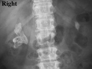 Struvite and staghorn calculi. Plain abdominal radiograph demonstrating a right staghorn calculus and a smaller left renal pelvic stone. The patient is a 72-year-old woman.
Struvite stones are also known as triple-phosphate (3 cations associated with 1 anion), infection (or infection-induced), phosphatic, and urease stones. Other, less common staghorn calculi can be composed of mixtures of calcium oxalate and calcium phosphate.
In the past, removal of large complex renal calculi required major open operations, with attendant morbidity and no guarantee of complete stone removal. The current approach favors minimally invasive modalities such as extracorporeal shockwave lithotripsy (SWL) and percutaneous nephrolithotomy (PNL). In a select group of patients with unilateral asymptomatic stones and minimal infection, a more conservative management strategy may be appropriate. [3] See Treatment.
History of the Procedure
The concept that urinary tract infections play a role in lithogenesis is not new. Hippocrates noted the relationship between renal calculi and loin abscesses. In 1817, Marcet recognized the association of phosphate calculi with infection, alkaline urine, and ammoniacal urine. Not until the early 20th century did Brown propose that urea-splitting bacteria were responsible for urinary ammonia, alkalinity, and stone formation. [4] The isolation of urease, the first enzyme ever purified, earned Sumner [5] the Nobel Prize for Chemistry in 1946. Urease-producing organisms are listed in Etiology.
Problem
Struvite stones are invariably associated with urinary tract infections. Specifically, the presence of urease-producing bacteria, including Ureaplasma urealyticum and Proteus species (most common), Staphylococcus species, Klebsiella species, Providencia species, and Pseudomonas species, leads to the hydrolysis of urea into ammonium and hydroxyl ions. Escherichia coli does not produce urease and is not associated with struvite stone formation. Other common bacteria that have not been shown to produce urea include Citrobacter freundii, enterococci, and streptococci.
The resulting increase in ammonium and phosphate concentrations combined with the alkalotic urine (pH >7.2) is necessary for struvite and carbonate apatite crystallization. Magnesium ammonium phosphate crystals (MgNH4 PO4 •6H2 O) are admixed with carbonate apatite (Ca10 (PO4) 6•CO3) in varying proportions along with matrix. The proportion of matrix, typically low molecular weight mucoproteins, is greater than in other types of calcium-based stones and is thought to protect the bacteria from antimicrobials.
Epidemiology
Frequency
Although calcium oxalate stones are most prevalent in the Western world, struvite calculi account for up to 30% of urinary tract stones worldwide. In the United States, 10-15% of all stones are composed of struvite. They are found more frequently in women and in persons older than 50 years, likely reflecting the population at increased risk of recurrent or persistent urinary tract infections. Accordingly, treatment of struvite stones must also address the source of these infections.
The natural history of struvite calculi mandates the complete removal of stones. First, infection stones generally grow rapidly, and any remaining stone material may serve as a nidus for future stone formation. Second, even after complete stone removal, struvite stones recur in approximately 10% of patients; if residual stones or fragments are left after treatment, recurrence rates approach 85%.
Third, struvite stones are a potential source of significant morbidity. Previously, it was believed that asymptomatic struvite stones could be managed expectantly; however, studies have demonstrated that 30% of patients treated conservatively (ie, no surgery to remove stones) died of renal failure or of pyelonephritis and sepsis.
In rare cases, chronic irritation, infection, and inflammation from staghorn calculi can cause squamous metaplasia, leading to squamous cell carcinoma of the renal collecting system. These malignancies carry a very poor prognosis, with a 5-year survival rate of less than 10%. [6]
Priestley and Dunn reported a 41% 5-year survival rate in patients with untreated unilateral struvite stones. [7] These data underscore the importance of approaches, primarily surgical, to completely remove the stone material.
Etiology
Gram-positive bacteria that cause struvite stones are as follows:
-
Staphylococcus aureus
-
Staphylococcus epidermidis
-
Corynebacterium species (ie, C ulcerans, C renale, C ovis, C hofmannii, C murium, C equi)
-
Mycobacterium rhodochrous group
-
Micrococcus varians
-
Bacillus species
-
Clostridium tetani
-
Peptococcus asaccharolyticus
Gram-negative bacteria that cause struvite stones are as follows:
-
Bacteroides corrodens
-
Helicobacter pylori
-
Bordetella pertussis
-
Bordetella bronchiseptica
-
Haemophilus influenzae
-
Haemophilus parainfluenzae
-
Proteus species (ie, P mirabilis, P morganii, P rettgeri)
-
Providencia stuartii
-
Klebsiella species ( K pneumoniae, K oxytoca)
-
Pasteurella species
-
Pseudomonas aeruginosa
-
Aeromonas hydrophilia
-
Yersinia enterocolitica
-
Brucella species
-
Flavobacterium species
-
Serratia marcescens
-
Ureaplasma urealyticum
-
Mycoplasma T-strain
Yeasts that cause struvite stones are as follows:
-
Cryptococcus species
-
Rhodotorula species
-
Sporobolomyces species
-
Trichosporon cutaneum
-
Candida humicola
Pathophysiology
Two conditions must coexist for the formation of struvite calculi. These are (1) alkaline urine (pH >7.2) and (2) the presence of ammonia in the urine. This leads to magnesium ammonium phosphate and carbonate apatite crystallization. The conversions of urea to ammonia, ammonia to ammonium, and acidification from carbon dioxide are as follows:
H2 NCONH2 + H2 O → 2NH3 + CO2 2NH3 + H2 O → 2NH4+ + 2OH- (increase pH >7.2)CO2 + H2 O → H+ + HCO3 → 2H+ + CO32-
Presentation
The clinical presentation of patients with struvite stones can be variable. Consider struvite stones in patients with risk factors for developing urinary tract infections (eg, prior urinary diversion or urologic surgery, presence of indwelling catheters, neurogenic bladder, vesicoureteral reflux, other anatomic abnormalities).
Infections may result in pyelonephritis, pyonephrosis, or perinephric abscess. Symptoms may include flank pain classic for renal colic, fever, urinary symptoms (eg, frequency, dysuria), and hematuria (either gross or microscopic). However, struvite stones rarely manifest as a solitary ureteral stone with acute renal colic in the absence of prior intervention. Concomitant urinary obstruction and hydronephrosis may be present and can result in nausea or vomiting.
In institutionalized patients susceptible to infection stones, the ability to elicit symptoms may be limited; sepsis may be the only evidence of an underlying struvite staghorn calculus. Note that patients with struvite calculi can be asymptomatic, even when calculi occupy the entire renal collecting system. Even with progression to xanthogranulomatous pyelonephritis, 25% of patients may remain completely free of symptoms. Systemic manifestations of large struvite stones and associated chronic infection include generalized fatigue, malaise, and weight loss.
Indications
Staghorn calculi represent a less-common nephrolithiasis subgroup so named because the significant stone burden that fills the renal pelvis and calyces forms a shape on radiographs that resembles a deer's horns. Most staghorn stones in Western society are composed of struvite and can cause significant morbidity and mortality if left untreated; therefore, large struvite stones must typically be removed.
Interestingly, an article investigating the structural analysis of renal calculi in northern India reported that over 90% of staghorn stones were composed of oxalates. [8] In a study from southern Thailand, the most common component of staghorn calculi was uric acid; struvite was found in only 11.6% of cases. [9] A US study, published in 2011, reported that slightly over half of complete staghorn calculi were metabolic in origin, consisting of calcium phosphate (55%), uric acid (21%), calcium oxalate (14%), or cystine (10%). [10]
Unlike other urinary stones that commonly produce symptoms (eg, renal colic) that necessitate intervention, treatment of struvite stones often occurs in patients without classic signs of nephrolithiasis; this is because large staghorn calculi may not cause acute renal or ureteral dilatation and resultant pain.
Relevant Anatomy
A comprehensive discussion of renal anatomy is beyond the scope of this article; however, several points relevant to endourologic techniques are discussed.
First, the kidneys are retroperitoneal organs enclosed within several layers, including the adjacent adherent renal capsule and the renal Gerota fascia surrounding the perinephric fat. Severe renal infections associated with struvite stones may lead to abscess formation, both within the kidney and within the Gerota fascia (ie, perinephric abscess).
Second, the kidneys are intimately associated with many nearby organs. On the right side, the liver may be posterolateral to the kidney at the level of the superior pole; on the left side, the spleen resides in an analogous position. These organs may be injured during percutaneous renal access. On both sides, the colon has retroperitoneal portions that can be located posterior to the kidneys. Studies have demonstrated that retrorenal colon positions are present in up to 10% of patients.
A single kidney contains 5-14 calyces, each of which drains a renal papilla. These minor calyces may coalesce to form major calyces, all of which subsequently drain into an infundibulum.
The placement of percutaneous tubes into the kidney should be guided by the following three principles:
- Access should not be placed through an infundibulum, because of greater risks of vascular injury
- In all areas of the kidney (both superior and inferior), access should be gained near the fornix of the calyx
- Entry into a posterior calyx allows the greatest ability to examine and remove stones in the renal pelvis and in additional infundibula and calyces
Contraindications
The presence of an active, untreated urinary tract infection is a contraindication to stone removal. Patients with struvite stones have chronic bacteriuria, and their urine is never sterilized by antibiotics alone; however, appropriate antibiotics should be administered prior to surgical intervention in an attempt to minimize the potential for sepsis during treatment. Similarly, if concomitant urinary obstruction and purulent infection exist (ie, pyonephrosis), percutaneous drainage and antibiotics are necessary before further manipulation of the stone and urinary tract.
If pyonephrosis is seen at the time of percutaneous renal access, percutaneous drainage should be left in place to maximally drain the infected collecting system, and nephrolithotomy should be deferred. The patient should be left on antibiotics and admitted for monitoring for urosepsis. Stone removal should be deferred to a later time to minimize infectious complications.
-
Struvite and staghorn calculi. Plain abdominal radiograph demonstrating a right staghorn calculus and a smaller left renal pelvic stone. The patient is a 72-year-old woman.
-
Struvite and staghorn calculi. Plain abdominal radiograph of a 72-year-old woman. She underwent right percutaneous nephrolithotomy, with the path of renal access demonstrated by the remaining nephrostomy tube. She was rendered stone free in the single-session procedure.


