Practice Essentials
Cardiac tamponade is a clinical syndrome caused by the accumulation of fluid in the pericardial space, resulting in reduced ventricular filling and subsequent hemodynamic compromise. The condition is a medical emergency, the complications of which include pulmonary edema, shock, and death.
Signs and symptoms
Symptoms vary with the acuteness and underlying cause of the tamponade. Patients with acute tamponade may present with dyspnea, tachycardia, and tachypnea. Cold and clammy extremities from hypoperfusion are also observed in some patients. Other symptoms and signs may include the following:
-
Elevated jugular venous pressure
-
Pulsus paradoxus
-
Chest pressure
-
Decreased urine output
-
Confusion
-
Dysphoria
See Clinical Presentation for more detail.
Diagnosis
Prompt diagnosis is key to reducing the mortality risk for patients with cardiac tamponade. Although echocardiography provides useful information, cardiac tamponade is a clinical diagnosis. Echocardiography can be used to visualize ventricular and atrial compression abnormalities as blood cycles through the heart. The following may be observed with 2-dimensional (2-D) echocardiography:
-
An echo-free space posterior and anterior to the left ventricle and behind the left atrium
-
Early diastolic collapse of the right ventricular free wall
-
Late diastolic compression/collapse of the right atrium
-
Swinging of the heart in the pericardial sac
-
Left ventricular pseudohypertrophy
-
Inferior vena cava plethora with minimal or no collapse with inspiration
-
A greater than 40% relative inspiratory augmentation of blood flow across the tricuspid valve
-
A greater than 25% relative decrease in inspiratory flow across the mitral valve
See Workup for more detail.
Management
Removal of pericardial fluid is the definitive therapy for tamponade and can be done using the following three methods:
-
Emergency subxiphoid percutaneous drainage
-
Pericardiocentesis (with or without echocardiographic guidance)
-
Percutaneous balloon pericardiotomy
The role of medication therapy in cardiac tamponade is limited.
See Treatment and Medication for more detail.
Background
Cardiac tamponade is a clinical syndrome caused by the accumulation of fluid in the pericardial space, resulting in reduced ventricular filling and subsequent hemodynamic compromise. The condition is a medical emergency, the complications of which include pulmonary edema, shock, and death. (See Pathophysiology, Etiology, and Prognosis.)
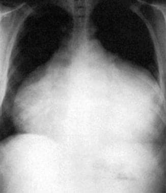
The overall mortality risk depends on the speed of diagnosis, the treatment provided, and the underlying cause of the tamponade. Untreated, the condition is rapidly and universally fatal (see the image below). (See Presentation, Workup, Treatment, and Medication.)
 This anteroposterior-view chest radiograph shows a massive, bottle-shaped heart and conspicuous absence of pulmonary vascular congestion. Reproduced with permission from Chest, 1996: 109:825.
Pathophysiology
The pericardium, which is the membrane surrounding the heart, is composed of 2 layers. The thicker parietal pericardium is the outer fibrous layer; the thinner visceral pericardium is the inner serous layer. The pericardial space normally contains 20-50mL of fluid.
Reddy et al describe 3 phases of hemodynamic changes in tamponade, as follows [1] :
-
Phase I - The accumulation of pericardial fluid impairs relaxation and filling of the ventricles, requiring a higher filling pressure; during this phase, the left and right ventricular filling pressures are higher than the intrapericardial pressure
-
Phase II - With further fluid accumulation, the pericardial pressure increases above the ventricular filling pressure, resulting in reduced cardiac output (see the Cardiac Output calculator)
-
Phase III - A further decrease in cardiac output occurs, which is due to the equilibration of pericardial and left ventricular (LV) filling pressures
Pericardial effusions, which cause cardiac tamponade, can be serous, serosanguineous, hemorrhagic, or chylous.
The underlying process for the development of tamponade is a marked reduction in diastolic filling, which results when transmural distending pressures become insufficient to overcome increased intrapericardial pressures. Tachycardia is the initial cardiac response to these changes to maintain the cardiac output.
Systemic venous return is also altered during tamponade. Because the heart is compressed throughout the cardiac cycle due to the increased intrapericardial pressure, systemic venous return is impaired and right atrial and right ventricular collapse occurs. Because the pulmonary vascular bed is a vast and compliant circuit, blood preferentially accumulates in the venous circulation, at the expense of LV filling. This results in reduced cardiac output and venous return.
The amount of pericardial fluid needed to impair diastolic filling of the heart depends on the rate of fluid accumulation and the compliance of the pericardium. Rapid accumulation of as little as 150mL of fluid can result in a marked increase in pericardial pressure and can severely impede cardiac output, [2] whereas 1000 mL of fluid may accumulate over a longer period without any significant effect on diastolic filling of the heart. This is due to adaptive stretching of the pericardium over time. A compliant pericardium can allow considerable fluid accumulation over a long time period without hemodynamic compromise.
Etiology
For all patients, malignant diseases are the most common cause of pericardial tamponade. Among etiologies for tamponade, Merce et al reported the following incidence rates:
-
Malignant diseases - 30-60% of cases
-
Uremia - 10-15% of cases
-
Idiopathic pericarditis - 5-15%
-
Infectious diseases - 5-10%
-
Anticoagulation - 5-10%
-
Connective tissue diseases - 2-6%
-
Dressler or postpericardiotomy syndrome - 1-2%
Tamponade can occur as a result of any process that causes pericarditis. Pericarditis can result from the following [3] :
-
Human immunodeficiency virus (HIV) infection
-
Infection - Viral, bacterial (tuberculosis), fungal
-
Drugs - Hydralazine, procainamide, isoniazid, minoxidil
-
Postcoronary intervention - Ie, coronary dissection and perforation
-
Acupuncture [4]
-
Postcardiac percutaneous procedures - Including mitral valvuloplasty, atrial septal defect (ASD) closure, left atrial appendage occlusion
-
Trauma to the chest
-
Cardiovascular surgery - Postoperative pericarditis [5]
-
Postmyocardial infarction - Free wall ventricular rupture, Dressler syndrome
-
Connective tissue diseases - Systemic lupus erythematosus, rheumatoid arthritis, dermatomyositis
-
Radiation therapy to the chest, radiofrequency ablation [6]
-
Iatrogenic [7] - After sternal biopsy, transvenous pacemaker lead implantation, pericardiocentesis, or central line insertion
-
Uremia
-
Anticoagulation treatment
-
Idiopathic pericarditis
-
Complication of surgery at the esophagogastric junction - Eg, antireflux surgery
-
Pneumopericardium - Due to mechanical ventilation or gastropericardial fistula
-
Hypothyroidism
-
Still disease
-
Duchenne muscular dystrophy
-
Type A aortic dissection
-
Streptococcus constellatus infection (very rare; only 3 cases in the literature) [8]
In patients undergoing heart valve surgery, cardiac tamponade has been associated with any amount of pericardial effusion at the first postoperative transthoracic echocardiography as well as with mechanical valve replacement of the aortic or mitral valve. [9]
Epidemiology
Occurrence in the United States
The incidence of cardiac tamponade is 2 cases per 10,000 population in the United States. Approximately 2% of penetrating injuries are reported to result in cardiac tamponade.
Sex- and age-related demographics
In children, cardiac tamponade is more common in boys than in girls, with a male-to-female ratio of 7:3. In adults, cardiac tamponade appears to be slightly more common in men than in women. A male-to-female ratio of 1.25:1 was observed at the author's referral center, based on the International Classification of Diseases (ICD) code 423.9. However, a male-to-female ratio of 1.7:1 was observed at another level 1 trauma center.
Cardiac tamponade related to trauma or HIV is more common in young adults, whereas tamponade due to malignancy and/or renal failure occurs more frequently in elderly individuals.
Prognosis
Cardiac tamponade is a medical emergency. The prognosis depends on prompt recognition and management of the condition and the underlying cause of the tamponade. Untreated, cardiac tamponade is rapidly and universally fatal.
Haneya et al retrospectively (2005-2011) evaluated the impact of timing and indication of reexploration for bleeding or tamponade following cardiac surgery in 209 patients and found that reexploration was associated with higher rates of mortality and morbidity. [10] Multivariate analysis indicated it was the adverse effects of reexploration (eg, blood loss, transfusion requirements) rather than the procedure itself that were independent risk factors for death. Adverse outcomes were more likely in those whose reexploration was delayed and who suffered from cardiac tamponade. [10]
In a separate study, Le et al indicated that following cardiac surgery, there is no advantage for the use of multiple mediastinal chest tubes over a single chest tube in preventing return to the operating room for bleeding or tamponade. [11]
In addition to treatment for the tamponade, all patients should also receive treatment for the condition’s underlying cause in order to prevent recurrence.
In a study of patients with cardiac tamponade, Cornily et al reported a 1-year mortality rate of 76.5% in patients whose tamponade was caused by malignant disease, compared with 13.3% in patients with no malignant disease. The investigators also noted a median survival of 150 days in patients with malignant disease. [12]
-
This anteroposterior-view chest radiograph shows a massive, bottle-shaped heart and conspicuous absence of pulmonary vascular congestion. Reproduced with permission from Chest, 1996: 109:825.
-
A 12-lead electrocardiogram showing sinus tachycardia with electrical alternans. Reproduced with permission from Chest, 1996; 109:825.
-
Early diastolic collapse of right ventricular free wall (subcostal view).
-
Early diastolic collapse of right ventricular free wall (parasternal short-axis view at aortic valve).
-
Late diastolic collapse of right atrium (subcostal view).
-
Dilated inferior vena cava.


