Practice Essentials
Penetrating neck trauma is an important area of trauma care that is continually evolving. A remarkable number of changes have occurred in the treatment paradigm as new technologies have developed and as surgeons have explored the outcomes from different treatment protocols. Therapy has evolved from no treatment (before effective anesthesia and instrumentation), to nonoperative management, to routine exploration, to selective exploration and adjunctive invasive or noninvasive assessment. Penetrating neck injuries remain challenging, as there are a number of important structures in a small area and injury to any of these structures may not be readily apparent. See the image below.
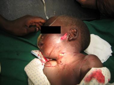 A Zone II penetrating neck injury in a young boy. This child fortunately had no other documented injuries.
Signs and symptoms
Evidence of significant injury to vital structures of the neck may be indicated by the following clinical manifestations:
-
Dysphagia – Tracheal and/or esophageal injury
-
Hoarseness – Tracheal and/or esophageal injury (especially recurrent laryngeal nerve)
-
Oronasopharyngeal bleeding – Vascular, tracheal, or esophageal injury
-
Neurologic deficit – Vascular and/or spinal cord injury
-
Hypotension – Nonspecific; may be related to the neck injury or may indicate trauma elsewhere
See Presentation for more detail.
Diagnosis
Laboratory studies
The following laboratory studies may be useful:
-
Hemoglobin concentration
-
A blood specimen for typing
-
A toxicologic screen
-
Coagulation studies
Imaging studies
In penetrating neck injuries, computed tomography (CT) angiography of the neck is the preferred imaging procedure to evaluate the extent of injury. Catheter-based arteriography is useful for further evaluation and an x-ray barium swallow single contrast may be considered in conjunction with direct visualization techniques if there remains a concern for aerodigestive injury.
See Workup for more detail.
Management
The standard of care is immediate surgical exploration for patients who present with signs and symptoms of shock and continuous hemorrhage from the neck wound. The type of incision depends on the neck zone and the structures at risk for injury.
See Treatment for more detail.
History of the Procedure
For centuries, carotid ligation was the only reliable treatment of severe penetrating neck injury. In 1552, Ambrose Pare ligated both common carotid arteries and the jugular vein of a soldier with a traumatic neck injury. The patient survived but developed aphasia and hemiplegia. In 1803, Fleming ligated a lacerated common carotid artery and reported a successful outcome with a 5-month follow-up. The noted author George Orwell suffered a penetrating neck injury causing a unilateral vocal fold paresis in 1936 as a result of his involvement in Spain's Civil War.
Nonoperative management of penetrating neck wounds was the standard until World War I.
During World War II, a more aggressive approach to neck exploration was adopted. The types of injuries seen on the battlefields of World War II and the then available diagnostic armamentarium are significantly different from those in the modern civilian trauma center. The changes associated with improved imaging modalities and nonmilitary injuries have resulted in a dramatic change in the treatment paradigm for penetrating neck injury. Continual advances in anesthesia and perioperative management since World War II have improved the care and the outcome of these patients.
Problem
Penetrating neck trauma involves a missile or sharp object penetrating the skin and violating the platysma layer of the neck. This includes gunshot wounds, stab or puncture wounds, and impalement injuries. [1, 2, 3]
Although penetrating injury to the neck represents a relatively small portion of all trauma cases (5-10%), it produces injuries with a high degree of mortality as well as complex clinical situations that can be challenging for providers. [4]
Pathophysiology
Two factors in the mechanism of injury or kinematics in penetrating neck trauma determine the extent of damage to the tissue: (1) weapon characteristics and (2) the location of injury and human tissues involved.
Weapon characteristics
The amount of kinetic energy delivered by the wounding agent has to be considered together with its interaction with the involved tissue. Kinetic energy (KE) is described by the following equation: KE = 1/2 mass × velocity (squared).
Low-energy weapons include hand-driven weapons, such as knives or ice picks, which damage with only their sharp point or cutting edge. Firearms may be classified as medium-energy (ie, handguns) and high-energy weapons (ie, military assault weapons), with the latter usually defined as having 461 joules or more.
Projectiles (ie, bullets, missiles) often are differentiated by mass, velocity, shape, and construction because these characteristics affect the extent of tissue disruption. Bullet velocity is the most important characteristic considered, with high velocity defined as greater than 2500 ft/s.
Location of injury and human tissues involved
Tissue injury results from either a direct impact by the penetrating projectile or tissue displacement from temporary cavitation.
Wound sites and, if present, the wounding agent in the neck provide an indication of the likely injury complex.
Etiology
Penetrating neck injuries, like any trauma, may be classified as intentional or nonintentional. The objects causing these injuries can be divided into stabbing instruments (eg, knives, cutting instruments, puncturing objects, impaling objects) and shooting instruments (eg, missiles, projectiles). Wounding instruments have specific characteristics that affect surgical findings. For example, stab wounds typically have a 10% higher rate of negative exploration than injuries from projectiles. Conversely, the higher energy and ballistic force associated with a gunshot wound to the neck should cause clinical suspicion and thorough investigation to determine if injury is present.
Epidemiology
The current mortality rate in civilians with penetrating neck injuries ranges from 3-6%. During World War II, the mortality rate was 7%, and, in World War I, it was 11%. Higher mortality rates occur with injuries to large vessels, such as the carotid or subclavian arteries and veins.
More recent experience in the treatment of casualties from the Iraq War at Walter Reed Army Medical Center reported the common carotid artery as the most frequently injured cervical vessel. [5]
Presentation
Evidence of significant injury to vital structures of the neck may be indicated by the following clinical manifestations:
-
Dysphagia – Tracheal and/or esophageal injury
-
Hoarseness – Tracheal and/or esophageal injury (especially recurrent laryngeal nerve)
-
Oronasopharyngeal bleeding – Vascular, tracheal, or esophageal injury
-
Neurologic deficit – Vascular and/or spinal cord injury
-
Hypotension – Nonspecific; may be related to the neck injury or may indicate trauma elsewhere
Proposed hard signs of airway injury include the following:
-
Subcutaneous emphysema – Tracheal, esophageal, or pulmonary injury
-
Air bubbling through the wound
-
Stridor or respiratory distress – Laryngeal and/or esophageal injury
Several so-called hard signs that strongly indicate vascular injury are as follows:
-
Hematoma (expanding) – Vascular injury
-
Active external hemorrhage from the wound site – Arterial vascular injury
-
Bruit/thrill – Arteriovenous fistula
-
Pulselessness/pulse deficit
-
Distal ischemia (neurologic deficit in this case)
The evaluation of a patient with penetrating neck trauma always should start with advanced trauma life support (ATLS), a paradigm that begins with a directed primary survey emphasizing airway, breathing, and circulation (ABC). After patients are stabilized, they undergo a secondary survey that includes a complete history and a thorough physical examination. These steps, together with the studies discussed in Workup, are used to identify the likely injury complex and to direct further treatment or diagnostic testing.
There is evidence to suggest that the hard signs of airway injury are more reliable and result in less negative operative explorations compared with hard signs of vascular injury. The rate of negative exploration for patients with hard signs of vascular injury varies widely, but it may be estimated at 10%. However, series that report these cases as "nonsignificant" injury or as negative explorations lack clear definition, and it is difficult to draw any useful conclusion from the data.
Relevant Anatomy
In few other regions of the body are so many vital structures (that would be of immediate concern following injury) located in so small a volume. An injury is not considered to have penetrated the neck unless the injury penetrates the platysma muscle layer. Injuries through the platysma and injuries crossing the midline usually cause a greater degree of damage. The sternocleidomastoid muscle delineates the posterior and anterior regions of the neck. The area of the neck posterior to the cervical vertebral body and the scalene muscles is composed mainly of muscle, bone, and nonvital vessels and lymphatics. Most of the vital structures are located in the anterior or lateral regions.
The neck may be divided into 3 zones using anatomic landmarks. Each zone has a group of vital structures that can be injured and may determine the kind of trauma management.
-
Zone I is the horizontal area between the clavicle/suprasternal notch and the cricoid cartilage encompassing the thoracic outlet structures. The proximal common carotid, vertebral, and subclavian arteries and the trachea, esophagus, thoracic duct, and thymus are located in zone I. This area is considered to be fairly well protected by the bony thorax but is vulnerable to penetrating injuries through the upper thoracic aperture.
-
Zone II is the area between the cricoid cartilage and the angle of the mandible. It contains the internal and external carotid arteries, jugular veins, pharynx, larynx, esophagus, recurrent laryngeal nerve, spinal cord, trachea, thyroid, and parathyroids. This area is at particular risk for injuring in penetrating trauma, as it contains many vital structures without significant overlying protective tissue.
-
Zone III is the area that lies between the angle of the mandible and the base of the skull. It has the distal extracranial carotid and vertebral arteries and the uppermost segments of the jugular veins.
Tight fascial compartments of neck structures may limit external hemorrhage from vascular injuries, minimizing the chance of exsanguination. However, these tight fascial boundaries may increase the risk of airway compromise because the airway is relatively mobile and compressible by an expanding hematoma.
Indications
The standard of care is immediate surgical exploration for patients who present with signs and symptoms of shock and continuous hemorrhage from the neck wound. The type of incision depends on the neck zone and the structures at risk for injury.
The following specific injuries must be confirmed and treated during neck exploration:
-
Carotid artery injuries
-
Vertebral artery injuries
-
Jugular vein injury
-
Laryngotracheal injuries
-
Esophageal injuries
-
Nerve injuries
-
Thoracic duct injuries
-
Thyroid injuries
Contraindications
No role exists for probing or local exploration of the neck in the trauma bay or emergency department because this may dislodge a clot and initiate uncontrollable hemorrhage. If no significant injuries requiring urgent intervention are present, the patient may proceed to diagnostic workup as indicated.
Prognosis
Vascular trauma is present in 25% of penetrating neck injuries, with mortality rates approaching 50% in some studies. Tracheobronchial injuries may have an incidence of less than 10% to as high as 20% and a mortality rate of as high as 20%. The injured cervical esophagus can result in devastating complications and eventual outcomes, such as leakage of saliva, bacteria, refluxed acid, pepsin, and even bile. Undiagnosed, this can produce early suppurative infection and an intense necrotizing inflammatory response in the neck, as well as a more devastating outcome if it descends to the mediastinum. An 11-17% increase in the overall mortality rate has been observed after delays of 12 hours in the diagnosis of esophageal injuries.
Two reports demonstrate the importance of the setting in which penetrating neck injuries occur, particularly treatment protocols in combat zones. Sarkar et al presented 2 cases from Western Baghdad, [6] and Ramasamy et al performed a retrospective medical record review of British military casualties from Iraq and Afghanistan who sustained penetrating neck injuries to determine the need for prehospital cervical immobilization, given current ATLS protocols requiring spinal precautions when a significant mechanism of injury may damage the cervical spine. [7]
In the study by Ramasamy et al, of 90 patients with a penetrating neck injury, 66 (73%) were from explosions and 24 (27%) were from gunshot wounds. In 20 (22%) patients, cervical spine injuries were present; only 6 (7%) survived to reach the hospital, and 4 of these 6 died within 72 hours of their injuries. [7] Of 56 survivors that reached a surgical facility, only 1 (1.8%) had an unstable cervical spine injury requiring surgical stabilization, and this patient subsequently died due to a concomitant head injury.
The investigators determined a high mortality rate is associated with penetrating ballistic trauma to the neck. [7] Furthermore, it appears unlikely that survivors of penetrating ballistic trauma to the neck will have unstable cervical spines; therefore, not only is the risk/benefit ratio of mandatory spinal immobilization unfavorable, but cervical collars may also hide potential life-threatening conditions, in addition to putting medical teams at prolonged personal risk. [7]
Complications
Missed injuries or delayed diagnosis can occur after any injury to the neck, particularly in patients presenting with minimal manifestations.
-
Persistent hemorrhage - Usually from a missed arterial or venous injury, particularly in zone I and zone III
-
Pseudoaneurysms - A later sequela from a missed vascular injury, which often is not bleeding actively during treatment
-
Arterial dissection - Incomplete transmural vessel injury may cause this disruption between the layers of the arterial wall.
-
Fistulas - Esophagocutaneous, esophagotracheal, tracheocutaneous, venoarterial
-
Infections - Most often occur from missed esophageal or laryngotracheal injuries; severe inflammation, abscess formation, or mediastinitis may result.
-
Stenosis or obstruction of luminal structures - May happen due to the inflammatory response and scarring around the injured esophagus, larynx, trachea, or vessels
-
Neurologic deficits - May occur due to the direct injury to a peripheral nerve or to ischemic infarct caused by arterial injury
-
Anastomotic or repair disruption - About 1% of surgical repairs leak and result in hemorrhage, infection, or fistula formation.
-
Luminal stenosis or obstruction - The surgical repair and the inflammation can cause the narrowing of the lumen of the injured esophagus, larynx, trachea, or vessels.
-
Infectious complications - Occurring particularly with injuries to the trachea and esophagus, severe inflammatory response in the neck, abscess formation, fistulas, or mediastinitis may result.
-
Neurologic complications - Can occur as strokes related to major vascular injuries or directly to peripheral nerves
-
Thrombosis of an internal jugular vein - Can occur regardless of the method of venorrhaphy
-
Massive air emboli - May result from major venous injuries and is an important cause of bilateral, diffuse stroke identified as hypodense lesions on CT scan of the brain
-
A Zone II penetrating neck injury in a young boy. This child fortunately had no other documented injuries.
-
Tribal violence in Kenya resulted in this zone II arrow injury.
-
Same patient as in Media file 2. Note the entry point and the palpable tip of the arrow in the posterior triangle.
-
Same patient as in Media file 2. The arrow has been removed after negative neck exploration.
-
Surgical cricothyroidotomy Seldinger. Video courtesy of Therese Canares, MD, and Jonathan Valente, MD, Rhode Island Hospital, Brown University.


