You are in: eMedicine Specialties > Vascular Surgery > MEDICAL TOPICS Thoracic Outlet ObstructionArticle Last Updated: May 23, 2007AUTHOR AND EDITOR INFORMATIONAuthor: Mark K Eskandari, MD, Associate Professor, Departments of Radiology and Division of Vascular Surgery, Feinberg School of Medicine, Northwestern University; Attending Surgeon, Division of Vascular Surgery, Northwestern Memorial Hospital; Consulting Staff, Division of Vascular Surgery, Northwestern Medical Faculty Foundation; Consulting Staff, Department of Surgery, Jesse Brown Veterans Affairs Medical Center; Consulting Staff, Department of Surgery, Evanston Northwestern Healthcare Mark K Eskandari is a member of the following medical societies: American College of Surgeons, American Medical Association, Association for Academic Surgery, Association of VA Surgeons, Central Surgical Association, International Society of Endovascular Specialists, Peripheral Vascular Surgery Society, Society for Clinical Vascular Surgery, Society for Vascular Surgery, Society of Interventional Radiology, Society of University Surgeons, and Western Surgical Association Coauthor(s): Nicholas D Garcia, MD, Section of Vascular Surgery, Medical Director, Exeter Health Resources Vascular Lab, Portsmouth Regional Hospital, Exeter Hospital, Merrimack Valley Hospital; Hassan Tehrani, MBBCh, Assistant Professor of Surgery, Department of Surgery, Division of Vascular Surgery, Jackson Memorial Hospital/University of Miami; Mark D Morasch, MD, Clinical Practice Director, Division of Vascular Surgery, Assistant Professor of Surgery, Department of Surgery, Northwestern University Feinberg School of Medicine Editors: Jeffrey Lawrence Kaufman, MD, Associate Professor, Department of Surgery, Division of Vascular Surgery, Tufts University School of Medicine; Francisco Talavera, PharmD, PhD, Senior Pharmacy Editor, eMedicine; Vincent Lopez Rowe, MD, Assistant Professor of Surgery, Department of Surgery, Division of Vascular Surgery, University of Southern California Medical Center; Paolo Zamboni, MD, Professor of Surgery, Chief of Day Surgery Unit, Chair of Vascular Diseases Center, University of Ferrara, Italy; William H Pearce, MD, Chief, Division of Vascular Surgery, Violet and Charles Baldwin Professor of Vascular Surgery, Department of Surgery, Northwestern University School of Medicine Author and Editor Disclosure Synonyms and related keywords: venous thoracic outlet syndrome, venous TOS, arterial thoracic outlet syndrome, neurogenic thoracic outlet syndrome, Paget-Schroetter syndrome INTRODUCTIONHistory of the ProcedureThoracic outlet syndrome (TOS) has been an important clinical entity for more than a century. In 1821, Sir Astley Cooper first described axillary–subclavian artery symptoms due to compression from a cervical rib. In 1875, James Paget described the clinical symptoms resulting from subclavian vein thrombosis (eg, arm swelling, pain). In 1884, von Schroetter correctly attributed these upper extremity venous symptoms to thrombosis or compression of the subclavian vein at the thoracic outlet. Consequently, venous thrombosis at the thoracic outlet is known as venous TOS or Paget-Schroetter syndrome. ProblemTOS has distinct clinical pictures (ie, neurogenic, arterial, venous) caused by compression of the neurovascular structures at the thoracic outlet. The classification is based on which structure is primarily involved. FrequencyNeurogenic TOS is the most common presentation, occurring in approximately 95% of patients. Arterial TOS is the next most common presentation and occurs in about 2-3% of patients who are affected. Venous TOS is the least common, representing only 1-2% of patients with TOS. Approximately 70% of patients with neurogenic TOS are females aged 20-50 years. Venous TOS occurs with a male-to-female ratio of 2:1. EtiologyThe potential for either neurologic or vascular compression exists at the thoracic outlet. When compression occurs, one of the TOSs may develop. Neurogenic and arterial TOS result from compression that occurs in the scalene triangle (see Image 1), and venous TOS results from compression in the costoclavicular space (see Image 2).1 PathophysiologyNeurogenic TOS most commonly is associated with a history of neck trauma. Swollen and scarred muscles or aberrant scalene anatomy can irritate cords of the brachial plexus locally and lead to the neurologic symptoms. Arterial TOS often is associated with cervical ribs or a rudimentary first rib. This aberrant anatomy leads to repeated intermittent arterial compression coinciding with arm movement. This repetitive localized trauma leads to intimal lesions, focal arterial stenosis, poststenotic dilatation, aneurysmal change, and subsequent thromboembolic complications. The second portion of the subclavian artery, which has a retroscalene position, often is the site of positional compression and stricture. Venous TOS usually results from compression of the subclavian vein by the subclavius muscle and costoclavicular ligament. When local structures are placed in abnormal or unaccustomed positions by extremes of activity or injury, vein compression and subsequent vein thrombosis can result. Venous TOS tends to occur in the more active dominant extremity. Arterial and venous TOS usually are associated with certain predisposing anatomic abnormalities, while neurogenic TOS is more likely to result following traumatic injury. ClinicalNeurogenic TOS is a clinical diagnosis that only is made when objective findings are supported by subjective symptoms and physical findings. Other diagnoses that should be considered include tendonitis, fibromyalgia, cervical disc herniation, spinal stenosis, carpal tunnel syndrome, repetitive motion syndrome, and epicondylitis. A history of a motor vehicle collision (MVC) or other neck trauma usually is elicited, and patients may report a variety of symptoms, such as neck, shoulder, and arm pain. Symptoms of compression from all cords of the brachial plexus are the most common neurologic pattern noted with TOS. Previous authors have written that ulnar nerve involvement is the typical pattern of symptoms related to neurogenic TOS, but this is not the case. Paresthesias and weakness in an arm, occipital headaches, and paraspinal muscle pain are common. The paraspinal muscle pain and occipital headaches are secondary to referred nerve pain. As expected, specific symptoms coincide with the area of the brachial plexus that is compressed. On physical examination, symptoms usually can be reproduced with pressure on the scalenes and with abduction/external rotation (AER) of the arms. Coldness and color changes in the hand usually are secondary to sympathetic nerve involvement rather than arterial involvement. Focus the examination on the neurologic and vascular findings. Carefully examine the scalene muscles and supraclavicular fossa for abnormal pulsations, bruits, and pain with palpation. Examine all upper extremity pulses, and perform provocative positioning during the vascular examination. Also seek petechiae and other evidence of embolic events. The mean age of patients with arterial TOS at presentation is approximately 10 years older than patients with neurogenic symptoms. Occasionally, arterial TOS is recognized during workup for other pathologies. An incidental pulsatile mass or a supraclavicular bruit may be noted during thorough physical examination. Unfortunately, arterial TOS usually remains unrecognized until a thromboembolic complication occurs. Patients who embolize may present with hand claudication, gangrene, and other embolic stigmata. In 80% of cases of venous TOS, the dominant extremity is involved. The diagnosis is based on the clinical presentation of upper extremity swelling, venous engorgement, and pain. These signs and symptoms, in association with radiologic documentation of venous compression at the thoracic outlet, confirm the diagnosis of Paget-Schroetter syndrome. Sometimes, venous compression cannot be demonstrated, and the diagnosis is made clinically and by the pattern of venous thrombosis. Pulmonary embolism has been reported in patients with primary venous thrombosis, but this more commonly occurs in patients presenting with secondary venous thrombosis. Secondary venous thrombosis occurs due to processes such as malignancy, polycythemia vera, heart failure, infection, drug abuse, thrombocytosis, estrogens, and most commonly, central venous catheters and indwelling cardiac pacing wires. Upper extremity deep venous thrombosis (DVT) accounts for 1-4% of all DVTs, and primary venous thrombosis accounts for 25% of these cases. Catheter-related DVTs account for most upper extremity DVTs. INDICATIONSThe indication for surgical treatment of neurogenic thoracic outlet syndrome (TOS) is the failure of conservative treatment in a patient with disability so severe that the patient is unable to work or live comfortably. The indication for surgical treatment of venous TOS is controversial but based on symptomatology and venographic evidence of compression at the thoracic outlet. Arterial TOS, however, in most circumstances should be treated surgically with 1st rib resection and arterial repair. RELEVANT ANATOMY
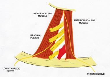
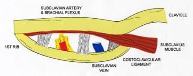
The relevant anatomy of TOS focuses on the scalene triangle and the costoclavicular space. Reports of compression of the neurovascular bundle at the area of the pectoralis minor space exist, but this is very uncommon and shall not be discussed further. Neurogenic and arterial TOS result from compression that occurs in the scalene triangle, which is defined by the first rib, the anterior scalene muscle, and the middle scalene muscle. The subclavian artery and the branches of the brachial plexus pass through the borders of this triangle (see Image 1). Venous TOS occurs secondary to compression that occurs in the costoclavicular space. The borders of the costoclavicular space are the first rib, costoclavicular ligament, subclavius muscle, and the anterior scalene. As the subclavian vein passes through this space, it is susceptible to compression by these structures (see Image 2). Other important local structures include the phrenic nerve, the lateral thoracic nerve, and the thoracic duct. The phrenic nerve passes from lateral to medial along the anterior border of the anterior scalene muscle, and the lateral thoracic nerve passes through the body of the middle scalene muscle. The thoracic duct joins cervical lymphatics and drains into the superior aspect of the jugulosubclavian vein confluence behind the left sternocleidomastoid muscle. Be careful to avoid injury to these structures during surgery. Search for cervical, rudimentary, or broad first ribs. Resect these structures during surgical therapy. Rudimentary ribs usually arise higher in the neck than normal first ribs and typically articulate with the second rib rather than with the sternum. Cervical ribs and rudimentary first ribs occur in less than 0.5% of the population. Abnormal scalene muscle anatomy also has been identified and may be a cause of some symptoms. For example, these muscles have been noted to interdigitate around the cords of the brachial plexus and, thus, have been implicated in the irritation of the cords of the brachial plexus. In a study of 98 meticulously dissected cadavers, the authors noted a number of abnormalities of the thoracic outlet fibrous bands and cervical ribs, and other abnormalities were found in most of the patients. Only 10% of the dissected cadavers were found to have normal anatomy bilaterally. CONTRAINDICATIONSNo absolute contraindication to the treatment of patients with thoracic outlet syndrome (TOS) exists. An individual patient may have significant comorbidities that outweigh the benefits of surgical repair and thus have a relative contraindication to surgery. WORKUPLab Studies
Imaging Studies
Other Tests
Histologic FindingsMuscle biopsy specimens show that type I fibers predominate and type II fibers atrophy in patients with neurogenic TOS. Increased fibrosis also has been noted in the scalene muscles of these patients. These findings suggest that repetitive trauma may have a role in the development of neurogenic symptoms. TREATMENTMedical therapyPhysical therapy has an important role in the initial treatment of neurogenic thoracic outlet syndrome (TOS). Postural exercises, stretching, abdominal breathing, and medications used to relieve muscular tension and pain are beneficial. Abdominal breathing and postural exercises relax the neck musculature, which helps to relieve symptoms. Approximately 60% of patients improve significantly with conservative treatment alone. The indication for surgical treatment of neurogenic TOS is the failure of conservative treatment in a patient with disability so severe that the patient is unable to work or live comfortably. Most physicians prescribe 3-12 months of physical therapy prior to considering surgical decompression of the thoracic outlet. No satisfactory medical treatment for arterial TOS exists. These patients usually present with a history of thromboembolic complications and require surgical repair. Arterial TOS requires prompt surgical intervention to treat or prevent acute thromboembolic events. Confusion may occur when a patient presents with an upper extremity thromboembolic event and no identifiable source. If a cervical rib or an aberrant first rib is identified under these circumstances, opening the artery and examining for intimal lesions has been proposed. If an intimal lesion is found, then the patient should undergo thoracic outlet decompression and repair of the artery. Endovascular repair of subclavian arterial aneurysms has been described, but this treatment modality does not abolish the need for surgical decompression. Aneurysm resection and arterial replacement remains the preferred treatment. Endovascular treatment of large arterial aneurysms may be useful when a difficult exposure is anticipated. Treatment for venous TOS–related effort thrombosis that relies on anticoagulation and arm elevation leaves 74% of patients with residual disability and 12% with significant complication. Thrombolytic therapy generally is preferred over venous thrombectomy; however, thrombectomy still may have a role in some cases with low surgical risk and contraindication to thrombolytic therapy. Although thrombolytic therapy alone is superior to simple anticoagulation in patients who present with venous TOS, the patients who achieve the best results are those who are treated with thrombolytics and surgical decompression. Unfortunately, most primary care or emergency department (ED) physicians do not appreciate or share this view, so most patients with venous TOS are not treated aggressively. Surgical therapyVenous thoracic outlet obstruction Surgical treatment of venous TOS consists of releasing the extrinsic compression and restoring luminal patency. Traditional therapy suggested that a staged approach would be more beneficial, with surgical decompression deferred until several weeks after thrombolytic intervention. The presumed advantage allowed for resolution of the inflammatory response before embarking on the surgical procedure. More recent data suggest that a more uniform approach with treatment completed during a single hospitalization may be a better option. If a residual lesion is less than 2 cm long, perform a thrombectomy with vein patch angioplasty and venolysis during decompression surgery. An alternative option is to perform vein angioplasty in a staggered fashion following decompressive surgery or, as Schneider et al have recently suggested, at the same time as open decompressive surgery. Other authors have cautioned against vein angioplasty at the time of decompressive surgery out of concern that a bleeding complication is more likely. A lesion longer than 2 cm may require venous bypass or a jugular vein turndown procedure. A consensus statement favored conservative treatment with anticoagulation under these circumstances and concluded that venous bypass should be reserved for only those patients with disabling symptoms and serious complications. Neurogenic/arterial thoracic outlet obstruction Thoracic outlet decompression can be performed through an axillary, supraclavicular, or posterior approach, and the choice usually is based on surgeon preference. In neurogenic TOS, results are equal, and the approach or operation performed for TOS may be selected irrespective of the presenting neurologic symptoms. For venous TOS, the operation can be performed using the axillary, supraclavicular, or combined supraclavicular/infraclavicular approach. If the supraclavicular approach is utilized, an infraclavicular counter-incision always can be performed for added exposure. The supraclavicular approach is becoming more popular and may be superior for total surgical decompression of the thoracic outlet. Thoracic outlet decompression may entail anterior and middle scalenectomy, first rib resection, or scalenectomy plus first rib resection. Reports of scalenectomy versus first rib resection have noted similar results for both procedures irrespective of the procedure performed. Sanders et al noted no difference in results when the procedure was rib resection only, anterior and middle scalenectomy, or combined first rib resection plus scalenectomy. A transclavicular approach via resection of the mid clavicle or medial two thirds of the clavicle also has been reported for repair of arterial pathology. An alternative approach utilizes both supraclavicular and infraclavicular incisions to achieve the necessary exposure. Plan arterial reconstruction when an arterial aneurysm or mural thrombus is identified; either autogenous or prosthetic repair can be performed, although autogenous repair with the saphenous vein usually is preferred. Vein graft aneurysms in the subclavian position may occur over time and with greater frequency than in other positions. Treat embolic events causing ischemia with embolectomy and reconstruction as necessary. Clinical results are good if initial surgical management has been appropriate. COMPLICATIONSNerve injuries, lymph leak, and bleeding are the most common complications following surgery. Phrenic, long thoracic, and sympathetic nerves are at risk of injury during this procedure. Injury to the sympathetic nerves results in Horner syndrome. Persistent lymph leak may follow injury to the thoracic duct and is more common following operations on the left side. Less than 1% of lymph leaks require reoperation for treatment. Of note, postoperative hemorrhage may be difficult to control, especially following transaxillary decompression because of poor exposure of vascular structures. OUTCOME AND PROGNOSISMost patients do not experience full relief of symptoms following surgery for neurogenic thoracic outlet syndrome (TOS). A good result occurs when partial relief and nonprogression of symptoms occur. Approximately 40-80% of patients who undergo surgical treatment have some relief of symptoms. Approximately 10-15% of patients who initially experience symptomatic relief have recurrence of symptoms. If only first rib resection was performed, these patients may benefit from anterior and middle scalenectomy along with neurolysis. If only scalenectomy was performed, a second operation with first rib resection and neurolysis may be considered. Only 40% of patients experience long-term symptomatic relief following reoperation for TOS. Patients with arterial and venous TOS usually do well if workup and treatment have been appropriate. FUTURE AND CONTROVERSIESContinued clinical investigation may help better define the timing of thoracic outlet decompression following thrombolytic therapy for Paget-Schroetter syndrome. In addition, better patient selection may improve the results for neurogenic thoracic outlet syndrome (TOS). A randomized prospective study would be helpful in determining optimal treatment for patients with neurogenic TOS; however, because of the relative rarity of this condition, multi-institutional participation would be required. MULTIMEDIA
REFERENCES
Thoracic Outlet Obstruction excerpt Article Last Updated: May 23, 2007 | |||||||||||||||||||||||||||||||||||||||||||||||||||||||||||||||||||||||||||