Background
This article discusses the management of chronic wounds. This topic is naturally diverse and far-reaching. Wound care in general and in terms of specific etiologies is considered. The images below depict a sacral pressure ulcer.
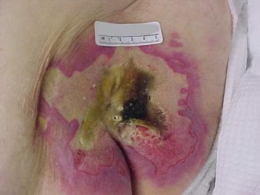 Image of advanced sacral pressure ulcer shows the effects of pressure, shearing, and moisture.
Epidemiology
Pressure ulcers are a commonly encountered condition in both acutely hospitalized and long-term institutionalized patients. They are estimated to occur in approximately 9% of hospitalized patients, usually during the first 2 weeks of hospitalization. This is in spite of adopting methods to reduce the development of pressure ulcers, such as repositioning, cushioning bony prominences, and using specialized mattresses. In high-risk hospitalized patient populations, the prevalence of pressure sores is even higher, with studies quoting incidence rates as high as 38%. [1] One study found that even with the use of a pressure-reducing bed and early nutritional support, 3% of patients in a surgical intensive care unit who were in the study developed pressure ulcers. [2] The annual risk of pressure ulceration in patients with neurologic impairment is 5-8%, with a lifetime risk of approximately 85% and a mortality rate of 8%.
In another prospective study, Campbell et al determined the incidence of heel pressure ulcers in 150 orthopedic patients. The cohort consisted of patients who were admitted to an acute care hospital either for elective orthopedic surgery or for the treatment of a fractured hip. The incidence of heel pressure ulcers in all patients was 13.3%, and the incidence in patients with hip fractures was higher than that of elective surgery patients (16% vs 13%, respectively). However, patients with hip fractures in whom pillows and rolled sheets were used to relieve heel pressure had a significantly lower ulcer rate than did the other hip fracture patients. When all patients in the study were taken into account, the presence of respiratory disease was the only factor significantly associated with the development of pressure ulcers. [3]
The prevalence of pressure ulcers among patients residing in long-term care facilities has been reported as anywhere from 2.3-28% and has been an increasingly common reason for litigation. [4, 5, 6, 7, 8] The presence of a pressure ulcer increases a nurse's workload for that patient by 50% and adds approximately $20,000 to the hospital bill. The treatment of pressure ulcers in the United States is estimated to cost more than $1 billion annually.
Venous ulcers make up 70% of chronic lower extremity ulcers. [9] The incidence of venous ulcers in the United States is approximately 600,000 cases annually. The recurrence rate is up to 90%.
According to the Centers for Disease Prevention National Diabetes Statistics Report, [10] an estimated 26.9 million Americans (8.2% of the population) are known to have diabetes and millions more are considered to be at risk. Of those at risk, diabetes is undiagnosed in 8.1 million individuals. Diabetic foot lesions are one of the most frequent causes of hospitalization secondary to a complication of diabetes. Among patients with diabetes, 15% will develop a foot ulcer and 12-24% of those with a foot ulcer will require amputation. Indeed, diabetes is the leading cause of nontraumatic lower-extremity amputations in the United States, accounting for 60% of these amputations.
In diabetic patients in the United States, the lifetime incidence of foot ulcers is 19-34%. More than 50% of these ulcers become infected at some point, and up to 20% of ulcers with moderate-to-severe infection result in amputation. This means that 1-2% of all patients with diabetic foot ulcers will at some point require an amputation. [11] Diabetic peripheral neuropathy confers the greatest risk of foot ulceration; microvascular disease and suboptimal glycemic control also contribute to this total. Even with successful treatment and ulcer healing, the recurrence rate in that patient population is 66% and the amputation rate rises to 12%. [12]
Etiology
In general, factors that adversely affect wound healing can be remembered by using the mnemonic device DIDN'T HEAL, as follows:
-
D = Diabetes: The long-term effects of diabetes impair wound healing by diminishing sensation and arterial inflow. In addition, even acute loss of diabetic control can affect wound healing by causing diminished cardiac output, poor peripheral perfusion, and impaired polymorphonuclear leukocyte phagocytosis.
-
I = Infection: Infection potentiates collagen lysis. Bacterial contamination is a necessary condition but is not sufficient for wound infection. A susceptible host and wound environment are also required. Foreign bodies (including sutures) potentiate wound infection.
-
D = Drugs: Steroids and antimetabolites impede proliferation of fibroblasts and collagen synthesis.
-
N = Nutritional problems: Protein-calorie malnutrition and deficiencies of vitamins A, C, and zinc impair normal wound-healing mechanisms.
-
T = Tissue necrosis, resulting from local or systemic ischemia or radiation injury, impairs wound healing. Wounds in characteristically well-perfused areas, such the face and neck, may heal surprisingly well despite unfavorable circumstances. Conversely, even a minor wound involving the foot, which has a borderline blood supply, may mark the onset of a long-term, nonhealing ulcer. Hypoxia and excessive tension on the wound edges also interfere with wound healing because of local oxygen deficits. See, for example, the pressure ulcers shown in the image below.
-
H = Hypoxia: Inadequate tissue oxygenation due to local vasoconstriction resulting from sympathetic overactivity may occur because of blood volume deficit, unrelieved pain, or hypothermia, especially involving the distal extent of the extremities.
-
E = Excessive tension on wound edges: This leads to local tissue ischemia and necrosis.
-
A = Another wound: Competition between several healing areas for the substrates required for wound healing impairs wound healing at all sites.
-
L = Low temperature: The relatively low tissue temperature in the distal aspects of the upper and lower extremities (a reduction of 1-1.5°C [2-3°F] from normal core body temperature) is responsible for slower healing of wounds at these sites.
Specific etiologies
Arterial insufficiency
See Infrainguinal Occlusive Disease.
Venous insufficiency
Patients with varicose veins or nonfunctional venous valves after deep vein thrombosis develop ambulatory venous hypertension, that is, distal venous pressure remains elevated despite ambulation. This constant venous hypertension seems to cause white cell and fibrin buildup, which impairs capillary blood flow or traps growth factors. Macromolecules pass into the dermis and eventually cause the hemosiderin deposition and brawny induration in the distal leg (gaiter area) characteristic of chronic venous insufficiency.
Lymphedema
Although not typically a cause of ulceration, extremity ulcers may fail to heal because of untreated lymphedema. Nocturnal leg elevation and elastic wraps or support hose are appropriate adjuncts to the treatment of recalcitrant wounds in edematous extremities. For advanced and nonresponsive lymphedema, complex decongestive physiotherapy is a useful treatment option.
Neuropathy
Sensory neuropathy involving the feet may lead to unrecognized episodes of trauma caused by ill-fitting shoes. This is compounded by motor neuropathy causing intrinsic muscle weakness and splaying of the foot on weight bearing. The result is a convex foot with a rocker-bottom appearance. Multiple fractures go unnoticed, until bone and joint deformities become marked. This is termed a Charcot foot (ie, neuropathic osteoarthropathy) and is observed most commonly in people with diabetes mellitus, affecting approximately 2% of persons with diabetes.
Pressure (decubitus) ulcers
Pressure (decubitus) ulcers occur because of prolonged ischemia-producing external pressure, usually to a soft tissue region overlying a bony prominence. Tissue ischemia results when external pressure exceeds capillary closing pressure (ie, 25-32 mm Hg in healthy individuals), the minimum pressure that causes collapse of the capillary when applied to a capillary bed.
Shearing forces, exposure to constant moisture, and heat buildup also are major contributing factors. For example, the stratum corneum, the outer layer of skin, becomes 25 times more fragile at a relative humidity of 100% when compared with a relative humidity of 25%, and it becomes 4 times more fragile at 95°F (35°C) than at 86°F (30°C).
Neoplasms
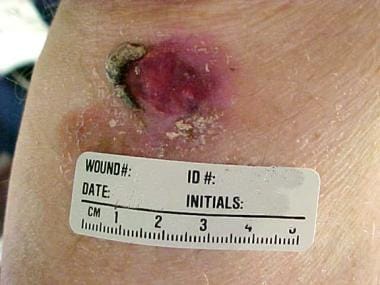
Neoplasms strongly suggest malignancy in any chronic nonhealing wound, particularly if the wound appears to have occurred spontaneously. [13]
Basal cell carcinoma appears smooth, pearly, and elevated above the skin surface, as illustrated in the image below, whereas squamous cell cancer is often somewhat erythematous and scaly and almost always occurs on sun-exposed areas. Particularly pertinent in wound care is the so-called Marjolin ulcer, a squamous cell carcinoma originating in a chronic wound, such as a burn scar or sinus tract. [14] This implies that even a wound that is decades old is not necessarily benign. Patients with Kaposi sarcoma typically present with multifocal violaceous lower extremity lesions. Patients with cutaneous lymphoma present with a single nodule or a group of papules from one to several centimeters in diameter, and these almost always occurs above the waist.
It is recommended to perform a biopsy of most wounds suggestive of a neoplasm. However, it is important to remember that biopsy findings are diagnostic only if an adequate representative specimen is obtained.
Radiation damage
The adverse effects of prolonged or excessive electromagnetic radiation vary with the wavelength. Wavelengths of electromagnetic radiation are as follows:
-
Gamma rays - Less than 0.01 nm
-
X-rays - 0.01-10 nm
-
Ultraviolet C - 10-280 nm
-
Ultraviolet B - 280-320 nm
-
Ultraviolet A - 320-400 nm
-
Visible light - 400-760 nm
-
Infrared - 760 nm to 1 mm
-
Microwave - 1 mm to 30 cm
-
Radio waves - Centimeters to meters
Gamma radiation and x-ray exposure cause a zone of stasis, in which local blood supply is impaired by coagulative necrosis due to thrombotic occlusion of smaller arteries. Gamma and x-ray radiation also spawn ionized oxygen that adversely affects DNA. The long-term result is the inhibition of regeneration of skin cells from dividing basal cells. This may cause recalcitrant, painful skin ulcers. The surrounding skin is atrophic, with atrophy of hair follicles and a paucity subcutaneous fat.
Ultraviolet radiation exposure, particularly ultraviolet B, causes sunburn initially and subsequently conveys a continuing risk of skin malignancy (eg, basal cell carcinoma, squamous cell carcinoma, melanoma).
Excessive exposure to infrared radiation, which induces repeated or persistent skin hyperthermia of 43-47°C, may cause erythema ab igne. Patients with this skin condition present with telangiectasia, erythematous patches, scaling, and hyperpigmentation.
Atheroembolism syndrome
Patchy areas of ischemia involving the feet, especially in the presence of palpable pedal pulses, suggest the possibility of atheroembolism of plaque fragments from ulcerated, although nonocclusive, proximal atherosclerotic plaques or from thrombi lining the wall of an infrarenal aortic aneurysm.
Pyoderma gangrenosum
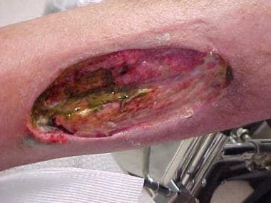
Pyoderma gangrenosum usually starts as a small, painful papule or nodule, which is often erroneously presumed to be the result of an insect bite. The lesion enlarges, becomes ulcerated, and develops overhanging, violaceous borders, as shown in the image below.
The histologic findings often are nonspecific. Associated underlying systemic problems, which occur in one half of patients with pyoderma gangrenosum, are often the best clues to the diagnosis. Examples of such systemic diseases include various arthritides, inflammatory bowel disease, hepatitis, myeloproliferative disorders, myeloma, primary biliary cirrhosis, systemic lupus erythematosus, and Sjögren syndrome. An important clue is a paradoxical response in which debridement exacerbates the wound, particularly near the areas debrided. When myofascial and osseous tissues become involved, the only choice may be surgical debridement to try to save the extremity. [15, 16] In extreme cases, this may result in amputation of the extremity.
Sickle cell
Patients with sickle cell–associated leg ulcers typically present with painful small ulcers that start as crusting nodules in the distal one third of the leg, often near the malleoli. The surrounding skin demonstrates absence of hair follicles, hyperpigmentation, and atrophy of subcutaneous fat. Radiograph findings may reveal periosteal thickening of underlying bone; true osteomyelitis is rare. Sickle cell ulcers are more common in males than in females and occur predominantly in persons aged 10-50 years. Patients with sickle cell anemia can also develop leg ulcers because of other etiologies; the physical examination should exclude arterial and venous insufficiency.
Calciphylaxis
Calciphylaxis is an unusual and often fatal syndrome of cutaneous necrosis that tends to develop in patients with chronic renal failure, particularly those with diabetes. The average time of onset is 3 years after the start of dialysis. The female-to-male ratio is 3:1. The initial finding of calciphylaxis may be that of livedo reticularis, followed by painful erythematous areas of thickening of the skin and subcutaneous tissues. The most common site is the thigh, though the condition may also occur in the legs or the upper extremities. [17, 18]
Panniculitis signaling the onset of calciphylaxis may be precipitated by trauma, such as the site of an injection. Proximal painful myopathy, muscle weakness, and elevated serum creatine kinase (CK) levels may occur. Laboratory testing may demonstrate a high serum phosphate level and an elevated parathyroid hormone level. Skin biopsy reveals calcification of the arterial media and luminal stricture of small-to-medium blood vessels in the subcutaneous fat. Muscle biopsy shows patchy necrosis and atrophy. [19]
Necrobiosis lipoidica
Necrobiosis lipoidica, a necrotizing skin lesion characterized by collagen degeneration and a granulomatous response, usually involves the anterior tibial areas, though it can also occur in the face, arms, and chest. Patients present with well-circumscribed, shiny, reddish-brown, oval, painless nodules or papules that have a thick shiny surface. Over several months or a year, the lesions may gradually expand and develop a waxy yellow color. Trauma may lead to infected ulcerations, and involvement of adjacent cutaneous nerves may precipitate considerable pain. The exact cause is unknown. Necrobiosis lipoidica is more common in women and in persons with diabetes than in others, but it may also occur in persons without diabetes and before the diagnosis of diabetes. [20] Long-standing necrobiosis lipoidica may harbor a squamous cell carcinoma.
Vasculitic wounds
Vasculitic wounds tend to occur throughout the lower legs as multiple, small, painful, erythematous nodules. Scars resulting from previous vasculitic lesions may be a useful clue. Any of the disparate systemic manifestations of the diseases of cellular immunity associated with atypical skin lesions, including unexplained fevers, jaw claudication, malaise, Raynaud phenomenon, myalgias, neurologic abnormalities, and craniofacial pain syndromes, suggest the possibility of vasculitis. These lesions are rare.
The differential diagnosis of wounds with these features includes other uncommon problems, such as anticoagulant-induced skin necrosis, atheroembolism syndrome (ie, trash foot), and Buerger disease. Leukocytoclastic vasculitides represent a disparate group of acquired connective tissue problems; patients present with palpable purpuric skin lesions, petechiae, and ecchymoses, usually involving the lower extremities. These syndromes include Wegener granulomatosis, Sjögren syndrome, cryoglobulinemia, systemic lupus erythematosus, rheumatoid arthritis, dermatomyositis, and hepatitis B. The common factor among these syndromes is a hypersensitivity angiitis. [21]
Skin biopsy demonstrates cuffing of the dermal microcirculation by granulocytes, which are found in diverse stages of viability, including complete cellular disintegration (ie, nuclear dust). The various disorders in this group are differentiated by clinical and serologic criteria. The presence of asymptomatic palpable purpura without thrombocytopenia suggests a drug adverse effect, such as those caused by iodides, penicillin, aspirin, chlorothiazides, oxytetracycline, isoniazid, or benzoic acid.
Anticoagulant-induced skin necrosis
Anticoagulant-induced skin necrosis is an unusual complication of anticoagulant therapy. [22] It can occur with the use of heparin or warfarin, though it is more common with warfarin. Warfarin-induced skin necrosis manifests as painful hemorrhagic skin lesions, usually in an area having abundant adipose tissue, such as the thighs, abdomen, or breasts. The female-to-male ratio is 4:1.
This complication is often attributable to hereditary coagulation abnormalities. Warfarin (Coumadin) depletes vitamin K–dependent coagulation factors, such as protein C. Therefore, during the first several days of warfarin therapy, a period of transient hypercoagulability may occur, particularly in patients with hereditary coagulation abnormalities, such as protein C deficiency or protein S deficiency, antithrombin 3 deficiency, or activated protein C resistance. [22]
Actinomycosis
Actinomyces israelii is a fastidious anaerobic bacterium that is relatively common and usually nonpathogenic. In rare individuals, particularly hosts who are immunocompromised, the bacterium can become pathogenic and cause chronic, draining, painless skin ulcers and sinuses, usually in the head and neck. False-negative tissue cultures are common because the organism is often difficult to culture in vitro. However, microscopic examination of wound exudates may demonstrate characteristic sulfur granules. Actinomycosis is responsive to penicillin but requires long-term therapy.
Yaws
Yaws is a treponematosis caused by Treponema pertenue, which is endemic in humid regions near the equator. Approximately 3-4 weeks after exposure, a pruritic sore that resembles a raspberry (the mother yaw) develops at the site where the spirochete enters the skin. This lesion eventually opens to form an ulcer. Scratching spreads the organism and results in multiple tubercles and ulcerations elsewhere, including the hands, feet, and genitals. These ulcers may have a caseous crust. Results of serologic testing for syphilis may be positive.
Treatment is with a single large dose of penicillin. Untreated yaws can erode to bone and joints and can become deforming and crippling.
The lesions of pinta, caused by Treponema carateum, are similar to those of yaws, but, unlike yaws, no ulceration is present. Pinta typically begins as a papule on the dorsum of the foot or leg. The papule enlarges and becomes a pruritic plaque, which changes from a copper to gray to bluish color over time. Regional lymphadenopathy may occur. Pinta is also responsive to penicillin.
Mucormycosis
Mucormycosis is an acute and sometimes rapidly progressive, even fatal, fungal infection that may occur in patients who are immunocompromised, especially following a burn. The primary lesions are plaques, ulcerations and abscesses, or painful ecchymotic nodules, which may ulcerate and then become necrotic and form eschars. The diagnosis is confirmed by demonstrating fungal elements of the black discharge in KOH preparations and by culturing on standard laboratory media.
Cutaneous anthrax
Cutaneous anthrax results from skin exposure to Bacillus anthracis, a gram-positive bacillus. Cutaneous anthrax evolves from a pruritic papule to an ulcerated wound in 1 or 2 days and then into a black eschar over the next week or so. Associated regional lymphadenopathy may be present. Findings on special stains and cultures of the wound exudate are diagnostic.
Anthrax is transmissible from specimens; therefore, laboratory personnel should be warned in the event of clinical suspicion of this disease. Of course, appropriate public health authorities must be notified. See Anthrax for details.
Pathophysiology
The phases of normal wound healing can be described as follows:
Hemostatic or inflammatory phase
This phase starts immediately and lasts 2-5 days. Tissue damage releases chemical mediators called cytokines (eg, transforming growth factor [TGF]-beta [interleukin-1beta]), which initiate a complex interrelated process that causes hemostasis and begins the healing process. Platelets aggregate to stem bleeding. They also release serotonin and other vasoconstrictors and activate the coagulation cascade. The result is conversion of fibrinogen into fibrin, which stabilizes the platelet plug. At that point, prostaglandins and activated complement cause vasodilation and increase capillary permeability. This allows plasma to leak into the tissue surrounding the wounded area. This is the inflammatory exudate.
Monocytes and neutrophils are attracted to the site of injury. Neutrophils trap and kill bacteria immediately, while monocytes become activated macrophages, which produce growth factors and cytokines and scavenge nonviable tissue and bacteria. Angiogenic growth factors stimulate neovascularization of the wound bed.
Proliferative phase
This phase lasts from 2 days to 3 weeks. Macrophages recruit fibroblasts. These cells create a network of collagen fibers. When adequate oxygen and vitamin C are present, granulation of tissue forms. Oxygen is incorporated by 2 amino acids, proline and lysine, which are both required for collagen chain synthesis. Vitamin C is required for the hydroxylation of proline to hydroxyproline, an amino acid found in collagen.
During granulation, fibroblasts create a collagen bed to fill the defect and grow new capillaries. During contraction, myofibroblasts pull the wound edges closer together to decrease the size of the wound. During epithelialization, new epithelium migrates from the intact epidermis around the wound and can grow up to 3 cm over the granulation tissue. This process requires a moist surface.
Remodeling phase
This phase lasts from 3 weeks to 2 years. [23] An organized form of collagen gradually replaces the immature, soft, gelatinous collagen. The effect is to increase the tensile strength of the healed wound, but it is less than 80% as strong as the original tissue.
Types of wound healing
First intention, also termed primary healing, is the healing that occurs when a clean laceration or a surgical incision is closed primarily with sutures, Steri-Strips, or skin adhesive.
Second intention, also termed secondary healing, is the healing that occurs when a wound is left open to heal by granulation, contraction, and epithelialization.
Delayed primary closure is a combination of the aforementioned types of wound healing. It is often intentionally applied to lacerations that are not considered clean enough for immediate primary closure. The wound is left open for 5-10 days; then, it is sutured closed to decrease the risk of wound infection, while also allowing expedited wound healing. Improved blood flow at the wound edges, which develops increasingly over the first few days, is another benefit of this style of wound healing and can be associated with progressive increases in resistance to infections.
Indications
All chronic wounds require assessment. [24] Many heal with topical wound care; some require surgical intervention. The details vary widely with the nature of the wound, and often a combination of management strategies is required, along with constant reassessment of wound progression. This article provides information regarding wound care in general and specific wound etiologies in particular.
Relevant Anatomy
Life is a constant battle against entropy (ie, disorder). The skin provides the primary barrier between the human protoplasm and the entropy of the external environment. Histologically, the skin is divided into the epidermis and the dermis.
The epidermis consists of 5 histologic strata. From superficial to deep these layers are the (1) stratum corneum, (2) stratum lucidum, (3) stratum granulosum, (4) stratum spinosum, and (5) stratum germinativum. The keratinocyte, the preponderant epidermal cell, is generated in the stratum germinativum and eventually desquamates (sloughs) when it reaches the stratum corneum.
The dermis underlies the epidermis. A dermal vascular network functions in thermoregulation and provides metabolic support for the avascular epidermis. Fibroblasts synthesize supportive and structural polymers, including ground substance, collagen, and elastin.
Skin appendages include sebaceous glands, hair follicles, and sweat glands.
For more information about the relevant anatomy, see Skin Anatomy.
Prognosis
The prognosis for healing of chronic wounds varies with the etiology of the wound and the general health status of the patient.
-
Image of advanced sacral pressure ulcer shows the effects of pressure, shearing, and moisture.
-
Sacral pressure ulcer before and after flap closure.
-
Chronic ulcer of medial aspect of right leg due to pyoderma gangrenosum.
-
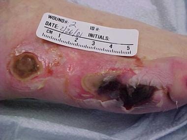
Pressure ulcers of the lateral aspect of the right foot.
-
Basal cell cancer manifesting as a chronic leg ulcer.
-
Heel pressure ulcer.
-
Sacral ulcer.
Tables
Category |
Examples |
Description |
Applications |
Alginate |
AlgiSite, Comfeel, Curasorb, Kaltogel, Kaltostat, Sorbsan, Tegagel |
Alginate dressings are made of seaweed extract contains guluronic and mannuronic acids that provide tensile strength and calcium and sodium alginates, which confer an absorptive capacity. Some can leave fibers in the wound if they are not thoroughly irrigated. These dressings are secured with secondary coverage. |
These dressings are highly absorbent and useful for wounds have copious exudate. Alginate rope is particularly useful to pack exudative wound cavities or sinus tracts. |
Hydrofiber |
Aquacel, Aquacel-Ag, Versiva |
An absorptive textile fiber pad, hydrofiber is also available as a ribbon for packing of deep wounds. This material is covered with a secondary dressing. The hydrofiber combines with wound exudate to produce a hydrophilic gel. Aquacel-Ag contains 1.2% ionic silver that has strong antimicrobial properties against many organisms, including methicillin-resistant Staphylococcus aureus and vancomycin-resistant enterococci. |
Hydrofiber absorbent dressings used for exudative wounds. |
Debriding agents |
Hypergel (hypertonic saline gel), Santyl (collagenase), Accuzyme (papain urea) |
Various products provide some chemical or enzymatic debridement. |
Debriding agents are useful for necrotic wounds as an adjunct to surgical debridement. |
Foam |
LYOfoam, Spyrosorb, Allevyn |
Polyurethane foam has absorptive capacity. |
These dressings are useful for cleaning granulating wounds with minimal exudate. |
Hydrocolloid |
CombiDERM, Comfeel, DuoDerm CGF Extra Thin, Granuflex, Tegasorb |
Hydrocolloid dressings are made of microgranular suspension of natural or synthetic polymers, such as gelatin or pectin, in an adhesive matrix. The granules change from a semihydrated state to a gel as the wound exudate is absorbed. |
Hydrocolloid dressings are useful for dry necrotic wounds, wounds with minimal exudate and for clean granulating wounds. |
Hydrogel |
Aquasorb, DuoDerm, Intrasite Gel, Granugel, Normlgel, Nu-Gel, Purilon Gel, KY Jelly |
Hydrogel dressings are water-based or glycerin-based semipermeable hydrophilic polymers; cooling properties may decrease wound pain. These gels can lose or absorb water depending upon the state of hydration of the wound. They are secured with secondary covering. |
These dressings are useful for dry, sloughy, necrotic wounds (eschar). |
Low-adherence dressing |
Mepore, Skintact, Release |
Low-adherence dressings are made of various materials designed to remove easily without damaging underlying skin. |
These dressings are useful for acute minor wounds, such as skin tears, or as a final dressing for chronic wounds that have nearly healed. |
Transparent film |
OpSite, Skintact, Release, Tegaderm, Bioclusive |
Transparent films are highly conformable acrylic adhesive films with no absorptive capacity and little hydrating ability. They may be vapor permeable or perforated. |
These dressings are useful for clean, dry wounds with minimal exudate. They also are used to secure an underlying absorptive material, to protect high-friction areas and areas that are difficult to bandage (eg, heels) and to secure intravenous catheters. |
Stage |
Definition |
Appearance |
Appropriate topical treatment |
Average healing time (d) |
I |
Nonblanchable erythema of intact skin |
Pink skin that does not resolve when pressure is relieved; discoloration; warmth; induration |
DuoDerm q2-3d |
14 |
II |
Partial-thickness skin loss involving epidermis and/or dermis |
Cracking, blistering, shallow crater, abrasion |
Cleanse with saline; DuoDerm/Tegaderm dressing |
45 |
III |
Full-thickness skin loss into subcutaneous fatty tissues or fascia |
Distinct ulcer margin; deep crater (in general, 2.075 mm or deeper [the thickness of a nickel]) |
Debride; irrigate with saline; apply DuoDerm/Tegaderm |
90 |
IV |
Full-thickness skin loss with extensive tissue involvement of underlying tissues |
Extensive necrosis; damage to underlying supporting structures, such as muscle, bone, tendon, or joint capsule |
Surgically debride; irrigate with saline (possibly under pressure); apply advanced topical dressings; consider antibiotics |
120 |
*When the overlying skin is necrotic, the staging cannot be accurate until debridement is performed. |
||||
Class |
Type |
Principle |
Examples |
I |
Simple |
Pressure-relieving pad or mat |
3- to 5-inch foam mattress, gel overlay, egg-crate mattress |
II |
Advanced |
Powered air* overlay for mattress with low air loss feature; nonpowered advanced pressure-reducing mattress replacement or powered air* flotation bed with or without low air loss feature |
Roho dry floatation mattress system, Pegasus Renaissance mattress |
III |
Air fluidized |
Flotation by filtered air* flow pumped through porcelain beads |
Clinitron bed |
*Long-term use of powered air devices is relatively contraindicated for patients with chronic obstructive lung disease, such as chronic bronchitis, emphysema, and asthma. |
|||
Type |
Description |
Examples |
Single layer |
Single-layer simple tubular woven nylon/elastic bandages may be imprinted with rectangles that stretch to squares when appropriate wrapping tension (30-40 mm Hg) is applied. |
ACE bandage, Comperm (Conco Medical), Setopress (Seton Healthcare Group) |
Three layer |
The layers include a padding absorption layer, a compression bandage layer, and a cohesive compression bandage. Bandages may be left in place for up to 1 week depending on wound exudate. |
Dyna-Flex (Johnson & Johnson) |
Four layer |
The layers include a nonwoven wound contact layer that is permeable to wound exudate and 4 overlying bandages. Bandages may be left in place for up to 1 week depending on exudate volume. |
Profore (Smith & Nephew) |
Impregnated wrap |
The porous flexible occlusive dressing is composed of stretchable gauze and a nonhardening zinc oxide paste. |
Unna boot (ConvaTec) |
*Compression wraps are contraindicated in severe arterial compromise. Some of these products are contraindicated in patients who are allergic to latex. |
||