You are in: eMedicine Specialties > Thoracic Surgery > Vascular Subclavian Artery ThrombosisArticle Last Updated: Jan 15, 2008AUTHOR AND EDITOR INFORMATIONAuthor: Mary C Mancini, MD, PhD, Director of Cardiothoracic Transplantation, Professor, Department of Surgery, Louisiana State University Health Sciences Center Mary C Mancini is a member of the following medical societies: American Heart Association, American Medical Association, American Thoracic Society, Association for Academic Surgery, Association for Surgical Education, International College of Surgeons, International Society for Heart and Lung Transplantation, New York Academy of Sciences, Phi Beta Kappa, and Southern Thoracic Surgical Association Editors: Jeffrey Lawrence Kaufman, MD, Associate Professor, Department of Surgery, Division of Vascular Surgery, Tufts University School of Medicine; Francisco Talavera, PharmD, PhD, Senior Pharmacy Editor, eMedicine; Daniel S Schwartz, MD, FACS, Clinical Assistant Professor of Cardiothoracic Surgery, New York University School of Medicine; Consulting Staff, Department of Surgery, Division of Thoracic Surgery, North Shore University Hospital/Long Island Jewish Medical Center; Paolo Zamboni, MD, Professor of Surgery, Chief of Day Surgery Unit, Chair of Vascular Diseases Center, University of Ferrara, Italy; John Geibel, MD, DSc, MA, Professor, Department of Surgery, Section of Gastrointestinal Medicine and Department of Cellular and Molecular Physiology, Yale University School of Medicine; Director of Surgical Research, Department of Surgery, Yale-New Haven Hospital Author and Editor Disclosure Synonyms and related keywords: subclavian artery thrombosis, subclavian artery occlusion, subclavian steal syndrome, cerebral steal syndrome, thoracic outlet syndrome, atherosclerosis, atherosclerotic disease, muscular compartment syndromes, hypercoagulable states, subclavian vein thrombosis, catheter-directed thrombolytic therapy, angioplasty, stents, stenting INTRODUCTIONSubclavian artery thrombosis is a condition in which the blood flow through the vessel is obstructed. The condition usually occurs secondary to some form of antecedent injury to the vessel, hypercoagulable state, or atherosclerotic changes. The condition is common in young athletic individuals who exert a significant amount of upper body activity. Sudden occlusion from emboli followed by thrombosis of the artery is common in the population with signs of significant atherosclerotic disease. The patient presenting with acute subclavian artery occlusion usually has a history of repetitive use and/or stress injury to the upper extremity on the affected side. A history of upper extremity claudication is common. In situations in which the occlusion is secondary to atherosclerosis, acute thromboses of the artery are generally asymptomatic. In fact, in 9% of autopsy series, the left subclavian artery was either stenotic or occluded. If symptoms are present, upper extremity claudication on the affected side is most common. The patient may also present with dizziness, vertigo, imbalance, visual disturbances, or hemisensory dysfunction indicative of a subclavian steal syndrome. However, note that subclavian steal is observed on 2% of cerebral angiograms and causes no symptoms. ProblemThe occlusion arises secondary to damage to the intima of the artery. This damage can occur as a result of external muscular compression and repetitive stress to the artery or because of atherosclerotic changes to the vessel. Embolic phenomena and hypercoagulable states are also contributing factors. Symptoms occur secondary to lack of blood flow to the affected extremity. To maintain blood supply to the extremity, blood is naturally rerouted from the vertebral, carotid, and internal mammary arteries, producing the various steal syndromes. FrequencySymptomatic lesions occur in less than 1% of the population. In autopsy series, 9% of the population demonstrate stenosis or occlusion of one subclavian artery, usually on the left. Two percent of cerebral angiograms demonstrate asymptomatic subclavian steal. EtiologyThe occlusion arises secondary to damage to the intima of the artery. This damage can occur as a result of external muscular compression and repetitive stress to the artery, atherosclerotic changes to the vessel, or inflammatory processes. Embolic or thrombotic occlusion of the artery occurs, particularly in the presence of atherosclerotic stenoses. Hypercoagulable states contribute to this scenario. PathophysiologyThe affected artery demonstrates detectable intimal damage, which is usually secondary to compressive forces exerted by the muscles of the shoulder girdle that compress the artery. Bony abnormalities in this area can also contribute to the process. As these muscles enlarge secondary to physical activity, they exert pressure on the artery. This pressure, coupled with exertional activity of the upper extremity, can stretch and compress the intima, thus disrupting its natural integrity. This disruption precipitates platelet deposition in the area, with resulting thrombosis. Atherosclerotic changes in the vessel occur secondary to the flow characteristics in the area. These depositions are accelerated by all of the dietary and sociological influences that affect the progression of atherosclerotic disease, including smoking, hypercholesterolemia, and hypertension. Occlusion secondary to atherosclerosis is more insidious and often causes no symptoms. At times, the symptom complex of claudication precedes the actual loss of blood flow. Patients with hypercoagulable states, either intrinsic or secondary to dehydration complicated by concomitant cardiac arrhythmias and systemic inflammatory processes, comprise a small subset of individuals who may exhibit this pathology. ClinicalA patient with an acute occlusion presents with a cold, painful, pulseless upper extremity. Axillary, brachial, and radial pulses are generally absent. When the occlusion is secondary to atherosclerotic changes, various prodromes and manifestations may be observed. The patient may present with no symptoms or upper extremity claudication secondary to exertion. If the condition has precipitated a steal syndrome, no symptoms are typically present. The examining physician should be aware of the rare presentation of various neurological symptoms and findings that may be associated with the steal syndromes, including syncope, vertigo, ataxia, sensory loss, visual changes, and stroke, depending on the vessels involved in the steal. The affected upper extremity may or may not demonstrate diminished pulses. Blood pressure differences between the affected and unaffected sides may be noted. INDICATIONSTherapeutic intervention is indicated in any symptomatic patient once the etiology of the symptoms has been defined. For instances of upper extremity claudication or acute thrombosis in which the problem has been attributed to the subclavian artery, intervention should be planned and executed. For patients in whom cerebrovascular symptoms predominate, a careful neurological evaluation must be undertaken in order to isolate the problem. Once the anatomical aberration has been defined, intervention is indicated if the subclavian artery is involved. RELEVANT ANATOMY
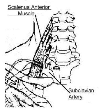
In patients with subclavian artery occlusion secondary to variations in the thoracic outlet, 2 areas can undergo vascular compression during hyperabduction of the extremity. One site is where the axillary artery passes posterior to the pectoralis minor muscle and beneath the coracoid process. The other point is where the artery courses between the clavicle and the first rib. Fibrous tissue proliferation in this area can impose extrinsic compression on the vessel. Aberrant origins of the subclavian artery off the aortic arch can be a cause of subclavian artery occlusion. In atherosclerotic disease, the carotid-subclavian junction or carotid-vertebral junctions are areas that appear to be predisposed to atheromata formation and calcification. Subsequently, this region is most likely to be involved in the occlusive process. Areas of the subclavian artery that are exposed to repeated forms of injury resulting in intimal damage are predisposed to occlusion. CONTRAINDICATIONSContraindications to surgical intervention include inadequate distal runoff, inadequate vessel size, and marked collateralization of the occluded area. Concomitant medical problems that would endanger the patient during a surgical intervention are also contraindications to surgery. With the advent of stenting, patients with greater medical challenges can be treated successfully; however, the presence of appropriate arterial runoff and adequate artery size are imperative in order to ensure success of the procedure. WORKUPLab Studies
Imaging Studies
Other Tests
TREATMENTMedical TherapyEarly diagnosis and therapy of subclavian artery thrombosis are indicated to prevent disabling upper extremity ischemia and gangrene. As a temporizing measure, the use of catheter-directed thrombolytic therapy may be indicated for superimposed clot formation in an area of stenosis until definitive treatment of the obstruction can be undertaken. Prolonged anticoagulation therapy for an obvious mechanical problem is not indicated. Anticoagulation may be considered as supplemental therapy after surgical intervention. Surgical TherapySurgery to correct subclavian artery thrombosis is the treatment of choice. Interventions include catheter-based procedures and formal operative procedures. Angioplasty and stenting of stenotic and even occluded arteries have been undertaken successfully with adequate patency rates and minimal morbidity. These interventions are particularly appropriate for atherosclerotic arteries. Subclavian artery occlusion secondary to thoracic outlet syndrome or muscular compression is treated by excision of the anatomical structure compressing the artery, whether muscle or bone. The artery may or may not require additional reconstruction, depending on the presence or absence of intimal damage. The occluded artery may require a bypass procedure, depending on the location of the occlusion or the presence of a subclavian steal syndrome. The bypass options include subclavian-carotid, subclavian-subclavian, and axillary-axillary bypasses. Another possible bypass option is transposition of the subclavian artery to the ipsilateral carotid artery. In any operative procedure for the problem, care must be taken to protect the thoracic duct from damage. Preoperative DetailsAfter the appropriate diagnostic studies have been performed to define the problem, operative preparation should be made. Administer preoperative prophylactic antibiotic therapy in the form of a first-generation cephalosporin. Perform appropriate preoperative medical screening to assess for evidence of other atherosclerotic disease such as coronary, carotid, or peripheral vascular occlusions. Intraoperative DetailsFor patients in whom the occlusion is secondary to atherosclerotic disease, perform a bypass of the area. For subclavian steal syndrome, a carotid-subclavian bypass using a ribbed synthetic tube graft is the procedure of choice. Depending upon the location of the occlusion, subclavian-subclavian or axillary-axillary bypass can be undertaken using a ribbed synthetic conduit. Because of the position of the graft and the necessity to, in some cases, cross bony structures, autologous vein conduits have limited patency. For patients in whom thoracic outlet compression is the cause of the thrombosis, cervical rib resection via a supraclavicular incision appears to be adequate treatment. Resection of the midportion of the clavicle is sometimes needed for exposure. Undertake arterial resection because the intima of the vessel is damaged. Graft interposition may or may not be required. Redundancy of the normal adjacent artery may allow for end-to-end reconstruction. If a graft is required for arterial reconstruction, a large autogenous saphenous vein or expanded polytetrafluoroethylene or Dacron fabric grafts may be used. First rib resection may be indicated in these instances as well, depending on the size of the thoracic outlet. In all procedures, care must be taken to identify and preserve the thoracic duct. Postoperative DetailsCheck distal upper extremity pulses immediately after the operative procedure while the field is still sterile. Maintain careful observation for the development of muscular compartment syndromes, and perform compartment measurements if indicated. Concomitant fasciotomies may be indicated, depending on the length of ischemia suffered by the extremity prior to revascularization. Evaluate postoperative effusions for the presence of chyle, which would be indicative of thoracic duct injury. Undertake prompt therapeutic measures if this complication occurs. Follow-upPerform postprocedural angiography at prescribed times after the operative intervention in order to assess patency of the graft or stent. One suggested timing sequence for the studies is at 1 month and 6 months, provided the patient remains asymptomatic. Noninvasive Doppler imaging can be used to assess distal flow to the extremity in the interim. COMPLICATIONSPotential complications of subclavian artery thrombosis secondary to atherosclerotic disease include distal embolization to the digits and neurological symptoms, including stroke secondary to a steal syndrome. Complications from operative interventions to address the arterial occlusion can include graft occlusion (acute and chronic), stent migration, stent occlusion, bleeding, and infection. If the subclavian artery thrombosis is secondary to thoracic outlet problems, subclavian vein thrombosis can occur as well. Accompanying neurological symptoms resulting from brachial plexus compression can also be associated with the syndrome. Repair of the arterial thrombosis can again be complicated by graft thrombosis. Although rare, the possibility of postoperative bleeding should be reviewed with the patient before the operative intervention. Promptly address bleeding complications by reoperation in order to correct the problem. Complications of the operative procedure can include injury to the thoracic duct. Pleural effusions should be promptly evaluated and appropriately treated. OUTCOME AND PROGNOSISThe results from stenting procedures on the subclavian artery document an 87% patency rate after 3 years. Operative outcomes demonstrate about a 90% patency rate after 5 years. The prognosis for the patient with atherosclerotic disease is directly dependent upon the severity of the disease and the willingness of the patient to modify lifestyle, including cessation of tobacco use and regulation of diet. If these modifications are made, the progression of the atherosclerotic process slows and the chance for recurrence of thrombosis falls. For the patient whose occlusion is secondary to thoracic outlet problems, the prognosis after therapy is excellent. FUTURE AND CONTROVERSIESThe future therapy of subclavian artery thrombosis will most likely involve the use of endovascular stents. As technology improves and a better understanding of restenosis issues is achieved, stenting of the lesions will be more commonplace. Careful assessment of patients with thoracic outlet syndrome will be required because of the complexity of this problem, the multiple structures involved, and the high-profile medicolegal issues that arise with treatment of these patients. Operative treatment of the arterial complications of the thoracic outlet syndrome should be performed. However, careful evaluation of the potentially associated venous and neurological pathologies should be undertaken prior to any operative therapy. MULTIMEDIA
REFERENCES
Subclavian Artery Thrombosis excerpt Article Last Updated: Jan 15, 2008 | ||||||||||||||||||||||||||