Background
Cardiac cirrhosis (congestive hepatopathy) includes a spectrum of hepatic derangements that occur in the setting of right-sided heart failure. Clinically, the signs and symptoms of congestive heart failure (CHF) dominate the disorder. Unlike cirrhosis caused by chronic alcohol use or viral hepatitis, the effect of cardiac cirrhosis on overall prognosis has not been clearly established. As a result, treatment is aimed at managing the underlying heart failure. [1, 2]
Distinguishing cardiac cirrhosis from ischemic hepatitis is important. The latter condition may involve massive hepatocellular necrosis caused by sudden cardiogenic shock or other hemodynamic collapse. Typically, sudden and dramatic serum hepatic transaminase elevations lead to its discovery. Although cardiac cirrhosis and ischemic hepatitis arise from distinct underlying cardiac lesions (right-sided heart failure in the former and left-sided failure in the latter), in clinical practice they may present together.
Despite its name, cardiac cirrhosis (which usually implies congestive hepatopathy that results in liver fibrosis) rarely satisfies strict pathologic criteria for cirrhosis. The terms congestive hepatopathy and chronic passive liver congestion are more accurate, but the name cardiac cirrhosis has become convention. For the remainder of this chapter, the term cardiac cirrhosis will be used to mean congestive hepatopathy with or without liver fibrosis.
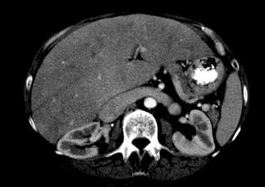 Cardiac cirrhosis and congestive hepatopathy. Congestive hepatopathy with large renal vein.
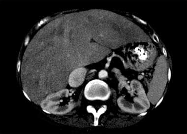 Cardiac cirrhosis and congestive hepatopathy. Congestive hepatopathy with large inferior vena cava.
Pathophysiology
Decompensated right ventricular or biventricular heart failure causes transmission of elevated right atrial pressure to the liver via the inferior vena cava and hepatic veins. At a cellular level, venous congestion impedes efficient drainage of sinusoidal blood flow into terminal hepatic venules. Sinusoidal stasis results in accumulation of deoxygenated blood, parenchymal atrophy, necrosis, collagen deposition, and, ultimately, fibrosis.
A separate theory proposes that cardiac cirrhosis is not simply a response to chronically increased pressure and sinusoidal stasis. Because intrahepatic vascular lesions are confined to areas of the liver with higher fibrotic burden, cardiac cirrhosis may require a higher grade of vascular obstruction, such as intrahepatic thrombosis, for its development. Thus, thrombosis of sinusoids and terminal hepatic venules propagates to medium-sized hepatic veins and to portal vein branches, resulting in parenchymal extinction and fibrosis.
Upon microscopic examination, sinusoidal engorgement, hepatocyte atrophy, and hemorrhagic necrosis is present in zone 3 of the hepatic acinus. [3] Also, there can be fatty change, cholestasis, and bile thrombi in the canaliculi.
Etiology
Causes of cardiac cirrhosis mirror the many etiologies of right-sided congestive heart failure (CHF), including congenital heart disease. Although inferior vena caval thrombosis and Budd-Chiari syndrome exhibit similar pathophysiology, they are categorized separately and are not included as causes of cardiac cirrhosis.
The most frequent causes of cardiac cirrhosis are the following:
-
Ischemic heart disease (31%)
-
Cardiomyopathy (23%)
-
Valvular heart disease (23%)
-
Primary lung disease (15%)
-
Pericardial disease (8%)
A study by Yoo et al suggested that the Fontan procedure is a significant risk factor for cardiac cirrhosis. The study included 46 patients with Fontan circulation, as well as 26 patients with right-sided heart failure and hepatic congestion. The Fontan patients were found, via transient elastography, to have a significantly higher liver-stiffness value than did the patients with right-sided heart failure. Moreover, a significant association was seen between liver stiffness in the Fontan patients and total bilirubin and albumin levels, white blood cell counts, and aspartate aminotransferase-to-platelet ratio indexes. The investigators found the age at which the Fontan procedure was completed and the total bilirubin level to be independent risk factors for hepatopathy. [4]
Epidemiology
United States data
Cardiac cirrhosis rarely occurs in the United States. Its true prevalence is difficult to estimate because the disease typically remains subclinical and undiagnosed. The incidence of cardiac cirrhosis at autopsy has decreased significantly over the past several decades. This may be due to lower rates of uncorrected rheumatic heart disease and constrictive pericardial disease.
Sex- and age-related demographic
Comparative sex data for cardiac cirrhosis do not exist. However, because CHF is more common in men than women in the United States, the same is likely for cardiac cirrhosis. [5]
No published data regard age and cardiac cirrhosis exist. However, the prevalence of cardiac cirrhosis in the United States, like that of congestive heart failure almost certainly increases with age.
Prognosis
Prognosis depends on the severity of the underlying heart disease and the degree of heart failure.
It has been proposed that use of the Fibrosis-4 index (Fib-4 index) (age [years] × aspartate aminotransferase [IU/L]/platelet count [109/L] × square root of alanine aminotransferase [IU/L]) can calculate a score to predict all-cause mortality in patients with liver cirrhosis. [6] A higher score on this index can predict a higher all-case mortality. [6]
Early recognition and medical treatment of the underlying heart disease is paramount in providing the best possible outcome to affected patients.
Morbidity/mortality/
The effect of cardiac cirrhosis on mortality and morbidity rates is unknown. The severity of the patient's underlying cardiac disease, which is typically advanced and chronic, is the major determinant of overall outcome.
-
Cardiac cirrhosis and congestive hepatopathy. Congestive hepatopathy with large renal vein.
-
Cardiac cirrhosis and congestive hepatopathy. Congestive hepatopathy with large inferior vena cava.


