Continually Updated Clinical Reference
 
 
  All Sources     eMedicine     Medscape     Drug Reference     MEDLINE
 
eMedicine - Pancreatic Pseudoaneurysm : Article by

Quick Find
Authors & Editors
Introduction
RELEVANT ANATOMY
Contraindications
Workup
Treatment
Complications
Outcome And Prognosis
Future And Controversies
Multimedia
References


Patient Education
Click here for patient education.


Author: Mazen S Itani, MD, Clinical Assistant Instructor, Department of Surgery/Division of Vascular Surgery, West Palm Beach VA

Mazen S Itani is a member of the following medical societies: American Medical Association

Coauthor(s): Nabil Sumrani, MD, Associate Professor, Department of Surgery, Division of Transplantation Surgery, State University of New York Health Science Center at Brooklyn

Editors: Alex Jacocks, MD, Program Director, Professor, Department of Surgery, University of Oklahoma School of Medicine; Francisco Talavera, PharmD, PhD, Senior Pharmacy Editor, eMedicine; Michael A Grosso, MD, Consulting Staff, Department of Cardiothoracic Surgery, St Francis Hospital; Paolo Zamboni, MD, Professor of Surgery, Chief of Day Surgery Unit, Chair of Vascular Diseases Center, University of Ferrara, Italy; John Geibel, MD, DSc, MA, Professor, Department of Surgery, Section of Gastrointestinal Medicine and Department of Cellular and Molecular Physiology, Yale University School of Medicine; Director of Surgical Research, Department of Surgery, Yale-New Haven Hospital

Author and Editor Disclosure

Synonyms and related keywords: pancreas, pancreatitis, biliopancreatic cancer, pseudocyst, pancreatic bleeding, peripancreatic bleeding, splenic vein thrombosis, angioembolization, angiography, esophagogastroduodenoscopy, alcoholism, alcohol abuse, hematemesis, hematochezia, hemoductal pancreatitis, hemosuccus pancreaticus, wirsungorrhagia

Pancreatitis with secondary pseudocyst formation is the most common cause of pancreatic pseudoaneurysms, although they are known to occur in the absence of a pseudocyst. Pancreatitis with secondary pseudocyst formation is a recently recognized complication after resection of biliopancreatic cancer and after transplantation.

Pancreatic or peripancreatic bleeding is one of the most life-threatening complications of pancreatitis. The standard of care in dealing with pseudoaneurysms has been surgical intervention; recently, many interventional radiologists have reported excellent outcome after angioembolization.

Problem

Pancreatic pseudoaneurysm is a malformation in the vessels of the pancreas and/or peripancreatic bed. These rather uncommon pseudoaneurysms are frequently accompanied by life-threatening complications, mainly rupture and bleeding. Better outcome requires accurate, timely, and appropriate diagnosis and medical and/or surgical intervention.

Frequency

  • Pseudoaneurysm formation in patients with chronic pancreatitis who undergo angiography may have an incidence as great as 10%.
  • Some of the factors associated with pancreatic pseudoaneurysms include the following:
    • Severity and duration of pancreatitis
    • Presence of pseudocyst and associated splenic vein thrombosis and endoscopically visualized varices
  • The splenic artery is the most commonly affected artery (30-50%) because of its proximity to the pancreas, followed by the gastroduodenal artery (10-15%), and the inferior and superior pancreaticoduodenal arteries (10%).
  • Other blood vessels mentioned in the literature include the superior mesenteric artery, hepatic artery, gastric artery, dorsal pancreatic artery, gastroepiploic artery, middle colic artery, aortic artery, and the portal vein.

Etiology

  • The etiologic requisites of the dysplastic and aneurysmal changes characteristic of pancreatic pseudoaneurysm formation include the following:
    • Acinar cell necrosis and/or ductal disruption with peripancreatic accumulations of exudative fluid containing activated proteolytic enzymes, which autodigest and weaken the arterial wall and lead to pseudoaneurysm formation
    • Severe inflammation of an infected pseudocyst leading to the same sequelae
  • Moderate-to-severe pancreatitis with or without pseudocyst/abscess is the major etiologic factor for pseudoaneurysm formation.
  • Pseudoaneurysm formation may occur after biliopancreatic resection for cancer.
    • Patients who have an anastomotic leak and develop intra-abdominal abscess may subsequently be prone to delayed arterial hemorrhage.
    • Focal sepsis erodes through vessels and causes pseudoaneurysm formation and delayed rupture and bleeding.
  • Pancreatic transplantation is an occasionally reported third cause of pancreatic pseudoaneurysm formation.

Pathophysiology

  • Pseudoaneurysms form when enzyme-rich peripancreatic fluid, often within a pseudocyst, leads to autodigestion and weakening of the walls of adjacent arteries.
  • These arteries then undergo aneurysmal dilatation, with the aneurysmal bulge most often contained within the pseudocyst. At this point, the dilated region is correctly termed an aneurysm rather than a pseudoaneurysm because the blood is still contained within the complete, although thinned, arterial wall.
  • Rupture of the aneurysm into the pseudocyst converts the pseudocyst into a pseudoaneurysm (defined as extravascular hematoma communicating with the intravascular space).
  • In some instances, a pseudocyst can erode into a nearby artery, causing the conversion of pseudocyst into a pseudoaneurysm.
  • Despite these distinctions, all of these forms are generally classified as pseudoaneurysm because the end result is the formation of a total or partial vascular cystic structure.
    • The literature confirms that differentiating a pseudoaneurysm from a bleeding pseudocyst is difficult.
    • This form of pseudoaneurysm should be distinguished from primary peripancreatic vessel aneurysm, which tends to occur more often in women. The rare rupture of primary aneurysm tends to occur in pregnancy and manifests as massive intraperitoneal bleeding with hemodynamic instability.

Clinical

  • Because pancreatitis is the most common underlying cause of pancreatic pseudoaneurysm, most patients are males with alcoholism (80-90%) who have histories of episodic chronic pancreatitis and secondary pseudocyst formation.
  • The diagnosis of visceral artery pseudoaneurysm should be considered in any patient with a pseudocyst and a significant abdominal bruit.
  • The pseudoaneurysm tends to enlarge when subjected to sufficient intracystic pressure and ultimately ruptures into the gastrointestinal tract, biliopancreatic ducts, pseudocyst, peritoneal cavity, or retroperitoneum.
  • Although highly variable, clinical symptoms are very suggestive and include the following:
    • Anemia of unexplained cause
    • Recurrent or intermittent hematemesis or hematochezia in patients who have pancreatitis, particularly when due to chronic alcohol abuse or trauma
    • Rapid enlargement of a pseudocyst or a pulsatile abdominal mass, especially in the presence of abdominal bruit and hyperamylasemia
    • The syndrome known as hemosuccus pancreaticus, characterized by bleeding from the ampulla of Vater, colicky pain, and jaundice
  • Patients with pancreatitis may have the following symptoms:
    • Persistent or abrupt increase in abdominal pain
    • Decreasing hematocrit values and/or hemodynamic instability and/or gastrointestinal bleeding with no obvious intraluminal cause
  • The clinical picture may vary widely.
  • The most common form of bleeding is probably rupture into a pseudocyst with eventual bleeding through the pancreatic duct and, subsequently, the ampulla of Vater if the pseudocyst is connected with the pancreatic duct.
    • This "wirsungorrhagia" (ie, hemosuccus pancreaticus) manifests as intermittent pain caused by sudden filling with blood and resultant distention of the pancreatic duct and may sometimes be accompanied by elevated levels of pancreatic enzymes.
    • Once the intraductal pressure reaches a certain level, the bleeding stops and a clot forms. The clot subsequently lyses at a later stage, leading the cycle to repeat itself.
  • On the other hand, if the pseudocyst does not communicate with the duct of Wirsung, then blood accumulates in the pseudocyst, leading to sudden enlargement and causing abdominal pain and a drop in the hematocrit value.


  • The splenic artery, which is most commonly involved in pancreatic pseudoaneurysm, is one of the 3 branches of the celiac artery; the other two are the common hepatic artery and the left gastric artery. Because the splenic artery runs along the pancreatic bed before reaching the spleen, it is the artery most commonly affected by the erosive effect of pancreatitis.
  • After giving off the proper hepatic artery, the common hepatic artery becomes the gastroduodenal artery, which gives rise to the superior pancreaticoduodenal artery, which anastomoses with the inferior branch coming off the superior mesenteric artery to supply the head of the pancreas and the duodenum.
  • In addition to encasing the distal end of the common bile duct, the pancreas, a retroperitoneal organ, is near the C-loop of the duodenum laterally and the lesser sac anteriorly. This explains why the pseudoaneurysm can erode and bleed into the bowel, biliary tree, retroperitoneum, or peritoneal cavity.


  • Endoscopic drainage is contraindicated. Drainage is an inadequate treatment of a pseudocyst that has bled.


Imaging Studies

  • Angiography
    • Angiography is currently the criterion standard for diagnosing pancreatic pseudoaneurysm.
    • If the patient is hemodynamically stable, performing a preoperative angiogram helps confirm the diagnosis.
    • Angiography defines the character—unique or otherwise—of the lesion and allows therapeutic planning.
    • Angiography greatly facilitates identification of the location and serves as a topographical guide for the pseudoaneurysm, which aids in operative proximal and distal control of the bleeding vessel.
    • Preoperative angiography might constitute an opportunity to gain temporary control over the bleeding vessel by performing transcatheter embolization, thus providing a time window for the surgeon to operate on a high-risk patient under optimum clinical conditions.


Medical therapy

  • Although occasional reports have alluded to the spontaneous thrombosis of some pancreatic pseudoaneurysms, the current consensus holds that all these malformations should be treated to prevent the complication of bleeding.
  • Nonsurgical management consists of transarterial catheter angioembolization with or without endoscopic stent placement.
    • Angioembolization is considered much less invasive than surgery. The procedure can be completed quickly and is comfortable for the patient. It also allows the performance of surgery under optimal conditions.
    • The interventional approach has a reported success rate of 67-100% over the past few years.
    • Most authorities agree that embolization is appropriate when bleeding is diffuse or emanating from the pancreatic head, for unsuccessful surgery, or during postoperative bleeding.
    • Failure results from an inability to selectively catheterize the bleeding vessel or the misplacement or poor placement of embolization material.
    • In addition to rebleeding, complications of this procedure include rupture of the pseudoaneurysm during embolization, arterial perforation by the catheter, intestinal necrosis, and aortic thrombosis.
  • Whether angioembolization should be used as a definitive procedure or as a prelude to operative management is still debatable.

Surgical therapy

  • Emergency exploratory laparotomy is always indicated once the diagnosis of bleeding pseudoaneurysm is confirmed arteriographically, or it is entertained in the case of hemodynamic instability caused by an exsanguinating hemorrhage that obviates the need for angiography.
  • In addition, a surgical approach is necessary in patients with bleeding vessels that are impossible to catheterize to perform embolization and in patients in whom angiographic attempts to control bleeding have failed.
  • A major controversy concerns the best approach to control the bleeding. Arterial ligation on both sides of the bleeding sites, pancreatic resection, and intracystic/extracystic multiple ligatures are all suggested therapies.
  • Some pseudocyst drainage procedures have been frequently performed concomitantly with the primary hemostatic surgery.

Preoperative details

  • Manage the hemodynamically unstable patient in an emergent fashion. Approach the patient in a manner similar to that for a trauma patient.
  • After having secured the airway and checked the breathing, place large-bore intravenous catheters and immediately start crystalloid infusions.
  • Send a type and cross of the blood to the blood bank while the need for O-negative and type-specific blood transfusion is being assessed.
  • The patient's hemodynamic status and comorbid medical issues dictate the necessity for invasive hemodynamic monitoring.
  • Patients should undergo emergent celiotomy to control the bleeding pseudoaneurysm as soon as possible.
  • If the diagnosis of a ruptured pseudoaneurysm has been seriously entertained and the patient is hemodynamically stable, some studies may be performed before the patient enters the operating theater.
    • Performing a preoperative angiogram has several benefits. Identifying the bleeding vessel during surgery is difficult because of the friability, necrosis, and severe inflammation caused by pancreatitis. Also, because preoperative angiography identifies the bleeding vessel, it might dictate the optimal therapy.
    • Performing arterial ligation or pancreaticoduodenectomy on bleeding vessels involving the pancreatic head has been demonstrated to carry a high mortality rate.
    • Furthermore, angiography might constitute an opportunity to gain temporary preoperative control over the bleeding vessel by performing transcatheter embolization, thus providing a time window for the surgeon to operate on a high-risk patient under optimum clinical conditions.

Intraoperative details

  • Once celiotomy and adequate exposure is performed, direct attention toward the most common source of bleeding, mainly the peripancreatic vasculature.
  • Multiple effective measures to gain rapid control of the actively bleeding pseudoaneurysm have been described. These include manual tamponade, gauze packing, digital compression of the bleeding pseudoaneurysm or pseudocyst, and even supraceliac infradiaphragmatic cross-clamping of the aorta for brisk bleeding.
  • Institute these measures, especially in the actively bleeding, hemodynamically unstable patient, while aggressive volume resuscitation is being undertaken by the anesthesia team.
  • After establishing these initial measures, a more delicate and precise dissection can be performed in order to obtain definitive control of the bleeding vessels.
    • As previously mentioned, exposure of the bleeding site can sometimes be challenging because the surrounding inflammation from pancreatitis obscures the visual field.
    • Several adjunctive techniques have been listed to gain operative access to the bleeding pseudoaneurysm; these include gastrotomy, duodenotomy, and major gastrectomy.
  • Once the bleeding vessel is identified, the surgeon may perform one of several surgical methods to control the bleeding.
    • Intracystic ligation without proximal/distal control or resection is not recommended because the friable tissues of the posterior pseudocyst wall do not hold sutures, and the feeding vessel that lies deep within the substance of the pancreas is still patent.
    • For treatment of the pseudocyst, several surgical options are available, ranging from resection to external or internal drainage methods.

Postoperative details

  • Carefully monitor the patient after surgery.
  • The occurrence of postresectional hemorrhage is well documented in the literature, with a reported incidence of 5-19% and a mortality rate of 6-58%. This may be the result of ongoing pancreatitis and continuous damaging of the arteries, iatrogenic trauma to the vessels, and/or inadequate control of the bleeding vessels.
  • While some surgeons have advocated surgical ligation of the bleeding vessel in the nonseptic patient and pancreatic resection in those with abscess or established fistula, interventional radiologists have strongly recommended angioembolization for postoperative hemorrhage.

Follow-up

  • Despite few reports of resolution of pseudocysts with embolization alone, little long-term follow-up care is available for patients treated angiographically, particularly for patients who have underlying pathology that predisposes them to recurrent complications.


  • As mentioned previously, the most serious and life-threatening complications of pseudoaneurysms are rupture and subsequent hemorrhage.
  • Bleeding is usually brisk but varies from short, repeated, and self-limiting episodes to massive hemorrhage requiring emergent laparotomy.
  • The frequency of arterial lesion hemorrhage during pancreatitis varies from 5-10%. However, when pseudocysts are present, the hemorrhage rate rises to 15-20% of cases.
  • The most common site of rupture is intracystic, and the incidence of spontaneous hemorrhage arising from a pancreatic pseudocyst reportedly ranges from 1.4-8.4%.
    • This bleeding can be localized in the cyst, causing sudden enlargement and abdominal pain, or bleeding can occur through the cyst into the pancreatic duct if a communication between these structures exists. (In this case, the patient will have gastrointestinal bleeding.)
    • Other sites of rupture include the biliopancreatic duct, peritoneal cavity, retroperitoneum, and sometimes direct erosion into the duodenum and other parts of the gastrointestinal tract.
  • Hemorrhage is a significant complication that carries a mortality rate of 13-40% and is almost always fatal if left unattended.
  • Other infrequent complications include arteriovenous fistula formation and extrahepatic biliary tract obstruction.


  • The mortality rate following surgical treatment for arterial hemorrhage of pancreatic origin ranges from 28-56%, primarily depending on the anatomic location of the pseudoaneurysm and not the surgical method.
    • Surgical intervention for treatment of pseudoaneurysm in the head of the pancreas has a 43% mortality rate but only a 16% mortality rate in the body or tail of the pancreas.
    • Patients treated with supportive measures have more than a 90% mortality rate.
  • The mortality rate with postoperative hemorrhage is nearly double (50-60%).
  • For pseudoaneurysms in the head of the pancreas, the surgical approach necessitates a Whipple procedure, which likely contributes to the higher mortality rate.
  • Embolotherapy has a high initial success rate (90-100%), although some reports indicate a recurrence rate of 37% and an overall mortality rate of 16%. Embolotherapy is the preferred initial therapy for bleeding originating from the head of the pancreas.


  • The major controversy surrounding the operative management of the bleeding pseudoaneurysm is whether to perform arterial ligation or pancreatic resection.
    • Some authors have strongly advocated resection because it is technically easier to perform than ligation in an inflammatory milieu.
    • Other authors have reported better outcome after performing proximal and distal arterial ligation and intracystic suture ligation.
  • Another major controversy is whether transarterial catheter angioembolization should be the definitive approach or if it should always be followed by surgical intervention, especially if bleeding is located in the tail or body of the pancreas and/or is associated with a pseudocyst.
    • Some authors have found no rebleeding after seemingly successful angioembolization of the pseudoaneurysm.
    • Other authors have found statistically significant rebleeding rates, which necessitated surgical resection after embolization.
  • A third controversy is the management of postoperative bleeding despite recent adoption of the operative option by more health care providers.
  • A recent article describes the percutaneous injection of thrombin into the head of the pancreas for treating pancreatic pseudoaneurysm, the significance, safety, and efficacy of which remain to be seen (Luchs, 1999).


Media file 1:  
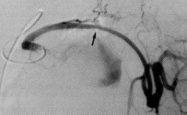
Splenic artery angiogram demonstrating contrast (white arrow) extravasating into a pseudoaneurysm (black arrow).
Click to see larger pictureClick to see detailView Full Size Image
Media type:  X-RAY

Media file 2:  A CT scan with intravenous contrast enhancement (arrow) within a pancreatic pseudocyst indicating the presence of a pseudoaneurysm.
Click to see larger pictureClick to see detailView Full Size Image
Media type:  CT

Media file 3:  Preembolization angiogram depicting a splenic artery pseudoaneurysm.
Click to see larger pictureClick to see detailView Full Size Image
Media type:  X-RAY

Media file 4:  Postembolization angiogram depicting successful coil embolization of the pseudoaneurysm.
Click to see larger pictureClick to see detailView Full Size Image
Media type:  X-RAY


  • Bender JS, Bouwman DL, Levison MA. Pseudocysts and pseudoaneurysms: surgical strategy. Pancreas. Mar 1995;10(2):143-7. [Medline].
  • Bender JS, Levison MA. Massive hemorrhage associated with pancreatic pseudocyst: successful treatment by pancreaticoduodenectomy. Am Surg. Oct 1991;57(10):653-5. [Medline].
  • Boudghene F, L''Hermine C, Bigot JM. Arterial complications of pancreatitis: diagnostic and therapeutic aspects in 104 cases. J Vasc Interv Radiol. Jul-Aug 1993;4(4):551-8. [Medline].
  • Bradley EL, Clements JL Jr, Gonzalez AC. The natural history of pancreatic pseudocysts: a unified concept of management. Am J Surg. Jan 1979;137(1):135-41. [Medline].
  • Bresler L, Boissel P, Grosdidier J. Major hemorrhage from pseudocysts and pseudoaneurysms caused by chronic pancreatitis: surgical therapy. World J Surg. Sep-Oct 1991;15(5):649-52; discussion 652-3. [Medline].
  • Brodsky JT, Turnbull AD. Arterial hemorrhage after pancreatoduodenectomy. The ''sentinel bleed''. Arch Surg. Aug 1991;126(8):1037-40. [Medline].
  • Cahow CE, Gusberg RJ, Gottlieb LJ. Gastrointestinal hemorrhage from pseudoaneurysms in pancreatic pseudocysts. Am J Surg. Apr 1983;145(4):534-41. [Medline].
  • Clay RP, Farnell MB, Lancaster JR. Hemosuccus pancreaticus. An unusual cause of upper gastrointestinal bleeding. Ann Surg. Jul 1985;202(1):75-9. [Medline].
  • De Ronde T, Van Beers B, de Canniere L et al. Thrombosis of splenic artery pseudoaneurysm complicating pancreatitis. Gut. Sep 1993;34(9):1271-3. [Medline].
  • Eckhauser FE, Stanley JC, Zelenock GB. Gastroduodenal and pancreaticoduodenal artery aneurysms: a complication of pancreatitis causing spontaneous gastrointestinal hemorrhage. Surgery. Sep 1980;88(3):335-44. [Medline].
  • Elton E, Howell DA, Amberson SM. Combined angiographic and endoscopic management of bleeding pancreatic pseudoaneurysms [see comments]. Gastrointest Endosc. Dec 1997;46(6):544-9. [Medline].
  • Frey CF, Child CG, Fry W. Pancreatectomy for chronic pancreatitis. Ann Surg. Oct 1976;184(4):403-13. [Medline].
  • Gadacz TR, Trunkey D, Kieffer RF Jr. Visceral vessel erosion associated with pancreatitis. Case reports and a review of the literature. Arch Surg. Dec 1978;113(12):1438-40. [Medline].
  • Gambiez LP, Ernst OJ, Merlier OA. Arterial embolization for bleeding pseudocysts complicating chronic pancreatitis. Arch Surg. Sep 1997;132(9):1016-21. [Medline].
  • Hofer BO, Ryan JA Jr, Freeny PC. Surgical significance of vascular changes in chronic pancreatitis. Surg Gynecol Obstet. Jun 1987;164(6):499-505. [Medline].
  • Huizinga WK, Kalideen JM, Bryer JV. Control of major haemorrhage associated with pancreatic pseudocysts by transcatheter arterial embolization. Br J Surg. Feb 1984;71(2):133-6. [Medline].
  • Khan TF, Ciancio G, Burke GW 3rd. Pseudoaneurysm of the superior mesenteric artery with an arteriovenous fistula after simultaneous kidney-pancreas transplantation. Clin Transplant. Jun 1999;13(3):277-9. [Medline].
  • Lee MJ, Saini S, Geller SC. Pancreatitis with pseudoaneurysm formation: a pitfall for the interventional radiologist. AJR Am J Roentgenol. Jan 1991;156(1):97-8. [Medline].
  • Lina JR, Jaques P, Mandell V. Aneurysm rupture secondary to transcatheter embolization. AJR Am J Roentgenol. Apr 1979;132(4):553-6. [Medline].
  • Luchs SG, Antonacci VP, Reid SK. Vascular and interventional case of the day. Pancreatic head pseudoaneurysm treated with percutaneous thrombin injection. AJR Am J Roentgenol. Sep 1999;173(3):830, 833-4. [Medline].
  • Mandel SR, Jaques PF, Sanofsky S. Nonoperative management of peripancreatic arterial aneurysms. A 10-year experience. Ann Surg. Feb 1987;205(2):126-8. [Medline].
  • Marshall GT, Howell DA, Hansen BL. Multidisciplinary approach to pseudoaneurysms complicating pancreatic pseudocysts. Impact of pretreatment diagnosis. Arch Surg. Mar 1996;131(3):278-83. [Medline].
  • Morita R, Muto N, Konagaya M. Successful transcatheter embolization of pseudoaneurysm associated with pancreatic pseudocyst. Am J Gastroenterol. Sep 1991;86(9):1264-7. [Medline].
  • Sato N, Yamaguchi K, Shimizu S. Coil embolization of bleeding visceral pseudoaneurysms following pancreatectomy: the importance of early angiography. Arch Surg. Oct 1998;133(10):1099-102. [Medline].
  • Shankar S, Russell RC. Haemorrhage in pancreatic disease. Br J Surg. Aug 1989;76(8):863-6. [Medline].
  • Shapiro N, Brandt L, Sprayregan S. Duodenal infarction after therapeutic Gelfoam embolization of a bleeding duodenal ulcer. Gastroenterology. Jan 1981;80(1):176-80. [Medline].
  • Sharara AI, Le TH, Suhocki P. Extrahepatic biliary obstruction resulting from a pancreatic pseudoaneurysm. Gastrointest Endosc. Aug 1995;42(2):176-7. [Medline].
  • Stabile BE, Wilson SE, Debas HT. Reduced mortality from bleeding pseudocysts and pseudoaneurysms caused by pancreatitis. Arch Surg. Jan 1983;118(1):45-51. [Medline].
  • Stanley JC, Frey CF, Miller TA. Major arterial hemorrhage: a complication of pancreatic pseudocysts and chronic pancreatitis. Arch Surg. Apr 1976;111(4):435-40. [Medline].
  • Starling JR, Crummy AB. Hemosuccus pancreaticus secondary to ruptured splenic artery aneurysm. Dig Dis Sci. Sep 1979;24(9):726-29. [Medline].
  • Stroud WH, Cullom JW, Anderson MC. Hemorrhagic complications of severe pancreatitis. Surgery. Oct 1981;90(4):657-65. [Medline].
  • Trojanowski JQ, Harrist TJ, Athanasoulis CA. Hepatic and splenic infarctions: complications of therapeutic transcatheter embolization. Am J Surg. Feb 1980;139(2):272-7. [Medline].
  • Tsiotos GG, Munoz Juarez MM, Sarr MG. Intraabdominal hemorrhage complicating surgical management of necrotizing pancreatitis. Pancreas. Mar 1996;12(2):126-30. [Medline].
  • Vanlangenhove P, Defreyne L, Kunnen M. Spontaneous thrombosis of a pseudoaneurysm complicating pancreatitis. Abdom Imaging. Sep-Oct 1999;24(5):491-3. [Medline].
  • Waltman AC, Luers PR, Athanasoulis CA. Massive arterial hemorrhage in patients with pancreatitis. Complementary roles of surgery and transcatheter occlusive techniques. Arch Surg. Apr 1986;121(4):439-43. [Medline].
  • Wolstenholme JT. Major gastrointestinal hemorrhage associated with pancreatic pseudocyst. Am J Surg. Apr 1974;127(4):377-81. [Medline].
  • el Hamel A, Parc R, Adda G. Bleeding pseudocysts and pseudoaneurysms in chronic pancreatitis [see comments]. Br J Surg. Sep 1991;78(9):1059-63. [Medline].
  • van Berge Henegouwen MI, Allema JH, van Gulik TM. Delayed massive haemorrhage after pancreatic and biliary surgery. Br J Surg. Nov 1995;82(11):1527-31. [Medline].

Pancreatic Pseudoaneurysm excerpt

Article Last Updated: Mar 12, 2005