Practice Essentials
Omental torsion is a condition in which the organ twists on its long axis to such an extent that its vascularity is compromised. Although it is rarely diagnosed preoperatively, knowledge of this entity is important to the surgeon because it mimics the common causes of acute surgical abdomen. [1] Since the first describtion of omental torsion by Eitel in 1899, only a few hundred cases have been reported. [2]
Omental torsion usually occurs in adults. The twisted portion of the omentum tends to be localized to a right-side segment, thereby giving rise to the sudden onset of pain and signs of peritoneal irritation on the right side of the abdomen. The condition may be associated with nausea, vomiting, or low-grade fever. An abdominal mass may be palpable.
Ultrasonography (US) and computed tomography (CT) may show a characteristic appearance of twisted omentum; however, because the disease may mimic other surgical emergencies, extensive radiologic studies are usually not indicated.
Treatment consists of resection of the affected portion of the omentum. Any disease process associated with secondary torsion should be corrected.
Pathophysiology
In omental torsion, the omentum twists around a pivotal point, usually in a clockwise direction (see the image below). Venous return is compromised, and the distal omentum becomes congested and edematous. Resultant hemorrhagic extravasation creates a characteristic serosanguineous fluid in the peritoneal cavity.
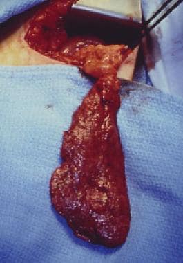 Example of primary torsion of omentum. Normal-appearing omentum can be seen above torsion point. Omentum below that point is edematous and congested.
As the torsion progresses, arterial occlusion leads to acute hemorrhagic infarction, and necrosis of the omentum eventually occurs. Spontaneous derotation may be possible and may explain omental adhesions in the right lower quadrant, which are often found during laparotomy and have no clear cause.
Etiology
Torsion of the omentum may be either primary or secondary. In primary torsion, a mobile segment of omentum rotates around a proximal fixed point in the absence of any associated intra-abdominal pathology. Although the precise cause is unknown, both predisposing and precipitating factors in the pathogenesis of the condition can be identified. [3, 4, 5, 6, 7]
Factors that predispose a patient to torsion include anatomic variations of the omentum itself, such as accessory omentum, bifid omentum, irregular accumulations of omental fat (in patients who are obese), and narrowed omentum pedicle. Any redundancy of omental veins may lead to kinking and twisting around the shorter and tenser arteries. The higher incidence of torsion on the right side of the omentum is related to the greater size and mobility of that side.
Precipitating factors are those causing displacement of the omentum, including trauma, violent exercise, and hyperperistalsis with resultant increased passive movement of the omentum.
Secondary torsion is more common than primary torsion and is associated with preexisting abdominal pathology, including cysts, tumors, [8] foci of intra-abdominal inflammation, postsurgical wounds or scarring, and hernial sacs. Most cases of secondary torsion occur in patients with inguinal hernias. [9]
-
Example of primary torsion of omentum. Normal-appearing omentum can be seen above torsion point. Omentum below that point is edematous and congested.


