Practice Essentials
Lipomas are the most common soft-tissue tumor. These slow-growing, benign fatty tumors form soft, lobulated masses enclosed by a thin, fibrous capsule. Although it has been hypothesized that lipomas may rarely undergo sarcomatous change, this event has never been convincingly documented. It is more probable that lipomas are at the benign end of the spectrum of tumors, which, at the malignant end, include liposarcomas (see Pathophysiology).
Lipomas are most often asymptomatic. Symptoms in other sites depend on the location (see Presentation).
The plasma D-dimer level may help in distinguishing lipoma from well-differentiated liposarcoma. For most subcutaneous lipomas, no imaging studies are required. For lipomas in atypical locations (or for which the differential diagnosis includes sarcoma), ultrasonography (US), computed tomography (CT), and magnetic resonance imaging (MRI) may be considered. (See Workup.)
Treatment consists of complete excision. No contraindications for removal exist, unless the patient is unfit for surgery or the anatomic location makes removal unfeasible. Benign lipomas are simply "shelled out," with complete removal of the capsule in an extracapsular plane; however, this is an inadequate operation for a liposarcoma. (See Treatment.) Nonoperative (endoscopic) therapy may be employed for some lipomas of the gastrointestinal (GI) tract. Liposuction is an alternative in some cases. Specific therapy depends on the location of the tumor.
Because more than half of lipomas encountered by clinicians are subcutaneous in location, most of this article will be devoted to that subgroup. Additional information about other locations (eg, intramuscular, retroperitoneal, gastrointestinal [GI]) will be included as appropriate.
Pathophysiology
Lipomas are common benign mesenchymal tumors. They may develop in virtually all organs throughout the body. The anatomy depends on the tumor site. Subcutaneous lipomas are usually not fixed to the underlying fascia. The fibrous capsule must be removed to prevent recurrence.
In the GI tract, lipomas present as submucosal fatty tumors. The most common locations include the esophagus, stomach, [63] and small intestine. Symptoms occur from luminal obstruction or bleeding.
Duodenal lipomas are mostly small but may become pedunculated with obstruction of the lumen. They may cause pain, obstructive jaundice, or intussusception in younger patients. [1] Mucosal erosion over the lipoma may lead to severe bleeding (see the image below). Small intestinal lipomas occur mainly in elderly patients. They tend to be pedunculated submucosal lesions. They are more common in the ileum than in the duodenum or jejunum. As with duodenal lipomas, severe hemorrhage or intussusception may occur.
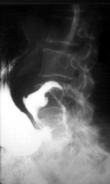 Upper gastrointestinal series shows duodenal lipoma with central ulceration where the overlying mucosa has thinned, ulcerated, and bled.
Colonic lipomas are usually discovered on endoscopy. Gentle palpation with a biopsy forceps reveals the soft nature of the submucosal mass. A biopsy specimen of the mucosa may reveal underlying fat, the so-called naked fat sign. As with lipomas in other locations, colonic lipomas may cause pain with obstruction or intussusception.
As noted above, a fatty protrusion of preperitoneal fat termed a "lipoma of the spermatic cord" is a common finding on groin exploration for hernia repair.
Numerous case reports document the presence of lipomas in other, rare locations, with these tumors having been found virtually everywhere in the body. [2, 3, 4, 5, 6] Lipomatous involvement of endocrine organs, including the thyroid, adrenal glands, pancreas, and parathyroid glands, has been described. Maxillofacial lipomas, including intralingual, parotid, orbitonasal, maxillary sinusoidal, and parapharyngeal space masses, have also been documented.
In rare instances, intraosseous and intra-articular involvement occurs. Involvement of the structural components of the mediastinum, including the airways and pleura, has also been reported. Gynecologic lipomas may occur in the uterus, ovaries, and broad ligament. Critical organ involvement of the heart (causing ventricular tachycardia), superior vena cava, brain, and spinal cord may pose a significant clinical challenge. [7, 8]
Mixed histologies, such as angiolipomas and fibrolipomas, are often encountered and are usually benign. Differentiation from liposarcoma may be difficult.
Other fatty tumors include lipoblastomas, hibernomas, atypical lipomatous tumors, and liposarcomas. Lipoblastomas occur almost exclusively in infants and children. They have a benign clinical course and a low recurrence rate after surgical excision. Hibernomas, also rare, derive their name from the morphologic resemblance to the brown fat of hibernating animals. They presumably arise from fat that may occur in the back, hips, or neck in adults and infants. Atypical lipomatous tumors are generally considered to be low-grade sarcomas, with a strong propensity to recurrence but little metastatic potential. Liposarcomas are true mesenchymal malignancies.
Etiology
Speculation exists regarding a potential link between trauma and subsequent lipoma formation. [9] One theory suggests that trauma-related fat herniation through tissue planes creates so-called pseudolipomas. It has also been suggested that trauma-induced cytokine release triggers pre-adipocyte differentiation and maturation. To date, no definitive link between trauma and lipoma formation has been prospectively demonstrated.
While the exact etiology of lipomas remains uncertain, an association with gene rearrangements of chromosome 12 has been established in cases of solitary lipomas, as has an abnormality in the HMGA2-LPP fusion gene. [10]
Epidemiology
Lipomas occur in 1% of the population. Most of these are small subcutaneous tumors that are removed for cosmetic reasons. These subcutaneous lipomas will be considered separately from lipomas in other locations in the discussion below. In the intestine, lipomas constitute 16% of benign, small neoplasms; this percentage is lower than that of leiomyomas (18%) and higher than that of adenomas (14%).
Prognosis
The outcome and prognosis are excellent for benign lipomas. Recurrence is uncommon but may develop if the excision was incomplete.
Pang et al compared outcomes in 238 patients who underwent total or near-total (T/NT) resection for dorsal, transitional, or chaotic spinal cord lipomas (with 16-year follow-up), along with complete reconstruction of the neural placode, with results from 116 patients who underwent partial resection for spinal cord lipomas (with 11-year follow-up). [11, 12]
Although in the T/NT and partial resection groups the rate of immediate symptom stabilization or improvement was similar (more than 95%), the combined cerebrospinal fluid leakage and wound complication rate was only 2.5% for T/NT resections, compared with 6.9% for partial resections. Moreover, the overall progression-free survival probability (Kaplan-Meier analysis) was 82.8% for T/NT resection patients at 16 years postoperative, compared with 34.6% for partial resection patients at 10.5 years post operation. Evidence indicated that the superior results in the T/NT resection patients were associated with the fact that lower cord-sac ratios were achieved in these patients than in the partial-resection group. [11, 12]
-
Upper gastrointestinal series shows duodenal lipoma with central ulceration where the overlying mucosa has thinned, ulcerated, and bled.
-
Duodenal lipoma resected through a duodenotomy. Overlying mucosa with central ulceration removed and lobulated fatty tumor shelled out intact with capsule. The mucosa was then sutured closed, and the duodenotomy closed. The stitch was placed to orient the specimen for pathologic examination.


