Continually Updated Clinical Reference
 
 
  All Sources     eMedicine     Medscape     Drug Reference     MEDLINE
 
eMedicine - Gastric Outlet Obstruction : Article by

Quick Find
Authors & Editors
Introduction
Indications
Relevant Anatomy
Contraindications
Workup
Treatment
Complications
Outcome And Prognosis
Future And Controversies
Multimedia
References


Patient Education
Click here for patient education.


Author: Andres E Castellanos, MD, Assistant Program Director, Surgery Residency Program, Assistant Professor, Department of Surgery, Drexel University College of Medicine

Andres Castellanos is a member of the following medical societies: American College of Surgeons

Coauthor(s): Elisa A Stein, MD, Staff Physician, Department of Surgery, Drexel University College of Medicine, Hahnemann University Hospital; Barry D Mann, MD, Program Director, Associate Professor, Department of Surgery, Division of General Surgery, MCP Hahnemann University; James de Caestecker, DO, Instructor, Department of Surgery, MCP Hahnemann University

Editors: Brian James Daley, MD, MBA, FACS, Associate Program Director, Professor, Department of Surgery, Division of Trauma and Critical Care, University of Tennessee School of Medicine; Francisco Talavera, PharmD, PhD, Senior Pharmacy Editor, eMedicine; Michael A Grosso, MD, Consulting Staff, Department of Cardiothoracic Surgery, St Francis Hospital; Paolo Zamboni, MD, Professor of Surgery, Chief of Day Surgery Unit, Chair of Vascular Diseases Center, University of Ferrara, Italy; John Geibel, MD, DSc, MA, Professor, Department of Surgery, Section of Gastrointestinal Medicine and Department of Cellular and Molecular Physiology, Yale University School of Medicine; Director of Surgical Research, Department of Surgery, Yale-New Haven Hospital

Author and Editor Disclosure

Synonyms and related keywords: GOO, gastric obstruction, proximal bowel obstruction, peptic ulcer disease, PUD, peripancreatic malignancy

Gastric outlet obstruction (GOO) is not a single entity; it is the clinical and pathophysiological consequence of any disease process that produces a mechanical impediment to gastric emptying.

Clinical entities that can result in GOO generally are categorized into 2 well-defined groups of causes—benign and malignant. This classification facilitates discussion of management and treatment. In the past, when peptic ulcer disease (PUD) was more prevalent, benign causes were the most common; however, one review shows that only 37% of patients with GOO have benign disease and the remaining patients have obstruction secondary to malignancy.1

Problem

GOO can be a diagnostic and treatment dilemma. As part of the initial workup, exclude the possibility of functional nonmechanical causes of obstruction, such as diabetic gastroparesis.

Once a mechanical obstruction is confirmed, differentiate between benign and malignant processes because definitive treatment is based on recognition of the specific underlying cause.

Carry out diagnosis and treatment expeditiously because delay may result in further compromise of the patient's nutritional status.

Frequency

The incidence of GOO has been reported to be less than 5% in patients with PUD, which is the leading benign cause of the problem.

The incidence of GOO in patients with peripancreatic malignancy, the most common malignant etiology, has been reported as 15-20%.

Etiology

The major benign causes of GOO are PUD, gastric polyps, ingestion of caustics, pyloric stenosis, congenital duodenal webs, gallstone obstruction (Bouveret syndrome), pancreatic pseudocysts, and bezoars.

PUD manifests in approximately 5% of all patients with GOO. Ulcers within the pyloric channel and first portion of the duodenum usually are responsible for outlet obstruction. Obstruction can occur in an acute setting secondary to acute inflammation and edema or, more commonly, in a chronic setting secondary to scarring and fibrosis. Helicobacter pylori has been implicated as a frequent associated finding in patients with GOO, but its exact incidence has not been defined precisely.

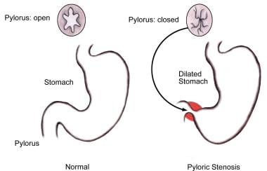
Within the pediatric population, pyloric stenosis constitutes the most important cause of GOO. Pyloric stenosis occurs in 1 per 750 births. It is more common in boys than in girls and also is more common in first-born children. Pyloric stenosis is the result of gradual hypertrophy of the circular smooth muscle of the pylorus.

Pancreatic cancer is the most common malignancy causing GOO. Outlet obstruction may occur in 10-20% of patients with pancreatic carcinoma. Other tumors that may obstruct the gastric outlet include ampullary cancer, duodenal cancer, cholangiocarcinomas, and gastric cancer. Metastases to the gastric outlet also may be caused by other primary tumors.

Pathophysiology

Intrinsic or extrinsic obstruction of the pyloric channel or duodenum is the usual pathophysiology of GOO; as previously noted, the mechanism of obstruction depends upon the underlying etiology.

Patients present with intermittent symptoms that progress until obstruction is complete. Vomiting is the cardinal symptom. Initially, patients may demonstrate better tolerance to liquids than solid food.

In a later stage, patients may develop significant weight loss due to poor caloric intake. Malnutrition is a late sign, but it may be very profound in patients with concomitant malignancy.

In the acute or chronic phase of obstruction, continuous vomiting may lead to dehydration and electrolyte abnormalities.

When obstruction persists, patients may develop significant and progressive gastric dilatation. The stomach eventually loses its contractility. Undigested food accumulates and may represent a constant risk for aspiration pneumonia.

Clinical

Nausea and vomiting are the cardinal symptoms of GOO. Vomiting usually is described as nonbilious, and it characteristically contains undigested food particles. In the early stages of obstruction, vomiting may be intermittent and usually occurs within 1 hour of a meal.

Early satiety and epigastric fullness are common. Weight loss is frequent when the condition approaches chronicity and is most significant in patients with malignant disease.

Abdominal pain is not frequent and usually relates to the underlying cause, eg, PUD, pancreatic cancer.

Physical examination often demonstrates the presence of chronic dehydration and malnutrition. A dilated stomach may be appreciated as a tympanitic mass in the epigastric area and/or left upper quadrant.

Dehydration and electrolyte abnormalities can be demonstrated by routine laboratory examinations. Increases in BUN and creatinine are late features of dehydration. Prolonged vomiting causes loss of hydrochloric (HCl) acid and produces an increase of bicarbonate in the plasma to compensate for the lost chloride and sodium. The result is a hypokalemic hypochloremic metabolic alkalosis. Alkalosis shifts the intracellular potassium to the extracellular compartment, and the serum positive potassium is increased factitiously. With continued vomiting, the renal excretion of potassium increases in order to preserve sodium. The adrenocortical response to hypovolemia intensifies the exchange of potassium for sodium at the distal tubule, with subsequent aggravation of the hypokalemia.


Once the diagnosis is suspected, request a surgical consultation.

Patients with GOO due to benign ulcer disease may be treated medically if results of imaging studies or endoscopy determine that acute inflammation and edema are the principle causes of the outlet obstruction (as opposed to scarring and fibrosis, which may be fixed). If medical therapy conducted for a reasonable period fails to alleviate the obstruction, then surgical intervention becomes appropriate. The choice of surgical procedure depends upon the patient's particular circumstances; however, vagotomy and antrectomy should be considered the criterion standard against which the efficacy of other procedures should be measured.

Weigh the extent of surgical intervention for the relief of GOO against the type and extent of malignancy and the patient's anticipated long-term prognosis. As a guiding principle, undertake major tumor resections in the absence of metastatic disease in a patient who can withstand such a procedure from a nutritional standpoint. In patients with largely metastatic disease, determine the degree of surgical intervention for palliation in light of the patient's realistic prognosis and personal wishes.


The stomach is located mainly in the left upper quadrant beneath the diaphragm and is attached superiorly to the esophagus and distally to the duodenum. The stomach is divided into 4 portions, the cardia, the body, the antrum, and the pylorus. Inflammation, scarring, or infiltration of the antrum and pylorus are associated with the development of GOO.

The duodenum begins immediately beyond the pylorus and mostly is a retroperitoneal structure, wrapping around the head of the pancreas. The duodenum classically is divided into 4 portions. It is intimately related to the gallbladder, liver, and pancreas; therefore, a malignant process of any adjacent structure may cause outlet obstruction due to extrinsic compression.


Contraindications for surgery relate to the underlying medical condition.

Most patients benefit from an initial period of gastric decompression, hydration, and correction of electrolyte imbalances. In patients who are severely malnourished, postponing surgical intervention until the nutritional status has been optimized may be wise. In selective cases, some patients may benefit from total parenteral nutrition (TPN) or distal tube feeding (eg, placed via a percutaneous jejunostomy).

One of the relative contraindications for surgery is the presence of advanced malignancy; in these cases, life expectancy may be limited to a few months.

Overall, every patient with GOO deserves evaluation by a surgeon. Even if the patient has unresectable disease, other palliative surgical measures may improve the quality of life.


Lab Studies

  • Obtain a CBC. Check the hemoglobin and hematocrit to rule out the possibility of anemia.
  • Obtain an electrolyte panel. As noted previously, identifying and correcting electrolyte abnormalities that tend to occur is essential.
  • Liver function tests may be helpful, particularly when a malignant etiology is suspected.
  • A test for H pylori is helpful when the diagnosis of PUD is suspected.

Imaging Studies

  • Plain abdominal radiographs, contrast upper GI studies (Gastrografin or barium), and CT scans with oral contrast are helpful.
  • Plain radiographs, including the obstruction series (ie, supine abdomen, upright abdomen, chest posteroanterior), can demonstrate the presence of gastric dilatation and may be helpful in distinguishing the differential diagnosis.

Diagnostic Procedures

  • Upper endoscopy can help visualize the gastric outlet and may provide a tissue diagnosis when the obstruction is intraluminal.
  • The sodium chloride load test is a traditional clinical nonimaging study that may be helpful.
    • The traditional sodium chloride load test is performed by infusing 750 cc of sodium chloride solution into the stomach via a nasogastric tube (NGT).
    • A diagnosis of GOO is made if more than 400 cc remain in the stomach after 30 minutes.
  • Nuclear gastric emptying studies measure the passage of orally administered radionuclide over time. Unfortunately, both the nuclear test and the saline load test may produce abnormal results in functional states.
  • Barium upper GI studies are very helpful because they can delineate the gastric silhouette and demonstrate the site of obstruction. An enlarged stomach with a narrowing of the pyloric channel or first portion of the duodenum helps differentiate GOO from gastroparesis.
  • The specific cause may be identified as an ulcer mass or intrinsic tumor.
  • In the presence of PUD, perform endoscopic biopsy to rule out the presence of malignancy.
  • In the case of peripancreatic malignancy, CT scan–guided biopsy may be helpful in establishing a preoperative diagnosis.
  • Needle-guided biopsy also may be helpful in establishing the presence of metastatic disease. This knowledge may impact the magnitude of the procedure planned to alleviate the GOO.

Histologic Findings

Histologic findings relate to the individual underlying cause.


Medical therapy

Initial management of GOO should be the same regardless of the primary cause. After a diagnosis is made, admit patients for hydration and correction of electrolyte abnormalities. Remembering that the metabolic alkalosis of GOO responds to the administration of chloride is important; therefore, sodium chloride solution should be the initial IV fluid of choice. Potassium deficits are corrected after repletion of volume status and after replacement of chloride.

Place a NGT to decompress the stomach. Occasionally, a large tube is required because the undigested food blocks tubes with small diameters.

Further treatment is tailored to the underlying cause; this is where the distinction between benign and malignant disease becomes important.

Surgical therapy

Management of benign disease

When acute PUD has been identified as a primary cause of GOO, focus treatment on the reduction of acid production. Histamine 2 (H2) blockers and proton pump inhibitors comprise the mainstay of treatment. Treat H pylori infection, when identified, according to current recommendations.  Although most patients improve temporarily with treatment, scarring and fibrosis may worsen over time. These patients are likely to present with recurrent GOO. More than 75% of patients presenting with GOO eventually require surgical intervention.2

Surgical intervention usually provides definitive treatment of GOO, but it may result in its own comorbid consequences. Operative management should offer both relief of obstruction and correction of the acid problem.

The most common surgical procedures performed for GOO related to PUD are vagotomy and antrectomy, vagotomy and pyloroplasty, truncal vagotomy and gastrojejunostomy, pyloroplasty, and laparoscopic variants of the aforementioned procedures. Of these, vagotomy and antrectomy with Billroth II reconstruction (gastrojejunostomy) seem to offer the best results. Vagotomy and pyloroplasty and pyloroplasty alone, although used with some success, can be technically difficult to perform due to scarring at the gastric outlet. A combination of balloon dilatation and highly selective vagotomy has been described, but it is associated with gastroparesis and a high recurrence rate.

The role of the laparoscopic approach in the treatment of GOO is under investigation and may represent a valid form of therapy with low morbidity. The experience of several international centers has been published. One group in China performed laparoscopic truncal vagotomy and gastrojejunostomy for GOO related to PUD with nearly complete resolution of symptomatology. They reported no conversions to open procedure or mortalities. Twenty-seven percent of patients did experience transiently delayed gastric emptying, which resolved with conservative measures.3

Pneumatic balloon dilatation of a chronic benign stricture can be performed via endoscopy. Published series using this technique report success rates over 76% after multiple dilatations.4 Patients treated with balloon dilatation, without treatment of H pylori infection, have a higher rate of failure and recurrent obstruction.5 Patients who are negative for H pylori do not respond favorably to balloon dilatation and should be considered for surgical treatment early in the process.6

Management of malignant disease

The management of GOO secondary to malignancy is controversial. Of patients with periampullary cancer, 30-50% present with nausea and vomiting at the time of diagnosis.7, 8 Most of these tumors are unresectable (approximately 40% of gastric cancers and 80-90% of periampullary cancers).9, 10 When tumors are found to be unresectable, 13-20% of patients eventually develop GOO before they succumb to their disease. The 1-year survival rate is poor.

Gastrojejunostomy remains the surgical treatment of choice for GOO secondary to malignancy. Although surgeons traditionally have preferred an antecolic anastomosis to prevent further obstruction by advancing tumor growth, a publication evaluating the retrocolic anastomosis in this setting challenges conventional wisdom.11 Results demonstrate that a retrocolic anastomosis may be associated with decreased incidences of delayed gastric emptying (6% vs 17%) and late GOO (2% vs 9%). Other groups have illustrated that partial stomach-partitioning gastrojejunostomy decreases the rates of delayed gastric emptying as compared to traditional gastrojejunostomy.12

Internationally, studies are underway using laparoscopic gastrojejunostomy instead of the open procedure. In the United States, critics cite a nearly 20% conversion rate and a delay in the return of gut function as reasons to not perform the procedure laparoscopically.  Comparisons of laparoscopic GI anastomosis versus the open procedure have revealed less morbidity and mortality, shorter hospital stays, fewer blood transfusions, and faster GI transit recovery time.13, 14 Researchers at Johns Hopkins Hospital have attempted endoscopic transgastric approaches to create a gastrojejunostomy in a porcine model.15 As natural orifice transluminal surgery gains more widespread interest, these novel approaches may become more popular.

Recently, self-expandable metallic stents have been used for the treatment of GOO in a malignant setting. Metallic stents have previously been used successfully to treat stenosis of such areas as the blood vessels, bile duct, esophagus, and trachea. With the development of newer stents and delivery systems, metallic stents may have a role in the nonsurgical treatment of gastroduodenal obstruction. Stents may allow the physician to avoid complicated surgical procedures. Currently, only the Wallstent has FDA approval for palliation in malignant gastroduodenal obstruction.  Significant complications include the following: malposition, misdeployment, tumor ingrowth or overgrowth, migration, bleeding, or perforation.16 

A review of 19 studies published in 2004 quoted clinical success rates of 80-90%.17 More recent multicenter trials using the enteral Wallstent in 176 patients with malignant GOO resulted in 89% of patients tolerating oral intake for a median of 219 days post procedure. Of the 84% in whom the stent was successful after the initial procedure, 22% required restenting to tolerate an oral diet. In addition, as other studies have demonstrated, chemotherapy was independently associated with an increased tolerance in oral intake.18 One proposed solution uses covered metallic stents that have a lower incidence of tumor ingrowth.  A 60% rate of tumor ingrowth in uncovered stents versus a 10% rate of tumor ingrowth in covered stents has been reported.  Furthermore, with the double stent technique, that is, simultaneous placement of both covered stents and uncovered stents, lower early restenosis rates have been achieved. A stent patency of 21.5 days for uncovered stents versus 150 days for double stents has been achieved.19

Several retrospective studies have been performed to compare the results of stenting versus surgical intervention. Survival rates are equivalent; however, costs, length of stay, and number of subsequent procedures are all decreased following stenting.20, 21 In addition, a delay of gastric emptying and morbidity decrease with the use of metallic stents.22 These promising results suggest that stents may eventually replace surgery as palliative intervention for unresectable periampullary malignancies.

Preoperative details

Perform standard preoperative evaluation in these patients. Correct fluid and electrolyte abnormalities prior to surgery.

Perform gastric decompression by NGT and suction and alert the anesthesiologist to the potential risk for aspiration upon induction.

Perform a preoperative nutritional evaluation and initiate appropriate nutritional therapy (ie, TPN or enteral feedings via a percutaneous jejunostomy placed distal to the obstruction) as soon as possible.

Intraoperative details

Intraoperative details depend upon the etiology of the underlying disease and the reason that the specific surgical procedure is undertaken.

Postoperative details

Admit patients to a monitor unit after the procedure. Pay special attention to fluid and electrolyte status.

Most surgeons agree that perioperative antibiotics are advisable but may be limited to use during the immediate perioperative period in the absence of intervening infection.

If a gastric reconstruction is performed, an NGT is recommended. The length of time that the NGT should remain in place is controversial; however, it is important to remember that a previously dilated stomach, the performance of a vagotomy, and the presence of metastatic cancer may all contribute to decreased gastric motility. An anatomically patent gastrojejunostomy may fail to empty for days. This syndrome of delayed gastric emptying is a well-known entity and requires surgical patience.

Aggressive pulmonary toilet, deep venous thrombosis (DVT) prophylaxis, and early ambulation are advisable.

Follow-up

Closely monitor patients after surgery and upon discharge. After relief of GOO, patients may continue to experience gastric dysmotility and may require medication to stimulate gastric emptying and motility.

In patients with malignancy, the potential for progressive and recurrent disease always remains. These patients should be monitored by a surgeon or an oncologist.

Closely monitor patients whose treatment consisted of balloon dilatation because most of these patients require subsequent dilatations to achieve satisfactory results.


Although the risk is small, patients undergoing endoscopic treatment with either balloon dilatation or stenting are at risk for perforation. Several literature reports exist regarding migration of the stents and reocclusion requiring further intervention.

Operative complications in patients undergoing surgery for GOO often are related to the nutritional status of the patients. Commencing nutritional support upon recognition of the presence of GOO is important. If surgery is anticipated, delaying the surgery or any intervention until TPN has been instituted for at least 1 week is often prudent.

Acute intervention may be technically difficult because of significant gastric dilatation and gastric wall edema. This circumstance may increase the rate of anastomotic leak. On occasion, delaying surgical intervention for several days while the stomach is decompressed by nasogastric suction may be prudent.

Alert patients undergoing gastric resection for benign or malignant disease to the possibility of well-known postgastrectomy syndromes, such as dumping, alkaline gastritis, and afferent loop syndrome. Severe symptoms may be present in 1-2% of patients.


GOO is a clinical condition that may result from a number of underlying causes, both benign and malignant. Despite medical advances in the acid suppression mechanism, the incidence of GOO remains a prevalent clinical problem in benign PUD. Also, an increase in the number of cases of GOO seems to be noted secondary to malignancy; this is possibly due to improvements in cancer therapy, which allow patients to live long enough to develop this complication.

Orient initial management to identification of the primary underlying cause and to the correction of volume and electrolyte abnormalities. Barium swallow studies and upper endoscopy are the main tests used to help make the diagnosis. Tailor treatment to the specific cause.


The role of prophylactic gastrojejunostomy in cases of malignant GOO is a question that has not been answered. Some surgeons argue that prophylactic gastrojejunostomy may increase postoperative morbidity, primarily due to delayed gastric emptying. Lillemoe and Cameron addressed this issue in a publication.23 Of patients with unresectable periampullary cancer, 87 were randomized to receive or not receive a prophylactic gastrojejunostomy. Although results demonstrated that no significant differences exist in morbidity, length of hospital stay, and survival rates, the group in which prophylactic gastrojejunostomies were performed had a 0% (0/44) incidence of GOO versus 19% (8/43) in the other group. The authors concluded that prophylactic gastrojejunostomy significantly decreased the incidence of late GOO and should be performed routinely when a patient is undergoing surgical palliation for periampullary cancer. Comparable data are supported by other series.


Media file 1:  Plain radiograph of the abdomen. Enlarged stomach with calcified content.
Click to see larger pictureClick to see detailView Full Size Image
Media type:  Image

Media file 2:  Contrast study demonstrating an enlarged stomach. The point of obstruction is visualized at the pyloric-duodenal junction (string sign).
Click to see larger pictureClick to see detailView Full Size Image
Media type:  Image


  1. Andersson A, Bergdahl L. Carcinoid tumors of the appendix in children. A report of 25 cases. Acta Chir Scand. 1977;143(3):173-5. [Medline].
  2. Doberneck RC, Berndt GA. Delayed gastric emptying after palliative gastrojejunostomy for carcinoma of the pancreas. Arch Surg. Jul 1987;122(7):827-9. [Medline].
  3. Siu WT, Tang CN, Law BK, Chau CH, Yau KK, Yang GP, et al. Vagotomy and gastrojejunostomy for benign gastric outlet obstruction. J Laparoendosc Adv Surg Tech A. Oct 2004;14(5):266-9. [Medline].
  4. Lam YH, Lau JY, Fung TM, et al. Endoscopic balloon dilation for benign gastric outlet obstruction with or without Helicobacter pylori infection. Gastrointest Endosc. Aug 2004;60(2):229-33. [Medline].
  5. Taskin V, Gurer I, Ozyilkan E, Sare M, Hilmioglu F. Effect of Helicobacter pylori eradication on peptic ulcer disease complicated with outlet obstruction. Helicobacter. Mar 2000;5(1):38-40. [Medline].
  6. Gouma DJ, van Geenen R, van Gulik T, de Wit LT, Obertop H. Surgical palliative treatment in bilio-pancreatic malignancy. Ann Oncol. 1999;10 Suppl 4:269-72. [Medline].
  7. Jaffin BW, Kaye MD. The prognosis of gastric outlet obstruction. Ann Surg. Feb 1985;201(2):176-9. [Medline].
  8. Khullar SK, DiSario JA. Gastric outlet obstruction. Gastrointest Endosc Clin N Am. Jul 1996;6(3):585-603. [Medline].
  9. Kurtz RC, Sherlock P. Carcinoma of the stomach. In: Bockus Gastroenterology. 4th ed. Philadelphia, Pa: WB Saunders Co; 1985.
  10. Lillemoe KD, Sauter PK, Pitt HA, Yeo CJ, Cameron JL. Current status of surgical palliation of periampullary carcinoma. Surg Gynecol Obstet. Jan 1993;176(1):1-10. [Medline].
  11. Lillemoe KD, Cameron JL, Hardacre JM, Sohn TA, Sauter PK, Coleman J, et al. Is prophylactic gastrojejunostomy indicated for unresectable periampullary cancer? A prospective randomized trial. Ann Surg. Sep 1999;230(3):322-8; discussion 328-30. [Medline].
  12. Arciero CA, Joseph N, Watson JC, Hoffman JP. Partial stomach-partitioning gastrojejunostomy for malignant duodenal obstruction. Am J Surg. Mar 2006;191(3):428-32. [Medline].
  13. Bergamaschi R, Marvik R, Thoresen JE, Ystgaard B, Johnsen G, Myrvold HE. Open versus laparoscopic gastrojejunostomy for palliation in advanced pancreatic cancer. Surg Laparosc Endosc. Apr 1998;8(2):92-6. [Medline].
  14. Alam TA, Baines M, Parker MC. The management of gastric outlet obstruction secondary to inoperable cancer. Surg Endosc. Feb 2003;17(2):320-3. [Medline].
  15. Kantsevoy SV, Jagannath SB, Niiyama H, Chung SS, Cotton PB, Gostout CJ, et al. Endoscopic gastrojejunostomy with survival in a porcine model. Gastrointest Endosc. Aug 2005;62(2):287-92. [Medline].
  16. Adler DG, Merwat SN. Endoscopic approaches for palliation of luminal gastrointestinal obstruction. Gastroenterol Clin North Am. Mar 2006;35(1):65-82, viii. [Medline].
  17. Baron TH. Surgical versus endoscopic palliation of malignant gastric outlet obstruction: big incision, little incision, or no incision?. Gastroenterology. Oct 2004;127(4):1268-9. [Medline].
  18. Telford JJ, Carr-Locke DL, Baron TH, Tringali A, Parsons WG, Gabbrielli A, et al. Palliation of patients with malignant gastric outlet obstruction with the enteral Wallstent: outcomes from a multicenter study. Gastrointest Endosc. Dec 2004;60(6):916-20. [Medline].
  19. Song GA, Kang DH, Kim TO, Heo J, Kim GH, Cho M, et al. Endoscopic stenting in patients with recurrent malignant obstruction after gastric surgery: uncovered versus simultaneously deployed uncovered and covered (double) self-expandable metal stents. Gastrointest Endosc. May 2007;65(6):782-7. [Medline].
  20. Yim HB, Jacobson BC, Saltzman JR, Johannes RS, Bounds BC, Lee JH, et al. Clinical outcome of the use of enteral stents for palliation of patients with malignant upper GI obstruction. Gastrointest Endosc. Mar 2001;53(3):329-32. [Medline].
  21. Del Piano M, Ballare M, Montino F, Todesco A, Orsello M, Magnani C, et al. Endoscopy or surgery for malignant GI outlet obstruction?. Gastrointest Endosc. Mar 2005;61(3):421-6. [Medline].
  22. Wong YT, Brams DM, Munson L, Sanders L, Heiss F, Chase M, et al. Gastric outlet obstruction secondary to pancreatic cancer: surgical vs endoscopic palliation. Surg Endosc. Feb 2002;16(2):310-2. [Medline].
  23. Quigley RL, Pruitt SK, Pappas TN, Akwari O. Primary hypertrophic pyloric stenosis in the adult. Arch Surg. Sep 1990;125(9):1219-21. [Medline].
  24. Chopita N, Landoni N, Ross A, Villaverde A. Malignant gastroenteric obstruction: therapeutic options. Gastrointest Endosc Clin N Am. Jul 2007;17(3):533-44, vi-vii. [Medline].
  25. Gibson JB, Behrman SW, Fabian TC, Britt LG. Gastric outlet obstruction resulting from peptic ulcer disease requiring surgical intervention is infrequently associated with Helicobacter pylori infection. J Am Coll Surg. Jul 2000;191(1):32-7. [Medline].
  26. Holt AP, Patel M, Ahmed MM. Palliation of patients with malignant gastroduodenal obstruction with self-expanding metallic stents: the treatment of choice?. Gastrointest Endosc. Dec 2004;60(6):1010-7. [Medline].
  27. Lillemoe KD, Cameron JL, Yeo CJ, Sohn TA, Nakeeb A, Sauter PK, et al. Pancreaticoduodenectomy. Does it have a role in the palliation of pancreatic cancer?. Ann Surg. Jun 1996;223(6):718-25; discussion 725-8. [Medline].
  28. Mauro MA, Koehler RE, Baron TH. Advances in gastrointestinal intervention: the treatment of gastroduodenal and colorectal obstructions with metallic stents. Radiology. Jun 2000;215(3):659-69. [Medline].
  29. Schwarz A, Beger HG. Biliary and gastric bypass or stenting in nonresectable periampullary cancer: analysis on the basis of controlled trials. Int J Pancreatol. Feb 2000;27(1):51-8. [Medline].
  30. Shyr YM, Su CH, Wu CW, Lui WY. Prospective study of gastric outlet obstruction in unresectable periampullary adenocarcinoma. World J Surg. Jan 2000;24(1):60-4; discussion 64-5. [Medline].
  31. Wade TP, Neuberger TJ, Swope TJ, Virgo KS, Johnson FE. Pancreatic cancer palliation: using tumor stage to select appropriate operation. Am J Surg. Jan 1994;167(1):208-12; discussion 212-3. [Medline].

Gastric Outlet Obstruction excerpt

Article Last Updated: Nov 20, 2007