Continually Updated Clinical Reference
 
 
  All Sources     eMedicine     Medscape     Drug Reference     MEDLINE
 
eMedicine - Chyle Fistula : Article by

Quick Find
Authors & Editors
Introduction
Indications
Relevant Anatomy
Contraindications
Treatment
Complications
Outcome and Prognosis
Future and Controversies
Acknowledgments
Multimedia
References


Patient Education
Click here for patient education.


Author: Deron J Tessier, MD, Staff Surgeon, Kaiser Permanente Medical Center, Fontana, CA

Deron J Tessier is a member of the following medical societies: American College of Surgeons and American Medical Association

Coauthor(s): Russell A Williams, MBBS, Program Director, Professor, Department of Surgery, University of California Medical Center at Irvine

Editors: Juan B Ochoa, MD, Assistant Professor, Department of Surgery, University of Pittsburgh; Francisco Talavera, PharmD, PhD, Senior Pharmacy Editor, eMedicine; David L Morris, MD, PhD, Professor, Department of Surgery, St George Hospital, University of New South Wales, Australia; Paolo Zamboni, MD, Professor of Surgery, Chief of Day Surgery Unit, Chair of Vascular Diseases Center, University of Ferrara, Italy; John Geibel, MD, DSc, MA, Professor, Department of Surgery, Section of Gastrointestinal Medicine and Department of Cellular and Molecular Physiology, Yale University School of Medicine; Director of Surgical Research, Department of Surgery, Yale-New Haven Hospital

Author and Editor Disclosure

Synonyms and related keywords: chyle fistulas, chylous ascites, chylothorax, chyloperitoneum, lymphatic fluid leakage

Chyle fistula is defined as a leakage of lymphatic fluid from the lymphatic vessels, typically accumulating in the thoracic or abdominal cavities, but occasionally manifesting as an external fistula. Chyle fistula is a rare but potentially devastating and morbid condition. First described in the 17th century as complications of trauma, chyle fistulas most commonly occur secondary to lymphatic disease or malignancy or following abdominal, neck, or thoracic operations. Chyle fistulas also can form as a result of venous hypertension, and they have been described in patients with superior vena cava syndrome or thrombosis of the vena cava, among others.

History of chyle fistulas

  • 17th century: Trauma is recognized as a primary cause of chylous fistulas.
  • 1627: Asellius first describes chyle from the mesenteric vessels of a dog.
  • 1691: Morton describes chylous ascites after performing paracentesis in an 18-month-old child.
  • 1854: Virchow publishes an account of chylous ascites in a newborn calf with occlusion of the subclavian vein.

History of the Procedure

Patients with chyle fistulas usually give histories of some comorbid conditions such as malignancy or prior operations in the chest, neck, or abdomen. In postoperative patients, symptoms become evident after the start of oral feeding and depend on the site of obstruction.

Problem

Chyle fistulas can be extremely morbid due to loss of fluids, electrolytes, and other nutrients. In addition, chyle fistulas can result in loss of lymphocytes and immune dysfunction. Finally, chyle fistulas are space-filling and exert pressure on surrounding tissues, creating symptoms that can range from minimal discomfort to life-threatening situations.

Frequency

Chyle fistulas are rare events. Typical causes include lymphatic disease; malignancy; trauma; and postoperative trauma following abdominal, neck, or thoracic operations. Postoperatively, approximately 75% of chyloperitoneum cases occur after abdominal aortic aneurysm repair, 19% after aortofemoral bypass, and 7% after resection of infected aortic grafts.

Etiology

The most common causes of chyle fistulas include subclavian vein thrombosis, malignant invasion of the lymphatics, inflammatory reactions (ie, tuberculosis, pancreatitis, cirrhosis, adhesions, pulmonary fibrosis), and prior surgeries near the cisterna chyli or thoracic duct.

Pathophysiology

Leakage of lymph from damaged lymph vessels is common after surgery or trauma. However, damaged lymphatics most often heal spontaneously or direct lymph centrally via rich interconnected lymphatic collaterals, without any significant morbidity. For chyle fistulas to form, either a scarcity of lymphatic collaterals must be present or the injury to the lymphatic channels must overwhelm the remaining lymphatic vessels. In addition, abnormal lymphatic vessels may be incapable of adequate lymph flow, leading to the accumulation of chyle.

Chyle flow varies dramatically depending on the quantity and quality of oral intake. During times of starvation, chyle flow is minimal. After meals, especially those with high contents of long-chain fatty acids, chyle flow increases dramatically. This basic knowledge provides the rationale for controlling dietary intake as part of the treatment of this disease.

Clinical

Because of the relatively low incidence of chyle fistulas, a high index of suspicion is required to make a timely diagnosis. Diagnosis usually is made after the patient has recovered from the injury and has started eating.

Symptoms of chyloperitoneum include (1) nausea, (2) vomiting, (3) early satiety or anorexia, (4) abdominal discomfort or pain, and (5) dyspnea due to chylous fluid causing abdominal distention and pressure on the visceral structures and diaphragm.

Symptoms of chylothorax include (1) shortness of breath, (2) pleural effusion, and (3) decreased cardiac preload due to a mediastinal shift from a large chylothorax.


Characteristics of thoracentesis or paracentesis aspirate

  • Odorless
  • Milky appearance that separates into a creamy layer when left to stand
  • Specific gravity greater than 1.012
  • Total fat composition of 0.4-4 g/L
  • Total protein greater than 30 g/L
  • pH greater than 7.0
  • Sterile fluid
  • Lipophilic globules when stained with Sudan III
  • WBC differential of predominately lymphocytes


The cisterna chyli and thoracic duct drain lymph from the entire body except the head, neck, arms, and right thorax (which instead use the right bronchomediastinal, jugular, and subclavian lymph trunks to form the right lymph duct). The anatomy is highly variable, with 50% of people not having an identifiable cisterna chyli. In addition, half of the 4 liters of lymph draining through the cisterna chyli and thoracic duct originates from the intestinal and hepatic lymphatics.

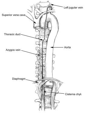
The cisterna chyli is found on the posterolateral edge to the right of the aorta at the level of vertebral bodies T12, L1, L2, and L3 as lymphatics from the mesentery, intercostal, and lumbar regions coalesce in the retroperitoneal space.

At approximately L1, the cisterna chyle ascends, becoming the thoracic duct. The duct then enters the posterior mediastinum, crosses at T4 into the left retropleural space, and continues in a cephalad direction. The thoracic duct then enters the venous system at the junction of the left subclavian and internal jugular veins (see Media file 1).


Contraindications to surgical correction of chyle fistulas are based on the patient's comorbidities and his or her ability to tolerate surgery.


Medical therapy

Treatment of chyle fistulas is determined by the following:

  • Etiology of the fistulas: Chyle fistulas secondary to malignancy are difficult to treat, while a definitive treatment is more successful after trauma or surgery.
  • Amount of output: Fistulas with higher output that cause more physiologic derangements may require earlier aggressive therapies.
  • Site of the fistulas: Fistulas arising in the neck are easier to access and identify than those occurring in the abdomen or thorax.

Nutritional intervention remains the mainstay of nonoperative treatment. Nutritional therapy includes the following:

  • Use of enteral diets with fat restriction or the use of medium chain triglycerides: Medium-chain triglycerides are absorbed directly from the gut into the portal venous circulation.
  • Use of total parenteral nutrition (TPN): TPN affords full caloric and nitrogenous support while allowing bowel rest. Bowel rest achieves a decrease in chyle flow, allowing healing to occur.

Several case reports and case series have reported the successful treatment of chylous leaks using octreotide in infants. One series reported 100% closure of chylous leaks from malignancy using 50 mcg of subcutaneous octreotide 3 times per day.

Transjugular intrahepatic portosystemic shunts (TIPS) have been reported to successfully treat chylous ascites due to cirrhosis.

A period of observation from one to several weeks should be allowed before determining whether these therapies are successful.

Repeated paracentesis/thoracentesis should be avoided because (1) results from this treatment modality are poor; (2) the chance of infecting chylous fluid is increased; (3) anasarca and cachexia may occur due to rapid loss of body protein, fat, and fluid; and (4) loss of lymphocytes may result in immune dysfunction.

In patients in whom accumulation of chyle leads to significant problems, such as shortness of breath and lung collapse, the use of drainage through a thoracostomy tube or paracentesis may be necessary despite the obvious adverse effects that this form of therapy may produce.

Surgical therapy

Surgery is undertaken when conservative therapies fail (approximately 40%). Surgical therapies include the following:

  • Surgical ligation of the leaking lymphatic vessels: Surgical ligation can be achieved successfully provided the site of the leak is identified and the primary pathology causing the leak has not caused disruption or blockage of other lymphatic vessels. Finding the leak may be aided by the use of scintigraphy. In some cases, the exact location of the disrupted lymphatic vessel is not identified and surgeons may use flaps (such as muscle flaps) to cover the area where the leak is occurring. Laparoscopic ligation of chyle leaks has been described.
  • Other forms of therapy to plug the leak have included fibrin glue or the use of chemical irritants such as tetracycline.
  • The use of peritoneovenous shunts (Denver or LeVeen) is a modality of treatment available when other treatments fail. Peritoneovenous shunts are especially useful for the management of chyle fistulas resulting from causes other than prior operations. The main objective of peritoneovenous shunts is to drain the chyle accumulated in the abdomen back to the venous circulation.

Preoperative details

If at all possible, ensure nutritional assessment and repletion before a surgical intervention because malnutrition increases morbidity significantly.

Administer gram-positive antibiotic coverage with a beta-lactamase inhibitor in the 24 hours prior to insertion of the shunt.

Because of the possible complication of disseminated intravascular coagulopathy, which is especially prevalent in patients with malignancy, include coagulation parameters in the preoperative evaluation.

Intraoperative details

Surgical approaches vary significantly depending on the site and etiology of the leak. Surgical approach to the thorax using a thoracoscopic approach may be tried, as long as adequate expertise is available. Denver and LeVeen shunts differ in the type of valve each uses.

  • Denver valve: These use a slit system that reduces flow by impedance. Insertion of these shunts can be achieved as a minimally invasive procedure, and they can be inserted under local anesthetic, sparing the severely ill patient the trauma of a major operation.
  • LeVeen valve: This is a closed-pressure system that opens at pressures of 3 cm of water or less. This prevents leakage of blood into the tubing, thereby decreasing the chances of the system clotting (with subsequent failure).

Postoperative details

Antibiotic coverage is continued empirically for 4 days after surgery. Any coagulopathy should be treated aggressively without delay.

Follow-up

Patients should be monitored routinely until the resolution of the fistula. Postoperative visits should commence approximately 1 week after the patient is discharged from the hospital.


Untreated, chyle fistulas can be fatal, with patients dying from severe fluid and electrolyte abnormalities, malnutrition, and overwhelming infections, including peritonitis and empyema. Iatrogenic complications also can occur. Complications from shunt placement, including disseminated intravascular coagulopathy, shunt failure, and fluid overload, also can occur.


Treatment of chyle fistula is very successful. However, unless the underlying etiology is reversed, the problem can be chronic and unrelenting.


The duration of medical therapies undertaken and the decision to proceed operatively are physician dependent. Because of the relative rarity of chyle fistulas, no definitive data are available to unequivocally direct the duration of therapy.


The authors and editors of eMedicine gratefully acknowledge the contributions of previous coauthor, Yale D Podnos, MD, MPH, to the development and writing of this article.


Media file 1:  Chyle fistula. Anatomy of the thoracic duct in relation to the aorta.
Click to see larger pictureClick to see detailView Full Size Image
Media type:  Image

Media file 2:  Chyle fistula. Management of chyloperitoneum.
Click to see larger pictureClick to see detailView Full Size Image
Media type:  Graph

Media file 3:  Chyle fistula. Management of chylothorax.
Click to see larger pictureClick to see detailView Full Size Image
Media type:  Graph


  • Browse NL, Allen DR, Wilson NM. Management of chylothorax. Br J Surg. Dec 1997;84(12):1711-6. [Medline].
  • Cerfolio RJ, Allen MS, Deschamps C, Trastek VF, Pairolero PC. Postoperative chylothorax. J Thorac Cardiovasc Surg. Nov 1996;112(5):1361-5; discussion 1365-6. [Medline].
  • de Vries GJ, Ryan BM, de Bievre M, Driessen A, Stockbrugger RW, Koek GH. Cirrhosis related chylous ascites successfully treated with TIPS. Eur J Gastroenterol Hepatol. Apr 2005;17(4):463-6. [Medline].
  • Gunnlaugsson CB, Iannettoni MD, Yu B, Chepeha DB, Teknos TN. Management of chyle fistula utilizing thoracoscopic ligation of the thoracic duct. ORL J Otorhinolaryngol Relat Spec. 2004;66(3):148-54. [Medline].
  • Hashim SA, Roholt JB, Babayan VK. Treatment of chyluria and chylothorax with medium chain triglycerides. N Engl J Med. 1964;270:276.
  • Hayden JD, Sue-Ling HM, Sarela AI, Dexter SP. Minimally invasive management of chylous fistula after esophagectomy. Dis Esophagus. 2007;20(3):251-5. [Medline].
  • Ikard RW. Iatrogenic chylous ascites. Am Surg. Aug 1972;38(8):436-8. [Medline].
  • Mincher L, Evans J, Jenner MW, Varney VA. The successful treatment of chylous effusions in malignant disease with octreotide. Clin Oncol (R Coll Radiol). Apr 2005;17(2):118-21. [Medline].
  • Nix JT, Albert M, Dugas JE. Chylothorax and chylous ascites: a trial of 302 selected cases. Amer J Gastroenterol. 1957;28:40.
  • Nyquist GG, Hagr A, Sobol SE, Hier MP, Black MJ. Octreotide in the medical management of chyle fistula. Otolaryngol Head Neck Surg. Jun 2003;128(6):910-1. [Medline].
  • Pabst TS 3rd, McIntyre KE Jr, Schilling JD, Hunter GC, Bernhard VM. Management of chyloperitoneum after abdominal aortic surgery. Am J Surg. Aug 1993;166(2):194-8; discussion 198-9. [Medline].
  • Podnos YD, Williams RA, Wilson SE. Management of chylothorax and chyloperitoneum after aortic reconstruction. Curr Ther Vascul Surg. 1999;3.
  • Press OW, Press NO, Kaufman SD. Evaluation and management of chylous ascites. Ann Intern Med. Mar 1982;96(3):358-64. [Medline].
  • Seelig MH, Klingler PJ, Oldenburg WA. Treatment of a postoperative cervical chylous lymphocele by percutaneous sclerosing with povidone-iodine. J Vasc Surg. Jun 1998;27(6):1148-51. [Medline].
  • Vasko JS, Tapper RI. The surgical significance of chylous ascites. Arch Surg. Sep 1967;95(3):355-68. [Medline].
  • Williams RA, Vetto J, Quiñones-Baldrich W, Bongard FS, Wilson SE. Chylous ascites following abdominal aortic surgery. Ann Vasc Surg. May 1991;5(3):247-52. [Medline].

Chyle Fistula excerpt

Article Last Updated: Jan 10, 2008