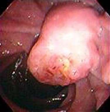
You are in: eMedicine Specialties > Medicine, Ob/Gyn, Psychiatry, and Surgery > Oncology
|
Carcinoma of the Ampulla of Vater Last Updated: September 1, 2005 |
|
| Synonyms and related keywords: periampullary carcinoma, periampullary malignancy, ampullary carcinoma, ampullary cancer, carcinoma of papilla of Vater, adenocarcinomas, neuroendocrine tumors, cystadenomas, adenomas, adenocarcinoma of the ampulla of Vater, Courvoisier sign, familial adenomatous polyposis, FAP, duodenal adenomas, endoscopic ultrasonography, EUS, endoscopic retrograde cholangiopancreatography, ERCP, percutaneous transhepatic cholangiography, PTC, kocherization, pancreaticoduodenectomy, pylorus-preserving pancreaticoduodenectomy, transduodenal excision
|
|   |
AUTHOR INFORMATION
| Section 1 of 11  |
|
| Author: Pankaj Chaturvedi, MBBS, MS, Assistant Surgeon, Department of Surgical Oncology, Tata Memorial Hospital, India Coauthor(s): Ronald S Chamberlain, MD, Chairman, Chief, Department of Surgery, St Barnabas Medical Center; Uma Chaturvedi, MD, Lecturer, Department of Pathology, KJ Somaiya Hospital and Research Center, India; Nafisa K Kuwajerwala, MD, Staff Physician, Department of General Surgery, North Oakland Medical Center, Wayne State University; Gunateet Goswami, MD, Consulting Staff, Department of Cardiology, St Joseph Mercy of Macomb Hospital |
| |
| Editor(s): Michael Perry, MD, Professor, Department of Internal Medicine, Nellie B Smith Chair of Oncology, Director, Division of Hematology and Oncology, University of Missouri at Columbia/Ellis Fischel Cancer Center; Francisco Talavera, PharmD, PhD, Senior Pharmacy Editor, eMedicine;
Benjamin Movsas, MD, Vice-Chairman, Department of Radiation Oncology, Fox Chase Cancer Center;
Rajalaxmi McKenna, MD, FACP, Consulting Staff, Department of Medicine, Southwest Medical Consultants, SC, Good Samaritan Hospital, Advocate Health Systems;
and John S Macdonald, MD, Professor of Medicine, New York Medical College; Chief, Division of Medical Oncology, St Vincent's Hospital and Medical Center; Medical Director, Saint Vincent's Comprehensive Cancer Center |
Disclosure
|   |
INTRODUCTION
| Section 2 of 11  |
|
Background: Carcinoma of the ampulla of Vater is a malignant tumor arising within 2 cm of the distal end of the common bile duct, where it passes through the wall of the duodenum and ampullary papilla. The common bile duct merges with the pancreatic duct of Wirsung at this point and exits through the ampulla into the duodenum. The most distal portion of the common bile duct is dilated (ie, forms the ampulla of Vater) and is surrounded by the sphincter of Oddi, which spirals upward around the terminal portion of the duct. Because of biliary outflow obstruction, carcinoma of the ampulla of Vater tends to manifest early, as opposed to other pancreatic neoplasms that often are advanced at the time of diagnosis.
Curative surgical resection is the only option for long-term survival. Surgical or radiologic biliary decompression, relief of gastric outlet obstruction, and adequate pain control may improve the quality of life but do not affect overall survival rate. Pathophysiology: Ninety percent of ampullary tumors are adenocarcinomas. Neuroendocrine tumors, cystadenomas, and adenomas represent additional, but uncommon, histologic types. Tumors originate from ductal epithelial cells and usually invade into the substance of the pancreas. In more advanced disease states, peripancreatic tissue and the adventitia of large neighboring vessels, such as the superior mesenteric and portal veins, may be involved.
Lymph nodes metastases are present in as many as half of patients. Pericanalicular lymph nodes usually are the first to be involved. Nodes along the superior mesenteric, gastroduodenal, common hepatic, and splenic arteries, as well as the celiac trunk, are the second station of lymph nodes. Perineural, vascular, and lymphatic invasion are associated with a poor prognosis. Liver is the most common site (66%) of distant metastasis, followed by lymph nodes (22%). In advanced cases, lung metastasis also may occur. Frequency:
- In the US: Carcinoma of the ampulla of Vater is an uncommon tumor; fewer than 2000 cases are diagnosed per year. Ampullary cancer accounts for approximately 0.2% of all gastrointestinal tract malignancies and about 7% of all periampullary carcinomas. Adenocarcinoma of the ampulla of Vater is the second most common periampullary malignancy.
- Internationally: Worldwide incidence is not known.
Mortality/Morbidity:
- Most of these tumors are resectable for cure at diagnosis; however, the 5-year survival rate is only 40%.
- Operative mortality rates have decreased significantly over the last decade because of increased surgical experience, improved anesthesia, better preoperative imaging, and better postoperative management.
- Surgical morbidity rates remain high, with a range of 25-65%, even in centers with experienced staff. Pancreatic fistulas, prolonged gastric emptying, wound complications, intraabdominal sepsis, thrombophlebitis, and marginal ulceration are the most common complications.
- Postoperative mortality rates in the best centers are 2-5%.
Race:
- No race predilection is seen.
Sex:
- No sex predilection is seen.
Age:
- Ampullary cancer most often is seen in the fifth through the seventh decades of life.
|   |
CLINICAL
| Section 3 of 11  |
|
History: - Jaundice is the means of presentation in three fourths of cases. Ampullary cancer has no additional classic early symptoms.
- Jaundice may intermittently wax and wane owing to central necrosis/sloughing or pressure opening of a minimally obstructed duct.
- Dyspepsia and vomiting: These may be present if the duodenal lumen is compromised.
- Epigastric pain: The abdominal pain usually is dull, aching midepigastric pain or right hypochondriac pain. Backache usually is a sign of advanced stage.
- Pancreatic cholera: Diarrhea is due to the absence of lipase within the gut caused by pancreatic duct obstruction.
- Hematemesis, melena, and hematochezia: These are uncommon features caused by tumor bleeding.
Physical: - The Courvoisier sign, painless jaundice associated with a palpable gallbladder, may be present. Unlike that due to a neoplasm, obstructive jaundice due to a stone causes scarring of the gallbladder, precluding its distension.
- Fever can occur in the setting of ascending cholangitis.
- Rarely, patients present with features of acute pancreatitis or migratory thrombophlebitis.
- Palpable fixed epigastric masses or supraclavicular nodes are signs of advanced disease and inoperability.
Causes: - The etiology of the disease is poorly understood.
- Patients with familial adenomatous polyposis (FAP) have an increased risk of both benign and malignant ampullary tumors (Burke, 1999).
- As many as 50-90% of patients with FAP develop duodenal adenomas, predominantly concentrated on or around the major papilla (Griffioen, 1998).
- Genomic anomalies may be a factor (Scarpa and Zamboni, 1999).
- K-ras mutations may be a factor (Berndt, 1998).
|   |
DIFFERENTIALS
| Section 4 of 11  |
|
Ascariasis Bile Duct Strictures Bile Duct Tumors Biliary Disease Biliary Obstruction Cholangiocarcinoma Choledocholithiasis Duodenal Ulcers Gallbladder Cancer
Gallbladder Tumors Lymphoma, Non-Hodgkin Pancreatic Cancer Pancreatitis, Chronic Papillary Necrosis Papillary Tumors
Other Problems to be Considered:
Duodenal carcinoma
Adenoma at the ampulla of Vater |
|
Patient Education
|
|
Click here for patient education.
|
|
|
|
|
|   |
WORKUP
| Section 5 of 11  |
|
Lab Studies:
- Test for anemia caused by bleeding from the ampullary mass.
- Test for hyperbilirubinemia (conjugated type) due to blockage of the biliary outflow.
- Test for a rise in alkaline phosphatase level, again due to blockage.
- Serum alanine aminotransferase (ALT) and aspartate aminotransferase (AST) rise in long-standing obstruction.
- Fecal occult blood testing results may be positive in ulcerated or bleeding tumors.
- In cases with complete obstruction and bleeding, the stool may be pale or silver white, so-called silver stools.
- A rise in serum amylase is not uncommon.
- Alteration in coagulation profile (eg, increased prothrombin time, decreased prothrombin time, prolonged bleeding and clotting times) is common.
- Urinalysis shows bile pigments.
- Absence of urinary urobilinogen signifies complete obstruction.
- Tumor markers: Currently, no tumor marker is sensitive or specific enough to serve as reliable screening tools for this carcinoma.
- Carbohydrate antigen (CA) 19-9 is the most studied and sensitive marker for pancreatic neoplasms at present. Unfortunately, CA 19-9 has almost no value in management of carcinoma of ampulla of Vater.
- Carcinoembryonic antigen (CEA), DU-PAN-2, alpha-fetoprotein (AFP) and pancreatic oncofetal antigen (POA) also have been evaluated and found inaccurate.
Imaging Studies:
- Abdominal ultrasonography
- Advantages
- Abdominal ultrasonography (US) is the most useful noninvasive initial investigation for distinguishing medical from surgical causes of jaundice. It is an inexpensive and readily available bedside procedure.
- Abdominal US can identify dilated ducts, liver metastasis (in almost 90% of cases), ascites, and nodal metastasis.
- Doppler US can be used to assess vascular involvement.
- The level of obstruction can be assessed in 90% patients.
- US-guided fine-needle aspiration (FNA) can be performed.
- Limitations
- Effectiveness is related to the skill of the user.
- Very superficial lesions and very deep lesions may be missed. Distinguishing a metastasis from a hemangioma may be difficult.
- Sensitivity is 80–90%, and information is inferior to that obtained by CT scan or MRI. Poor bowel preparation may obscure the important pathology.
- Endoscopic and laparoscopic ultrasonography
- Endoscopic ultrasonography (EUS) is performed through a peroral route.
- The test is highly sensitive in detecting major vascular involvement, which can prevent unnecessary surgery (Menzel, 1999).
- EUS may identify tumors less than 1 cm in size.
- Laparoscopic sonography can detect occult liver metastasis or peritoneal seeding missed by other imaging modalities.
- Staging laparoscopy with laparoscopic ultrasonography may be more specific and accurate in predicting tumor resectability than laparoscopy alone (88% and 89% vs 50% and 65%, respectively; John, 1995).
- Advantages
- This modality is most useful when US is equivocal or when visualization is obscured by gas or ascites.
- CT scan is superior to US, with an accuracy of more than 90%. CT scan findings correlate well with operative findings.
- CT scan is better in evaluating operability and preoperative staging. It gives better assessment of invasion or compression of vessels and adjacent organs.
- CT-guided biopsy may be obtained.
- Disadvantages
- Very ill patients may be unable to lie still or arrest respiration for the long periods required for high-quality imaging.
- CT scan is more expensive than US and requires expertise in interpretation.
- Potential radiation hazards exist for patients and staff.
- Rare contrast reactions may occur.
- Metal, stents, and clips may cause artifacts.
- Very small tumors (<1 cm) may be missed.
- Magnetic resonance imaging
- MRI is the most informative noninvasive method of evaluation currently available.
- MRI cholangiopancreatography (MRCP) provides 94% accuracy in identifying the cause and extent of the pathology.
- Results are reproducible.
- With growing expertise in the use of magnetic imaging, diagnostic endoscopic retrograde cholangiopancreatography (ERCP) is quickly becoming obsolete.
- The use of the hepatoiminodiacetic acid (HIDA) scan has declined in recent years.
- This scan is better used for assessing liver parenchyma lesions or for possible help in diagnosing Budd-Chiari syndrome.
- Use requires a qualified doctor and expensive equipment.
- Chest x-ray is performed to exclude pulmonary metastasis and other pulmonary diseases.
Other Tests:
- ECG is performed to assess cardiac status, since surgery will be considered as a means of treatment.
- Nutritional studies should be ordered in preparation for surgery.
Procedures:
- Endoscopic retrograde cholangiopancreatography
- Advantages
- ERCP allows diagnostic and therapeutic access to both the common bile duct and pancreatic duct.
- The procedure displays the details of ductal anatomy and accurately demonstrates the level and nature of the obstruction (see Image 1). Anatomical variations in ducts can be evaluated carefully.
- ERCP allows therapeutic procedures, eg, sphincterotomy, stenting, and nasobiliary drainage.
- It permits sampling of pancreatic juice, bile, and brush/grasp biopsy.
- Endoscopic excision of small periampullary tumors is gaining in popularity.
- Disadvantages
- ERCP is an invasive procedure, which requires an expert endoscopist/radiologist and a cooperative patient.
- Very small tumors (<1 cm) can be missed.
- ERCP is not possible if access to the duodenal papilla is difficult, eg, owing to diverticula, anatomical ductal variations, or prior surgical bypass.
- This procedure can precipitate pancreatitis and cholangitis.
- Perforation and hemorrhage are 2 of the more serious complications.
- Percutaneous transhepatic cholangiography
- Indications for this procedure, which is highly invasive, are very limited.
- Percutaneous transhepatic cholangiography (PTC) is most useful when ERCP is unavailable or technically not feasible.
- PTC can be useful in severely jaundiced patients when laparotomy or ERCP is not possible. Percutaneous transhepatic biliary drainage or transhepatic stenting may be the only option for some patients.
- Biliary leakage may lead to peritonitis. Excessive bleeding from the puncture site and pneumothorax represent significant, but uncommon, complications.
Histologic Findings: In cases of ampullary tumors, preoperative endoscopic biopsy should be attempted, and carcinoma should be confirmed histologically or cytologically, if possible. If the specimen is insufficient or not representative, or if the histologic examination is inconclusive, surgery may be performed if a clinical suspicion exists. Approximately 90% of these tumors are adenocarcinomas. Neuroendocrine tumors, cystadenomas, and adenomas represent additional uncommon histologic types.Staging: The tumor, node, metastases (TNM) classification and stage grouping is based on the Union Internationale Contre Cancrum (UICC) system, established in 1977, with separate classifications for pancreatic and periampullary carcinomas. The staging is important only to communicate a uniform definition of extent of disease. TNM classification and stage groups are as follows: - Tx - The primary tumor cannot be assessed
- T0 - No sign of primary tumor
- Tis - Carcinoma in situ
- T1 - Tumor limited to the ampulla or sphincter of Oddi
- T2 - Tumor invading the wall of the duodenum
- T3 - Tumor invasion into the pancreas 2 cm or less
- T4 - More than 2 cm tumor invasion into the pancreas or any other adjacent organ
Peripancreatic tissue includes the surrounding retroperitoneal fatty tissue (retroperitoneal soft tissue or retroperitoneal space), including the mesentery (mesenteric fat), mesocolon, greater and lesser omentum, and peritoneum. Direct invasion of the bile ducts and the duodenum includes involvement of the ampulla.
Adjacent large vessels include the portal vein, the celiac trunk, the superior mesenteric artery and the common hepatic artery and vein (not the splenic vessels).
- NX - Regional lymph nodes cannot be assessed
- N0 - No regional lymph node metastases
- N1 - Regional lymph node metastases
Subclassification of the category N1 into N1a (only 1 metastatic lymph node) and N1b (2 or more lymph nodes affected by metastases) is recommended, as the 2 categories appear to have marked prognostic differences. Total number of peripancreatic lymph nodes found in the surgical specimen must be mentioned.
- Stage grouping of periampullary carcinoma
- Stage 1 - T1 N0 M0
- Stage 2 - T2 N0 M0, T3 N0 M0
- Stage 3 - T1 N1 M0, T2 NI M0, T3 N1 M0
- Stage 4 - T4 every N and every M, Every T and N with M1
- Martin proposed a 4-stage system, as follows:
- Stage I - Vegetating tumor limited to the epithelium, with no involvement of the Oddi sphincter
- Stage II - Tumor localized in the duodenal submucosa without involvement of the duodenal muscularis propria but possible involvement of the sphincter of Oddi
- Stage III - Tumor involving the duodenal muscularis propria
- Stage IV - Tumor involving the periduodenal area or the pancreas, with proximal or distal lymph node involvement
|   |
TREATMENT
| Section 6 of 11  |
|
Medical Care: Hepatic metastasis, serosal implants, ascites, lymph node involvement outside the resectional field, and major vessel infiltration all are contraindications to surgical resection. Treatment options for advanced or unresectable stages are discussed below. - Willett and colleagues reported their experience with adjuvant radiotherapy (40-50 gray [Gy], with or without concurrent 5-fluorouracil as a radiosensitizer) for high-risk tumors of the ampulla of Vater. Compared to surgery alone, the radiotherapy group demonstrated a trend toward better locoregional control; however, no advantage in survival was seen.
- Barton and Copeland reported on the M. D. Anderson Cancer Center experience of using postoperative chemotherapy for carcinoma of the ampulla of Vater. No combination of drugs prolonged life.
- Yeung and colleagues used neoadjuvant chemoradiotherapy in 4 patients with duodenal/ampullary carcinomas. No residual tumor was found in pancreaticoduodenectomy specimens of these 4 patients.
- Gemcitabine has shown promise in cases of advanced ampullary carcinoma.
- Fractionated high-dose external beam radiotherapy (60-70 Gy) yields local tumor control in 35-50% of cases. Care should be taken to protect healthy tissue while delivering this radiotherapy (ie, conformal RT or brachytherapy).
- Pain can be relieved in as many as 65% of patients treated with external beam radiotherapy.
- Intraoperative radiotherapy of the tumor bed with fast electrons and doses of 20 Gy is a promising alternative for achieving locoregional control. Combining intraoperative irradiation with postoperative percutaneous irradiation (total dose 70 Gy) may further increase the duration of median survival.
- Two prospective randomized studies performed by the Gastrointestinal Tumor Study Group (GITSG) demonstrated that combining radiotherapy and chemotherapy prolonged median duration of survival compared to that achieved with chemotherapy or irradiation alone.
Surgical Care: - Surgical resection in an ampullary carcinoma is the primary modality of treatment. The highest cure rates are achieved if the tumor is localized to the ampullary region.
- Laparotomy should be performed to assess resectability in all cases for which sonography, CT scan, and laparoscopy do not show disseminated disease.
- With improvement in postoperative management and surgical technique, operative mortality rates are as low as 3-5% in most centers with experienced staff (Wagle, 2001).
- Extensive preoperative assessment of cardiac, respiratory, renal, and cerebral functions should be performed in older patients.
- Overall survival rates are better for ampullary carcinoma than for other periampullary malignancies, because the former disease typically manifests symptoms early.
- Tok et al reported 25 patients (13 men, 12 women) with a median age of 65 years who had an ampullary tumor. The resectability rate was 88%, with no operative mortality. The 5-year actuarial survival rate of patients who underwent radical resection was 49%. They concluded that local resection is recommended only for small, benign tumors and for patients who may be unfit for radical surgery; otherwise, pylorus-preserving pancreaticoduodenectomy is safe and the most effective procedure.
- Assessment of nutritional status and supplementation (Fortunately, most of these patients do not have any nutritional problems.)
- Standard mechanical and oral antibiotic bowel preparation
- Assessment of coagulation profile and correction of decreased prothrombin time by administration of vitamin K
- Intravenous antibiotic prophylaxis
- Preoperative nasobiliary drainage or stenting for preoperative biliary decompression in severely jaundiced patient
- Fluid and electrolyte correction
- Assessment of cardiac, renal, and pulmonary status
- Laparoscopic assessment is obtained for peritoneal metastasis, hepatic metastases, and extensive lymphatic, vascular, or surrounding organ invasion.
- Resectability of the primary tumor is determined by mobilizing the head of the pancreas (ie, kocherization), opening the lesser sac, and exposing and inspecting the confluence of the splenic vein and superior mesenteric vein (see Image 2). Invasion of the retropancreatic portal vein is not a universal contraindication, as this segment of portal vein may be resected en bloc and subsequent reanastomosis of the vein performed.
- Intraoperatively, a transduodenal FNA or core biopsy is the preferred method for pathologic confirmation of the diagnosis. In about 10% of cases, these methods do not permit intraoperative confirmation of carcinoma. Resection should be performed in such cases based on preoperative and intraoperative findings.
- Resectability may be a subjective phenomenon based on the experience and skill of the surgeon (Sohn, 1999).
- A feeding jejunostomy may be performed during the procedure to permit early resumption of enteral feeding. This rarely is necessary, as most patients can resume an oral diet within 2-3 days.
- This is the classic and standard resection procedure for ampullary carcinoma (see Image 3).
- In this operation, the pancreas is transected to the left of the portal vein, along with the uncinate process (in order to achieve lymph node dissection along the superior mesenteric artery). The lymph nodes along the common hepatic artery within the hepatoduodenal ligament and the precaval lymph nodes are removed. The gallbladder, along with the distal portion of the common bile duct and distal third of the stomach, is resected.
- Restoration of the gastrointestinal continuity is completed with pancreaticojejunostomy, hepaticojejunostomy, and gastrojejunostomy (see Image 4).
- Pylorus-preserving pancreaticoduodenectomy
- This preserves the entire pylorus, along with 1-2 cm of the first part of the duodenum. GI continuity is restored with a duodenojejunostomy.
- This represents a more physiologically acceptable procedure, with similar survival rates. Postgastrectomy complications, such as dumping and marginal ulceration, are reduced. Delayed gastric emptying may be exacerbated.
- Postprandial release of gastrin and secretin is nearly normal in patients who undergo this procedure.
- Transduodenal excision of ampullary tumors
- Transduodenal excision rarely is indicated and is reserved for elderly patients, patients with significant comorbid conditions, and those with favorable tumors (generally <2 cm, polypoid).
- This more limited resective technique is associated with compromised local control in many instances.
- Palliative surgery
- Palliative surgery is reserved for unresectable tumors or for patients who are unfit for curative surgery.
- The goal is to alleviate biliary obstruction, duodenal obstruction, or pain.
- Either cholecystojejunostomy or hepaticojejunostomy bypass is performed.
- Duodenal obstruction may require gastrojejunostomy. Prophylactic gastrojejunostomy should be done, even in a duodenum unobstructed at the time of laparotomy, because as many as one third of patients develop obstruction later. However, prophylactic gastrojejunostomy adds significant morbidity risk to the procedure.
- Chemical splanchnicectomy, using either 6% phenol or 50% ethanol, can be performed intraoperatively. This procedure controls pain in 80% of patients.
Consultations: - A nutrition specialist for tailoring the diet, when needed
- Physician in cases of postoperative fever, chest infection, or other problems
Diet: - Oral feeding usually can be started on the second postoperative day.
- The diet should be started with sips of water, which can be increased gradually over 48 hours to a liquid diet. Patients can have a semisolid diet by roughly the sixth day.
- Initially, the diet should be deficient in fat and protein.
Activity: - The patient should ambulate from the first postoperative day.
- Early ambulation and chest physiotherapy reduce morbidity.
|   |
MEDICATION
| Section 7 of 11  |
|
Prophylactic and postoperative antibiotics are given according to hospital protocol.
Drug Category: Chemotherapeutic agents -- Fluorouracil can be used as a radiosensitizer for high-risk tumors of the ampulla of Vater. Drug Name
| Fluorouracil (Adrucil) -- Fluorinated pyrimidine antimetabolite that inhibits thymidylate synthase and interferes with RNA synthesis and function. Has some effect on DNA. Useful in symptom palliation for patients with progressive disease. |
|---|
| Adult Dose | 500 mg/m2 IV |
|---|
| Pediatric Dose | Not established |
|---|
| Contraindications | Documented hypersensitivity; bone marrow suppression; serious infection |
|---|
| Interactions | Increased risk of bleeding with anticoagulants, NSAIDs, platelet inhibitors, thrombolytic agents; other immunosuppressive agents may enhance bone marrow toxicity |
|---|
| Pregnancy |
D - Unsafe in pregnancy
|
|---|
| Precautions | Nausea, oral and GI ulcers, depression of immune system, and hematopoiesis failure (bone marrow suppression) may occur; adjust dose in renal impairment |
|---|
Drug Category: Antibiotics -- Initial empiric antimicrobial therapy must be comprehensive and should cover both aerobic and anaerobic gram-negative organisms.Drug Name
| Cefoxitin (Mefoxin) -- Second-generation cephalosporin indicated for gram-positive cocci and gram-negative rod infections. Infections caused by cephalosporin- or penicillin-resistant gram-negative bacteria may respond to cefoxitin.
Any second-generation cephalosporin may be used instead of cefoxitin.| Adult Dose | Biliary stent present: 1 g IV preoperatively; continue until culture reported negative; if positive, adjust antibiotics on basis of culture sensitivity report
No biliary stent: 1 g IV preoperatively then 2 doses postoperatively| Pediatric Dose | Not established |
|---|
| Contraindications | Documented hypersensitivity |
|---|
| Interactions | Probenecid may increase effects aminoglycosides or furosemide may increase nephrotoxicity (closely monitor renal function) |
|---|
| Pregnancy |
B - Usually safe but benefits must outweigh the risks.
|
|---|
| Precautions | Adjust dose in severe renal insufficiency (high doses may cause CNS toxicity); superinfections and promotion of nonsusceptible organisms may occur with prolonged use or repeated therapy |
|---|
|
|---|
|
|---|
|   |
FOLLOW-UP
| Section 8 of 11  |
|
Further Inpatient Care:
- Employ broad-spectrum antibiotic coverage for 24 hours.
- Continuous nasogastric aspiration usually is maintained for the first 24 hours.
- Oral feeding usually is started on the second postoperative day.
- Subcutaneous heparin and pneumatic compression stockings are used to prevent deep vein thrombosis (DVT).
- Adequate blood replacement is necessary in cases of intraoperative blood loss.
- Early ambulation and chest physiotherapy reduce morbidity.
- Serum electrolytes, renal function, and liver function should be monitored.
- Abdominal drains can be removed after 3-5 days if no evidence of pancreatic fistulas exists.
- Tachycardia and tachypnea may at times be the earliest signs of a leak.
Further Outpatient Care:
- Recurrent disease usually is not curable; therefore, follow-up is limited principally to detection and treatment of secondary consequences, such as reducing pain and managing evident or latent exocrine or endocrine pancreatic insufficiency.
- Sonography, CT scan of the upper abdomen, and liver function tests may be used to detect recurrence and manage complications. These examinations should not be carried out on a general basis, however, as early diagnosis of recurrent disease apparently offers no therapeutic benefit.
Deterrence/Prevention:
- Those with FAP and their family members should be counseled about the possibility of acquiring ampullary carcinoma. As many as 50-90% of patients with FAP develop duodenal adenomas, concentrated predominantly on or around the major papilla (Griffioen, 1998). Such patients should receive close endoscopic surveillance.
Complications:
- Most complications that arise after conventional Whipple procedures are caused by dehiscence of the pancreatic anastomosis. Disruption of pancreatic anastomosis is at times a lethal complication.
- More than 40 reconstruction procedures are described in the literature to reduce the risk of pancreatic leak, eg, occlusion of the residual pancreas with Ethibloc or fibrin (as a means to avoid complications secondary to anastomosis) or temporary occlusion with a fibrin adhesive and subsequent anastomosis (in order to avoid 4-6 days of secretion and the risk of damage to the anastomosis).
- A dramatic reduction has been seen over the last decade in postoperative mortality following pancreaticoduodenectomy. This can be attributed to growing surgical experience, improved anesthesia, better preoperative imaging, and better postoperative management.
- Major postoperative complications occur in 25-65% of patients, depending upon the expertise of the surgical staff.
- Pancreatic fistula, prolonged ileus, intraabdominal sepsis, thrombophlebitis, marginal ulceration, and gastrointestinal motility disorder all can manifest as complications of the surgery.
- Dumping syndrome is seen in patients in whom a significant part of the stomach has been removed.
Prognosis:
- Most patients with carcinoma of the ampulla of Vater die of recurrent disease. Treatment fails in nearly three fourths of patients with poor prognostic features.
- Survival duration after surgical resection is related to the extent of local invasion of the primary lesion, lymph node involvement, vascular invasion, perineural invasion, cellular differentiation, uninvolved surgical margins, and perioperative blood transfusion.
- el-Ghazzawy et al reviewed their experience from 1987-1991 with 123 patients who had ampullary cancer. In the group that underwent surgical resection, survival was not influenced independently by perineural invasion, microlymphatic invasion, vascular invasion, or tumor differentiation when the tumors were controlled for stage.
- The surgical mortality rate following a Whipple operation or subtotal distal pancreatectomy ranges from 2-5% in centers with experienced staff.
- For ampullary carcinoma, the 5-year survival rate ranges from 30-50%, depending on the center and the extent of lymph node dissection. When tumor diameter is less than 2 cm (ie, early carcinoma), survival rates are 30% in patients with lymph node involvement and as high as 50% in patients without lymph node involvement.
Patient Education:
- Those with FAP, and their family members, should be counseled about the possibility of acquiring ampullary carcinoma.
|   |
MISCELLANEOUS
| Section 9 of 11  |
|
Medical/Legal Pitfalls:
- Failure to counsel FAP patients and their families regarding the possibility of acquiring ampullary carcinoma
|   |
PICTURES
| Section 10 of 11  |
|
| Caption: Picture 5. Double duct sign of periampullary cancers. Note the dilated common bile duct as well as the pancreatic duct. Liver metastatic lesion is also seen.
|  | View Full Size Image |
|
Picture Type: CT |
| Caption: Picture 6. Distended gall bladder with double duct sign in a patient with periampullary cancer.
|  | View Full Size Image |
|
Picture Type: CT |
|   |
BIBLIOGRAPHY
| Section 11 of 11 |
|
-
AJCC Cancer Staging Manual: Exocrine pancreas. In: American Joint Committee on Cancer Manual. 5th ed. Philadelphia, Pa: Lippincott-Raven; 1997:121-6.
-
Barton RM, Copeland EM 3rd: Carcinoma of the ampulla of Vater. Surg Gynecol Obstet 1983 Mar; 156(3): 297-301[Medline].
-
Berndt C, Haubold K, Wenger F, et al: K-ras mutations in stools and tissue samples from patients with malignant and nonmalignant pancreatic diseases. Clin Chem 1998 Oct; 44(10): 2103-7[Medline].
-
Burke CA, Beck GJ, Church JM, et al: The natural history of untreated duodenal and ampullary adenomas in patients with familial adenomatous polyposis followed in an endoscopic surveillance program. Gastrointest Endosc 1999 Mar; 49(3 Pt 1): 358-64[Medline].
-
Conlon KC: Carcinoma of the ampulla of vater: a distinct disease entity?. Ann Surg Oncol 2003 Dec; 10(10): 1136-7[Medline].
-
el-Ghazzawy AG, Wade TP, Virgo KS, et al: Recent experience with cancer of the ampulla of Vater in a national hospital group. Am Surg 1995 Jul; 61(7): 607-11[Medline].
-
Gastrointestinal Tumor Study Group: Further evidence of effective adjuvant combined radiation and chemotherapy following curative resection of pancreatic cancer. Cancer 1987 Jun 15; 59(12): 2006-10[Medline].
-
Griffioen G, Bus PJ, Vasen HF, et al: Extracolonic manifestations of familial adenomatous polyposis: desmoid tumours, and upper gastrointestinal adenomas and carcinomas. Scand J Gastroenterol Suppl 1998; 225: 85-91[Medline].
-
John TG, Greig JD, Carter DC, Garden OJ: Carcinoma of the pancreatic head and periampullary region. Tumor staging with laparoscopy and laparoscopic ultrasonography. Ann Surg 1995 Feb; 221(2): 156-64[Medline].
-
Martin ED: Anatomopathologie des tumeurs oddiennes. In: Les tumeurs oddiennes. 1978:35-52.
-
Menzel J, Hoepffner N, Sulkowski U, et al: Polypoid tumors of the major duodenal papilla: preoperative staging with intraductal US, EUS, and CT--a prospective, histopathologically controlled study. Gastrointest Endosc 1999 Mar; 49(3 Pt 1): 349-57[Medline].
-
Sarmiento JM, Nagomey DM, Sarr MG, Farnell MB: Periampullary cancers: are there differences? Surg Clin North Am 2001 Jun; 81(3): 543-55[Medline].
-
Sarr MG, Cameron JL: Surgical palliation of unresectable carcinoma of the pancreas. World J Surg 1984 Dec; 8(6): 906-18[Medline].
-
Scarpa A, Zamboni G: Genomic anomalies in pancreatic tumors other than common adenocarcinoma. Ann N Y Acad Sci 1999 Jun 30; 880: 179-90[Medline].
-
Sohn TA, Lillemoe KD, Cameron JL, et al: Reexploration for periampullary carcinoma: resectability, perioperative results, pathology, and long-term outcome. Ann Surg 1999 Mar; 229(3): 393-400[Medline].
-
Todoroki T, Koike N, Morishita Y, et al: Patterns and predictors of failure after curative resections of carcinoma of the ampulla of Vater. Ann Surg Oncol 2003 Dec; 10(10): 1176-83[Medline].
-
Toh SK, Davies N, Dolan P, et al: Good outcome from surgery for ampullary tumour. Aust N Z J Surg 1999 Mar; 69(3): 195-8[Medline].
-
Van Heek NT, De Castro SM, van Eijck CH, et al: The need for a prophylactic gastrojejunostomy for unresectable periampullary cancer: a prospective randomized multicenter trial with special focus on assessment of quality of life. Ann Surg 2003 Dec; 238(6): 894-902; discussion 902-5[Medline].
-
Wagle PK, Joshi RM, Mathur SK: Pancreaticoduodenectomy for periampullary carcinoma. Indian J Gastroenterol 2001 Mar-Apr; 20(2): 53-5[Medline].
-
Willett CG, Warshaw AL, Convery K, et al: Patterns of failure after pancreaticoduodenectomy for ampullary carcinoma. Surg Gynecol Obstet 1993 Jan; 176(1): 33-8[Medline].
-
Yeung RS, Weese JL, Hoffman JP, et al: Neoadjuvant chemoradiation in pancreatic and duodenal carcinoma. A Phase II Study. Cancer 1993 Oct 1; 72(7): 2124-33[Medline].
Carcinoma of the Ampulla of Vater excerpt |