Continually Updated Clinical Reference
 
 
  All Sources     eMedicine     Medscape     Drug Reference     MEDLINE
 
eMedicine - Pancreatic Pseudocysts : Article by

Quick Find
Authors & Editors
Introduction
Clinical
Differentials
Workup
Treatment
Medication
Follow-up
Miscellaneous
Multimedia
References

Related Articles
Pancreatic Cancer

Pancreatic Necrosis and Pancreatic Abscess

Pancreatic Pseudoaneurysm

Pancreatitis, Acute

Pancreatitis, Chronic

von Hippel-Lindau Disease


Patient Education
Liver, Gallbladder, and Pancreas Center

Esophagus, Stomach, and Intestine Center

Pancreatitis Overview

Pancreatitis Causes

Pancreatitis Symptoms

Pancreatitis Treatment

Abdominal Pain in Adults Overview


Author: Louis R Lambiase, MD, Associate Professor of Medicine, University of Florida College of Medicine; Chief, Division of Gastroenterology, Department of Internal Medicine, University of Florida Health Science Center/Jacksonville

Louis R Lambiase is a member of the following medical societies: American Gastroenterological Association, American Pancreatic Association, and American Society for Gastrointestinal Endoscopy

Editors: David Greenwald, MD, Fellowship Program Director, Associate Professor, Department of Medicine, Division of Gastroenterology, Montefiore Medical Center, Albert Einstein College of Medicine; Francisco Talavera, PharmD, PhD, Senior Pharmacy Editor, eMedicine; Simmy Bank, MD, Chair, Professor, Department of Internal Medicine, Division of Gastroenterology, Long Island Jewish Hospital, Albert Einstein College of Medicine; Alex J Mechaber, MD, FACP, Assistant Dean for Medical Curriculum, Associate Professor of Medicine, Division of General Internal Medicine, University of Miami Miller School of Medicine; Julian Katz, MD, Clinical Professor of Medicine, Drexel University College of Medicine; Consulting Staff, Department of Medicine, Section of Gastroenterology and Hepatology, Hospital of the Medical College of Pennsylvania

Author and Editor Disclosure

Synonyms and related keywords: pancreatic fluid collections, organized necrosis of the pancreas, pancreatic cysts, pancreatitis, abdominal trauma, pancreatic ducts, persistent abdominal pain, anorexia, abdominal mass, jaundice, sepsis, pleural effusion, scleral icterus, percutaneous catheter drainage

Background

Single or multiple fluid collections that look like cysts on pancreatic imaging are often observed during acute pancreatitis. Because of increasing sensitivity of imaging modalities and improvements in technology providing enhanced therapeutic abilities, the questions of when and whether drainage should be performed and what modality should be used to drain the cysts are often asked. Strictly defining the type of fluid collection is very important when reviewing pancreatic fluid collections. The therapeutic approach is different depending on the type of collection.

Pseudocysts are best defined as a localized fluid collection that is rich in amylase and other pancreatic enzymes, that has a nonepithelialized wall consisting of fibrous and granulation tissue, and that usually appears several weeks after the onset of pancreatitis.

These characteristics contrast with those of acute fluid collections, which are more evanescent and are serosanguinous inflammatory reactions to acute pancreatitis. These collections are noted in moderate-to-severe pancreatitis. Acute fluid collections usually have an irregular shape and lack a well-defined wall. In general, they resolve in about 65% of cases.

Two other types of fluid collection should be considered. First, organized necrosis is actually devitalized pancreatic tissue that appears cystlike on CT scan, but it appears to be solid on other imaging modalities. Second, an abscess is an infected area of necrosis or fluid.

Pathophysiology

Pancreatic pseudocysts can be single or multiple. Multiple cysts are more frequently observed in patients with alcoholism, and they can be multiple in about 15% of cases. Size varies from 2-30 cm. About one third of pseudocysts manifest in the head of the gland, and two thirds appear in the tail. The fluid in pseudocysts has been well characterized as clear or watery, or it can be xanthochromic. The fluid in pseudocysts usually contains very high amounts of amylase, lipase, and trypsin, though the amylase level may decrease over time.

The pathogenesis of pseudocysts seems to stem from disruptions of the pancreatic duct due to pancreatitis and extravasation of enzymatic material. Two thirds of patients with pseudocysts have demonstrable connections to the pancreatic duct. In the other third, an inflammatory reaction is supposed to have sealed the connection so that it is not demonstrable. The cause of pseudocysts parallels the cause of acute pancreatitis; 75-85% of cases are caused by alcohol or gallstone disease–related pancreatitis. In children, pseudocysts and trauma are frequently associated.

Sex

The male predominance in the incidence of pseudocysts mirrors the male predominance in the incidence of pancreatitis.

Age

Pseudocysts may occur after pancreatitis in any age group. In children, pseudocysts are most likely observed after abdominal trauma. In elderly persons, take care not to confuse cystic neoplasms with pseudocysts.


History

  • No specific set of symptoms is pathognomic of pseudocysts; however, consider the possibility of a pseudocyst in a patient who has persistent abdominal pain, anorexia, or abdominal mass after a case of pancreatitis.
  • Rarely, patients present with jaundice or sepsis from an infected pseudocyst.
  • Pleural effusion is also a common finding.

Physical

  • The sensitivity of physical examination findings is limited.
    • Patients very frequently have a tender abdomen.
    • Patients occasionally have a palpable mass in the abdomen.
  • Peritoneal signs suggest rupture of the cyst or infection.
  • Other possible findings include the following:
    • Fever
    • Scleral icterus
    • Pleural effusion

Causes

Acute or chronic pancreatitis or abdominal trauma causes pseudocysts. If no history of pancreatitis or trauma exists, the diagnosis must be carefully confirmed.


Pancreatic Cancer
Pancreatic Necrosis and Pancreatic Abscess
Pancreatic Pseudoaneurysm
Pancreatitis, Acute
Pancreatitis, Chronic
von Hippel-Lindau Disease

Other Problems to be Considered

Organized pancreatic necrosis
Acute pancreatic fluid collections
Serous cystadenoma of the pancreas
Mucinous cystadenoma of the pancreas
Mucinous cystadenocarcinoma
Pancreatic retention cyst


Lab Studies

  • Serum tests have limited use.  
    • Amylase and lipase levels are often elevated but may be within reference ranges.
    • Bilirubin and liver function test (LFT) findings may be elevated if the biliary tree is involved.
  • Analysis of the cyst fluid may help differentiate pseudocysts from tumors. Attempt to exclude tumors in any patient who does not have a clear history of pancreatitis.  
    • Carcinoembryonic antigen (CEA) and carcinoembryonic antigen-125 (CEA-125) tumor marker levels are low in pseudocysts and elevated in tumors.
    • Fluid viscosity is low in pseudocysts and elevated in tumors.
    • Amylase levels are usually high in pseudocysts and low in tumors.
    • Cytology is occasionally helpful in diagnosing tumors, but a negative result does not exclude tumors.
    • A CEA level of greater than 400 ng/mL within the cyst fluid strongly suggests malignancy.

Imaging Studies

  • Abdominal ultrasound: While cystic fluid collections in and around the pancreas may be visualized via ultrasound, the technique is limited by the operator’s skill, the patient's habitus, and any overlying bowel gas. As such, ultrasound is not the study of choice to establish a diagnosis.
  • Abdominal CT scan  
    • CT scan is the imaging criterion standard for pancreatic pseudocysts. It has a sensitivity of 90-100% and is not operator dependent.
    • The usual finding on CT scan is a large cyst cavity in and around the pancreas.
    • Multiple cysts may be present.
    • The pancreas may appear irregular or have calcifications.
    • Pseudoaneurysms of the splenic artery, bleeding into a pseudocyst, biliary and enteric obstruction, and other complications may be noted on CT scan.
    • The CT scan provides a very good appreciation of the wall thickness of the pseudocyst, which is useful in planning therapy.
  • Endoscopic retrograde cholangiopancreatography  
    • Endoscopic retrograde cholangiopancreatography (ERCP) is not necessary in diagnosing pseudocysts; however, it is useful in planning drainage strategy.
    • A study by Neil et al investigated the use of ERCP and the treatment of pseudocysts and acute pancreatitis and reported that a change in management occurred 35% of the time after the ERCP findings in pseudocysts were evaluated. Therefore, many authors recommend performing an ERCP before contemplated drainage procedures.
  • MRI  
    • MRI is not necessary to establish a diagnosis of pseudocysts; however, it is useful in detecting a solid component to the cyst and in differentiating between organized necrosis and a pseudocyst.
    • A solid component makes catheter drainage difficult; therefore, in the setting of acute necrotizing pancreatitis with resultant pseudocyst, an MRI may be very important before a planned catheter drainage procedure.
  • Endoscopic ultrasound  
    • Endoscopic ultrasound (EUS) is not necessary to establish a diagnosis but is very important in planning therapy, particularly if endoscopic drainage is contemplated.
    • A gastric wall with a thickness greater than 1 cm next to the cyst tends to predict a poor outcome with endoscopic drainage.
    • EUS may also be helpful in detecting small portal collaterals from otherwise undetected portal hypertension that may increase bleeding risks with transmural drainage.
    • Transmural drainage may be performed only when the symptomatic pseudocyst is positioned next to the gut wall.

Histologic Findings

Histologic findings vary with age because older cysts have thicker walls with more collagen. The etiology of the cyst does not change the histology.

  • Zone 1 - Hemosiderin pigment and loose connective tissue
  • Zone 2 - Inflammatory cells and capillary-rich fibrous tissue
  • Zone 3 - Hyalinized acellular connective tissue
  • Zone 4 - Capillary-rich fibrous stroma


Medical Care

The goal of therapy is avoidance of complications.

  • About 10% of pseudocysts become infected.
  • Pseudocysts can also rupture. A controlled rupture into an enteric organ can sometimes cause GI bleeding. A free rupture into the peritoneal cavity produces abdominal pain and, rarely, peritonitis or even death.
  • Most pseudocysts resolve without interference and only require supportive care.
  • Several studies have indicated that the size of the cyst and the length of time the cyst has been present are poor predictors of complications. In general, larger cysts are more likely to become symptomatic or cause complications. However, some patients with larger collections do well; therefore, the size of the pseudocyst alone is not an indication for drainage.
  • Indications for drainage include the following:
    • Complications
    • Symptoms
    • Concern about possible malignancy

Surgical Care

Drainage options are outlined below.

  • Catheter drainage
    • Percutaneous aspiration is useful only to establish a diagnosis or as a temporizing measure. It has a 54% failure rate and a 63% recurrence rate. This technique has a relatively high risk of infecting the pseudocyst. Percutaneous drainage may have a higher complication rate and inpatient mortality rate than surgical drainage.
    • Percutaneous catheter drainage is the procedure of choice for treating infected pseudocysts, allowing for rapid drainage of the cyst and identification of any microbial organism. A high recurrence and failure rate exist, but catheter drainage may be a good temporizing measure.
    • Percutaneous catheter drainage is contraindicated in patients who are poorly compliant and cannot manage a catheter at home. It is also contraindicated in patients with strictures of the main pancreatic duct and in patients with cysts containing bloody or solid material.
  • Endoscopic drainage may be either transpapillary (via ERCP) or transmural. Both modalities require careful patient selection to ensure success and safety.  
    • Transpapillary drainage, while safer and more effective than transmural drainage, requires cyst communication with the pancreatic duct. This technique may be technically challenging because it requires wire passage and stenting through the pancreatic duct to the pseudocyst. The success rate is about 80%. The recurrence rate is 10-14%, and, in most series, the complication rate (mainly pancreatitis) is approximately 13%.
    • Endoscopic transmural drainage is also possible. This involves performing an endoscopy and finding a bulge within the wall of the stomach or duodenum caused by compression of the pseudocyst. The cyst is generally entered using a needle knife to cut through the gastric or duodenum wall, and a series of pigtail stents are placed through the resulting communication. Some have adapted the technique to avoid diathermy, thus decreasing possible complications. The method has an 82-89% success rate in very experienced hands. The recurrent rate is 6-18%. The complication rate is 20%, with the most feared complication being bleeding.
    • One report suggests that the complication rate decreases and efficacy increases with experience. Weckman reported an approximately 86% success rate with endoscopic drainage with a 10% complication rate and a 14% failure rate.1 There appeared to be about a 15% recurrence rate. There was no real difference in outcome in patients treated with a transpapillary or transmural approach.
  • Surgical drainage is the criterion standard against which all therapies are measured.  
    • Internal drainage is the procedure of choice. A laparoscopic approach has been used in some cases with good results.
    • In most series, the mortality rate is 3%, and the complication rate is approximately 24%.
    • The success rate is 85-90%.
    • Recent work suggests that a laparoscopic approach to drainage has a high success rate and a low morbidity rate.2

Consultations

Management of pseudocysts requires a team approach. Gastroenterologists, surgeons, and invasive radiologists must work together to determine the necessity, timing, and method of intervention. If nonsurgical drainage is contemplated, it is important to elucidate the anatomy of the pancreatic duct beforehand. This may be done via ERCP or MRI. A large number of patients who fail or have complications with nonsurgical drainage have disruption or stenosis of the pancreatic duct.

Diet

Patients may eat a low-fat diet as tolerated. Patients in whom eating causes abdominal pain need parenteral or enteral nutrition through a percutaneously or endoscopically placed jejunal tube.

Activity

Patients may engage in activities as tolerated.


No medications are specific to the treatment of pancreatic pseudocysts. Antibiotics are an adjunct to drainage of infected pseudocysts. Octreotide can be useful as an adjunct to catheter drainage.

Drug Category: Somatostatin analogues

Used to reduce pancreatic exocrine secretion.

Drug NameOctreotide (Sandostatin)
DescriptionActs primarily on somatostatin receptor subtypes II and V. Inhibits GH secretion and has a multitude of other endocrine and nonendocrine effects, including inhibition of glucagon, VIP, and GI peptides.
Adult Dose200 mcg SC tid for 1 mo
Pediatric DoseNot established
ContraindicationsDocumented hypersensitivity
InteractionsMay reduce effects of cyclosporine; patients on insulin, oral hypoglycemics, beta-blockers, and calcium channel blockers may need dose adjustments
PregnancyB - Fetal risk not confirmed in studies in humans but has been shown in some studies in animals
PrecautionsAdverse effects are primarily related to altered GI motility (eg, nausea, abdominal pain, diarrhea, increased incidence of gallstones and biliary sludge); cholelithiasis may occur; because of alteration in counterregulatory hormones (eg, insulin, glucagon, GH), hypoglycemia or hyperglycemia may be observed; bradycardia, cardiac conduction abnormalities, and arrhythmias have been reported; because of inhibition of TSH secretion, hypothyroidism may also occur; caution in renal impairment


Further Outpatient Care

  • Patients who have endoscopically placed stents must be monitored via serial CT scans to observe resolution of the cyst. Stents may then be endoscopically removed after resolution.
  • Closely monitor patients with percutaneous drains for pain, infection, or catheter migration. Remove the drain when drainage ceases.

Complications

  • Bleeding is the most feared complication and is caused by the erosion of the pseudocyst into a vessel.
    • Consider the possibility of bleeding in any patient who has a sudden increase in abdominal pain coupled with a drop in hematocrit level or a change in vital signs.
    • Therapy is emergent surgery or angiography with embolization of the bleeding vessel.
    • Do not perform a percutaneous or endoscopic drainage procedure under any circumstances in patients with suspected bleeding into a pseudocyst.
  • Consider the possibility of infection of the pseudocyst in patients who develop fever or an elevated WBC count. Treat infection with antibiotics and urgent drainage.
  • GI obstruction, manifesting as nausea and vomiting, is an indication for drainage.
  • The pseudocyst can also rupture.
    • A controlled rupture into an enteric organ occasionally causes GI bleeding.
    • On rare occasions, a profound rupture into the peritoneal cavity causes peritonitis and death.

Prognosis

  • Most pseudocysts resolve without interference, and patients do well without intervention.
  • Outcome is much worse for patients who develop complications or who have the cyst drained. The presence of pancreatic necrosis is a poor prognostic sign.
  • The failure rate for drainage procedures is about 10%, the recurrence rate is about 15%, and the complication rate is 15-20%.

Patient Education


Medical/Legal Pitfalls

  • Misdiagnosis of a cystic neoplasm of the pancreas and treating it as a pseudocyst
  • Failure to recognize and treat complications


Media file 1:  
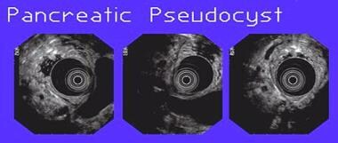
Three views of a pancreatic pseudocyst noted during endoscopic ultrasound. The concentric rings in the center of the images are the ultrasound transducer in the stomach. The cyst is observed as the large hypoechoic structure adjacent to the transducer.
Click to see larger pictureClick to see detailView Full Size Image
Media type:  Image

Media file 2:  These photographs show the endoscopic view of transpapillary pseudocyst drainage in a patient with pancreas divisum and a pseudocyst that communicates with the pancreatic duct. The image on the right shows the ampullary area. The middle image shows a wire placed in the minor papilla into the dorsal pancreatic duct. The left image shows a stent in place in the minor papilla.
Click to see larger pictureClick to see detailView Full Size Image
Media type:  Image


  1. Weckman L, Kylanpaa ML, Puolakkainen P, Halttunen J. Endoscopic treatment of pancreatic pseudocysts. Surg Endosc. Apr 2006;20(4):603-7. Epub 2006 Jan 19. [Medline].
  2. Aljarabah M, Ammori BJ. Laparoscopic and endoscopic approaches for drainage of pancreatic pseudocysts: a systematic review of published series. Surg Endosc. Nov 2007;21(11):1936-44. [Medline].
  3. Beckingham IJ, Krige JE, Bornman PC, Terblanche J. Long term outcome of endoscopic drainage of pancreatic pseudocysts. Am J Gastroenterol. Jan 1999;94(1):71-4. [Medline].
  4. Binmoeller KF, Seifert H, Walter A, Soehendra N. Transpapillary and transmural drainage of pancreatic pseudocysts. Gastrointest Endosc. Sep 1995;42(3):219-24. [Medline].
  5. Criado E, De Stefano AA, Weiner TM, Jaques PF. Long term results of percutaneous catheter drainage of pancreatic pseudocysts. Surg Gynecol Obstet. Oct 1992;175(4):293-8. [Medline].
  6. D'Agostino HB, vanSonnenberg E, Sanchez RB, Goodacre BW, Villaveiran RG, Lyche K. Treatment of pancreatic pseudocysts with percutaneous drainage and octreotide. Work in progress. Radiology. Jun 1993;187(3):685-8. [Medline].
  7. El Hamel A, Parc R, Adda G, Bouteloup PY, Huguet C, Malafosse M. Bleeding pseudocysts and pseudoaneurysms in chronic pancreatitis. Br J Surg. Sep 1991;78(9):1059-63. [Medline].
  8. Gershoni-Baruch R, Mandel H, Bar El H, Bar-Nizan N, Borochowitz Z, Dar H. Interstitial deletion (6)q13q15. Am J Med Genet. Apr 24 1996;62(4):345-7. [Medline].
  9. Heider R, Meyer AA, Galanko JA, Behrns KE. Percutaneous drainage of pancreatic pseudocysts is associated with a higher failure rate than surgical treatment in unselected patients. Ann Surg. Jun 1999;229(6):781-7; discussion 787-9. [Medline].
  10. Howell DA, Elton E, Parsons WG. Endoscopic management of pseudocysts of the pancreas. Gastrointest Endosc Clin N Am. Jan 1998;8(1):143-62. [Medline].
  11. Lewandrowski KB, Southern JF, Pins MR, Compton CC, Warshaw AL. Cyst fluid analysis in the differential diagnosis of pancreatic cysts. A comparison of pseudocysts, serous cystadenomas, mucinous cystic neoplasms, and mucinous cystadenocarcinoma. Ann Surg. Jan 1993;217(1):41-7. [Medline].
  12. Morton JM, Brown A, Galanko JA, Norton JA, Grimm IS, Behrns KE. A national comparison of surgical versus percutaneous drainage of pancreatic pseudocysts: 1997-2001. J Gastrointest Surg. Jan 2005;9(1):15-20; discussion 20-1. [Medline].
  13. Nealon WH, Walser E. Surgical management of complications associated with percutaneous and/or endoscopic management of pseudocyst of the pancreas. Ann Surg. Jun 2005;241(6):948-57; discussion 957-60. [Medline].
  14. Nguyen BL, Thompson JS, Edney JA, Bragg LE, Rikkers LF. Influence of the etiology of pancreatitis on the natural history of pancreatic pseudocysts. Am J Surg. Dec 1991;162(6):527-30; discussion 531. [Medline].
  15. Pitchumoni CS, Agarwal N. Pancreatic pseudocysts. When and how should drainage be performed?. Gastroenterol Clin North Am. Sep 1999;28(3):615-39. [Medline].
  16. Sahani DV, Kadavigere R, Saokar A, Fernandez-del Castillo C, Brugge WR, Hahn PF. Cystic pancreatic lesions: a simple imaging-based classification system for guiding management. Radiographics. Nov-Dec 2005;25(6):1471-84. [Medline].
  17. Soliani P, Franzini C, Ziegler S, Del Rio P, Dell'Abate P, Piccolo D, et al. Pancreatic pseudocysts following acute pancreatitis: risk factors influencing therapeutic outcomes. JOP. Sep 2004;5(5):338-47. [Medline].
  18. Vitas GJ, Sarr MG. Selected management of pancreatic pseudocysts: operative versus expectant management. Surgery. Feb 1992;111(2):123-30. [Medline].
  19. Warshaw AL, Compton CC, Lewandrowski K, Cardenosa G, Mueller PR. Cystic tumors of the pancreas. New clinical, radiologic, and pathologic observations in 67 patients. Ann Surg. Oct 1990;212(4):432-43; discussion 444-5. [Medline].
  20. Warshaw AL, Rutledge PL. Cystic tumors mistaken for pancreatic pseudocysts. Ann Surg. Apr 1987;205(4):393-8. [Medline].
  21. Yeo CJ, Bastidas JA, Lynch-Nyhan A, Fishman EK, Zinner MJ, Cameron JL. The natural history of pancreatic pseudocysts documented by computed tomography. Surg Gynecol Obstet. May 1990;170(5):411-7. [Medline].

Pancreatic Pseudocysts excerpt

Article Last Updated: Mar 18, 2008