Continually Updated Clinical Reference
 
 
  All Sources     eMedicine     Medscape     Drug Reference     MEDLINE
 
You are in: eMedicine Specialties > Medicine, Ob/Gyn, Psychiatry, and Surgery > Oncology

Malignant Carcinoid Syndrome

Last Updated: July 21, 2005
Email to a Colleague
Synonyms and related keywords: carcinoid tumors, metastases, gastroenteropancreatic tumors

  AUTHOR INFORMATION Section 1 of 11    Click here to go to the next section in this topic
Author Information Introduction Clinical Differentials Workup Treatment Medication Follow-up Miscellaneous Pictures Bibliography

Author: Luigi Santacroce, MD, Assistant Professor, Department of Dentistry and Surgery, Section of General Surgery, Medical and Dentistry School, State University at Bari, Italy

Coauthor(s): Lodovico Balducci, MD, Professor of Oncology and Medicine, University of South Florida College of Medicine; Division Chief, Senior Adult Oncology Program, H Lee Moffitt Cancer Center and Research Institute

Editor(s): Sanjiv S Agarwala, MD, Associate Professor of Medicine, University of Pittsburgh School of Medicine; Associate Chief, Division of Hematology and Oncology, University of Pittsburgh Cancer Institute; Francisco Talavera, PharmD, PhD, Senior Pharmacy Editor, eMedicine; Marc Jeffrey Kahn, MD, Program Director, Associate Professor, Department of Internal Medicine, Tulane University School of Medicine; Rajalaxmi McKenna, MD, FACP, Consulting Staff, Department of Medicine, Southwest Medical Consultants, SC, Good Samaritan Hospital, Advocate Health Systems; and John S Macdonald, MD, Professor of Medicine, New York Medical College; Chief, Division of Medical Oncology, St Vincent's Hospital and Medical Center; Medical Director, Saint Vincent's Comprehensive Cancer Center

Disclosure


  INTRODUCTION Section 2 of 11   Click here to go to the previous section in this topic Click here to go to the top of this page Click here to go to the next section in this topic
Author Information Introduction Clinical Differentials Workup Treatment Medication Follow-up Miscellaneous Pictures Bibliography

Background: Malignant carcinoid syndrome is the constellation of symptoms typically exhibited by patients with metastases from carcinoid tumors. These tumors usually secrete excessive amounts of the hormone serotonin. The carcinoid tumors arise from neuroendocrine cells, which are widespread in the human body, especially in the organs derived from the primitive intestine. In 1907, Oberndorfer called a group of small, benign-appearing tumors karzinoide tumoren (carcinoid). The name was chosen to separate these tumors from ordinary malignancies, but, by the 1950s, the fact that carcinoids could be malignant was obvious.

These intensely vascularized tumors follow the so-called "rule of one third," which states that one third of the tumors are multiple, one third of those in the GI tract are located in the small bowel, one third have a second malignancy, and one third metastasize. In fact, some of the tumors produce hormones excessively, causing a condition known as malignant carcinoid syndrome. This syndrome is characterized by hot red flushing of the face, severe and debilitating diarrhea, and asthma attacks. Malignant carcinoid syndrome occurs in less than 10% of patients with a carcinoid tumor. Typically, 90% of cases of carcinoid tumors originate from the distal ileum or appendix (the embryologic midgut) and represent 90% of appendiceal tumors.

Tumors arising from the foregut and hindgut are considered atypical; however, tumors can originate from any cell of the amine precursor uptake and decarboxylation system and, therefore, produce several intestinal hormones. Most of these tumors produce 5-hydroxytryptamine, which, in physiological conditions, is taken up and stored in the platelets while the excesses are inactivated from the liver and lung and transformed into 5-hydroxyindoleacetic acid (5-HIAA).

In order of frequency, carcinoids may occur in the appendix (35%), ileum (28%), rectum (13%), and bronchi (13%). Incidence is less than 1% in the pancreas, gallbladder, liver, larynx, testes, and ovaries; however, these tumors have a high incidence of metastases and spread through the mesenterial lymph nodes (see Image 3) and portal vein. Carcinoids do not produce the malignant carcinoid syndrome until they are no longer confined to the small bowel or mesentery, perhaps because of the liver breakdown of tumor products. After spreading to the liver, carcinoids can metastasize to the lungs, bone, skin, or almost any organ. Ovarian carcinoids may be considered exceptions. In fact, a patient with ovarian teratomas, whose secretory products enter into the systemic circulation, may present with this syndrome without liver metastasis.

If a patient is thought to have carcinoid syndrome, blood and urine tests must be performed to determine levels of bioactive substances secreted by carcinoid tumors. Imaging studies also must be performed to detect the sites of either primary tumors or metastases. Carcinoid tumors and related syndromes may be a part of multiple endocrine neoplasia.

Pathophysiology: Pathophysiology is closely related to the sites of the primary tumors. When these tumors spread to the liver, patients usually begin to develop malignant carcinoid syndrome. In fact, this syndrome develops when vasoactive substances produced by a carcinoid tumor escape hepatic degradation and gain access into the systemic circulation.

Carcinoids arising in the stomach usually are associated with low acid production, determining a condition termed hypochlorhydria or achlorhydria. Rarely does this condition become malignant, and it never causes metastases; yet, sometimes this condition may produce histamine. The carcinoid tumors arising in the lung generally produce serotonin, gastrin, adrenocorticotropic hormone (ACTH), and histamine. Carcinoids that primarily develop outside the appendix more often are malignant, while tumors developing in the appendix usually are benign if smaller than 2 cm in diameter. Rectal carcinoid tumors often produce polypeptides (PPs), polypeptide Y, neuropeptide Y, and other peptides, but none of the patients with this disease location has symptoms related to the production of these molecules. Few of these patients have liver metastases, and despite liver metastases, these patients do not have any hormone-related symptoms.

Tryptophan is an amino acid that is used by the body to build up niacin and several proteins. Physiologically, serotonin causes vasodilation and also determines increased blood clotting, stimulating platelet aggregation (diffuse intravascular coagulation [DIC]); however, serotonin is converted to 5-HIAA in the body. The carcinoids also may produce PPs and amines, as follows:

  • 5-HIAA

  • Chromogranin-A

  • Neurokinin-A

  • Bradykinin

  • Tachykinin

  • Several hormones affecting steroid production (ACTH, atrial natriuretic hormone)

  • Parathyroid and thyroid hormones

  • Gastrin

  • Motilin

  • Vasoactive intestinal polypeptide

  • Pancreatic PP

  • Insulin

  • Glucagon

The above reported molecules are responsible for the extreme symptoms of this condition. For example, the reason some patients develop a heart disease is not definitively known, but the serotonin produced by the tumor probably is involved. The bronchial constriction, which accounts for the asthmalike attacks, seems related to the tumoral tachykinins. Also, symptoms may relate to overproduction of PPs in the pro-opiomelanocortin family (eg, endorphin, enkephalin). Frequently, the enteric blood supply is impaired, which is caused by the desmoplastic reaction of mesenterial peritoneum and determines kinking and angulation of the loops of the small bowel, with consequent bowel obstruction.

Frequency:

  • In the US: The incidence of carcinoids is probably 7-8 cases per year, but this approximation is underestimated because many patients never develop the related syndrome.
  • Internationally: Carcinoid tumors account for 50-55% of all gastroenteropancreatic tumors. The reported incidence of new cases of malignant carcinoid syndrome found yearly is 7-13 (average is 5) cases per 1,000,000 individuals. This number probably is underestimated because a large number of patients do not develop the related syndrome. Carcinoid syndrome is discovered in approximately 1-2 appendectomy cases per 200-300 per year. These tumors also are observed in 0.5-0.75% of all autopsic dissections.

Mortality/Morbidity: Tumors that are smaller than 1 cm in diameter rarely metastasize, while lesions larger than 2 cm often metastasize. The presence of a few small metastases to the liver is associated with a longer life expectancy. Morbidity is related to vasoactive amine production. The survival rate usually correlates inversely with the levels of daily urinary 5-HIAA excretion. Death usually is caused by cardiac or hepatic failure and by complications associated with tumor growth. An increased risk of death is associated with high plasma levels in neuropeptide K and chromogranin A, the location of the tumor in the large bowel, the advanced stage of the disease, and a contemporary second malignancy. Mucus-producing tumors developing in the appendix also have some malignant characteristics.

Race: No racial prevalence is known.

Sex: This syndrome affects men and women equally, with a male-to-female ratio of 1:1.

Age: Carcinoids occur most frequently in patients aged 50-70 years. Age at diagnosis ranges from 10-93 years (mean age 55 y).


  CLINICAL Section 3 of 11   Click here to go to the previous section in this topic Click here to go to the top of this page Click here to go to the next section in this topic
Author Information Introduction Clinical Differentials Workup Treatment Medication Follow-up Miscellaneous Pictures Bibliography

History: Carcinoid tumors grow slowly. The symptoms are ill defined, occur after several years, and then may be neglected for a long time before being properly diagnosed. In fact, these tumors often are asymptomatic but may present as acute appendicitis or chronic pain of the right lower abdominal quadrant. For this reason, the condition frequently is misdiagnosed as an irritable bowel syndrome. Alcohol intolerance and weight loss also may be associated conditions. The patient's history is very important. Severity of symptoms varies. Symptoms may be spontaneous, although they can be precipitated by some foods and beverages (eg, alcohol), pharmacologic agents, and physical or emotional stress.

  • Most patients have diarrhea and flushing of the face and neck because of tumoral hormone production; however, even if a carcinoid tumor produces these molecules, some patients do not experience any symptoms.
  • Other patients develop right heart problems because the tricuspid valve is stenosed by serotonin action, causing shortness of breath after a few years.
  • Other common problems include the following:
    • Asthma
    • Wheezing
    • Dyspnea
    • Palpitations
    • Low blood pressure
    • Fatigue
    • Flushing
    • Dizziness
    • Asthenia
    • Diarrhea
  • Uncommon symptoms include the following:
    • Myopathy

    • Arthritis

    • Arthralgias

    • Changes in mental state
  • Flushing is a phenomenon of transient vasodilation causing reddening of the face, head, neck, and the upper chest and epigastric areas.
    • Flushing is the most frequent symptom and may be brief (eg, 2-5 min) or may last for several hours, usually in later disease stages.

    • Flushing may be accompanied by tachycardia, while the blood pressure usually falls or does not change.
    • Malignant carcinoid syndrome is not a cause of sustained hypertension, and a rise in blood pressure during flushing is rare.
    • In addition to cutaneous vasodilatation, some patients also develop telangiectasia, primarily on the face and neck, which is most marked in the malar area.
  • Intestinal obstruction may result from the primary tumor or from the sclerosing reaction in the surrounding mesentery. Necrosis of hepatic tumor masses may produce a typical acute syndrome with fever, abdominal pain, tenderness, and leukocytosis.

Physical: Wheezing, facial telangiectasis with cyanosis and edema, pallor, flushing, macular erythema, and periorbital edema, accompanied by hepatomegaly, pellagralike skin lesions, steatorrhea, and chronic diarrhea, hasten diagnosis.

  • During chest examination, a pulmonary systolic and diastolic heart murmur may be heard if cardiac involvement is present. Cardiac involvement is associated with pulmonic valve stenosis and/or tricuspid insufficiency.
  • Bronchospasms, which cause asthmalike attacks, generally are most pronounced during flushing attacks. Bronchospasms are a less common sign of malignant carcinoid syndrome but may be severe.
  • Any sign of niacin deficiency (pellagra) must be investigated carefully.
  • Debilitating diarrhea is common and may have a secretory component.
    • As many as 20 episodes of diarrhea per day are possible and cause marked debilitation due to fluid, electrolyte, and protein depletion.
    • The diarrhea persists with fasting, fails to disappear when the patient is fed intravenously, and usually is accompanied by flushing; however, diarrhea occurs alone in approximately 15% of patients.
    • Abdominal borborygmi and cramping or pain may be present. When severe, malabsorption may occur and may even cause death.
  • Primary tumors seldom cause gastrointestinal bleeding.
  • Hepatomegaly from metastases usually is present, but extensive metastatic liver involvement may occur before liver function test results become abnormal.
  • Carcinoid heart disease is reported in approximately 50-60% of all patients with malignant carcinoid syndrome and is severe in approximately 25%.
    • Carcinoid heart disease occurs primarily on the right side of the heart but may involve the left side to a minimal degree.
    • Fibrous deposits adhering to the surface of the valvular endocardium may occur as part of this condition.
    • Thickening of the endocardium of the cardiac chambers and papillary muscles and thickening and deformation of the valve cusps and chordae tendineae can lead to heart failure, influencing valvular function and causing regurgitation, stenosis, or combined functional lesions.
    • The tricuspid valve is affected most commonly.

Causes: As with many other cancers, the exact cause is unknown. Malignant carcinoid syndrome generally does not appear to be hereditary.
  DIFFERENTIALS Section 4 of 11   Click here to go to the previous section in this topic Click here to go to the top of this page Click here to go to the next section in this topic
Author Information Introduction Clinical Differentials Workup Treatment Medication Follow-up Miscellaneous Pictures Bibliography

Anaphylaxis
Angioedema
Intestinal Motility Disorders
Irritable Bowel Syndrome
Ogilvie Syndrome
Tumor Lysis Syndrome
Urticaria


Other Problems to be Considered:

Sprue, nontropical
Bowel obstruction
Pellagra

Quick Find
Author Information
Introduction
Clinical
Differentials
Workup
Treatment
Medication
Follow-up
Miscellaneous
Pictures
Bibliography

Click for related images.

Related Articles
Anaphylaxis

Angioedema

Intestinal Motility Disorders

Irritable Bowel Syndrome

Ogilvie Syndrome

Tumor Lysis Syndrome

Urticaria


Patient Education
Click here for patient education.


  WORKUP Section 5 of 11   Click here to go to the previous section in this topic Click here to go to the top of this page Click here to go to the next section in this topic
Author Information Introduction Clinical Differentials Workup Treatment Medication Follow-up Miscellaneous Pictures Bibliography

Lab Studies:

  • Measuring different hormones in blood and urine is used to follow the growth, activity, and eventual recurrence of the primary tumor.
  • The biochemical diagnosis of carcinoid tumors is based on the measurement of the serotonin metabolite 5-HIAA in a 24-hour urine collection (normal value [NV] = 0-8.9 mg/d, NV plasma serotonin is 0.04-0.2 mg/mL).
    • Consumption of foods that contain serotonin can complicate the biochemical diagnosis of malignant carcinoid syndrome; in fact, the following foods contain an amount of serotonin that can produce abnormally elevated excretion of urinary 5-HIAA after ingestion:

      • Spinach

      • Eggplant

      • Cheese (eg, Parmesan, Roquefort)

      • Wine (Chianti, Burgundy)

      • Caffeine

      • Tomatoes

      • Kiwi fruit

      • Bananas

      • Walnuts

      • Pineapples

      • Red plums

      • Avocados
    • The following drugs may have the same effects:

      • Isoniazid

      • Phenothiazine

      • Phenacetin

      • Monamine oxidase inhibitors

      • Acetaminophen

      • Fluorouracil

      • Iodine solutions
    • If dietary (or pharmaceutical) 5-hydroxyindoles are excluded, a urinary excretion of 5-HIAA of 25 mg/d is diagnostic of carcinoid. If the range value is 9-25 mg/d, the differential diagnosis includes carcinoid syndrome, nontropical sprue, or acute intestinal obstruction.
    • The measurement of other bioactive amines (eg, serotonin, catecholamines, histamine, histamine metabolites) in the platelets, plasma, and urine of patients with carcinoid tumors is of interest but has less diagnostic value than an assay of the major metabolite of serotonin in the urine.
    • Some authors have used high-performance liquid chromatography and gas chromatography mass spectrometry to characterize carcinoids in several patients. According to these authors, the platelet serotonin level seems to have a higher sensitivity for the detection of carcinoid tumors and is more consistently elevated than urinary 5-HIAA, especially with tumors that are characterized by a low rate of serotonin production. However, in patients with a high rate of serotonin secretion, the platelet serotonin level reaches a maximum, whereas urinary 5-HIAA does not, indicating that the platelet compartment is saturable. Differing from urinary 5-HIAA, platelet serotonin is not influenced by the consumption of a serotonin-rich diet; therefore, the measurement of platelet serotonin should be preferred for making the primary diagnosis.
  • Platelet serotonin levels also are followed during different treatments to evaluate the effect of therapy.
    • According to a recent study, the sensitivity and specificity of plasma chromogranin A elevations in the diagnosis of peptide-producing endocrine neoplasms is 81% and 100%, respectively.
    • In hindgut carcinoid tumors, chromogranin A, alpha human chorionic gonadotropin (HCG), beta HCG, and PP must be included in the follow-up study.
    • For lung carcinoids, gastrin, ACTH, growth hormone (GH), alpha HCG, and beta HCG are helpful, and levels of urinary 5-HIAA, chromogranin A, and tachykinins are useful in midgut carcinoid tumors.
  • Apart from measuring daily urinary 5-HIAA secretion, determining the presence of other bioactive amines enables more sensitive detection and also may indicate specific measures in particular patients.
  • Platelet aggregation test may show increased aggregation with certain agonists. This test helps to diagnose platelet dysfunction and to distinguish between inherited and acquired bleeding problems (eg, DIC occurring in some patients with malignant carcinoid syndrome). Platelet aggregation normally occurs within 3-5 minutes.
  • Total proteins and the amino acid tryptophan often are low because of malabsorption. The reference range is 6-8.3 g/dL.
  • Routine allergy tests usually are not positive in cases that simulate an anaphylactic attack.

Imaging Studies:

  • For the diagnosis of carcinoids, several diagnostic methods have been evaluated, including barium examinations, iodine-131 metaiodobenzylguanidine (MIBG) scanning and octreotide scanning, CT scan, angiography, and venous blood sampling with radioimmunoassay of tumor products.
  • Scintigraphy
    • Scintigraphy with indium-111 diethylenetriamine pentaacetic acid (DTPA) octreotide (In-111 DTPA Octr), or OctreoScan, localizes the primary carcinoid and eventual recurrences, as well as other neuroendocrine tumors, with high sensitivity and specificity.

    • The development of this diagnostic tool, with a half-life of 3 days, allows for a scan after 24, 48, and 72 hours.

    • This diagnostic tool also has obviated many of the problems of differential diagnosis with other neuroendocrine tumors that are frequent, using iodine-131 MIBG or iodine-123 tyrosine 3 octreotide scanning.

    • False-negative results are possible in 2% of cases (the mean percentage of carcinoids without receptors).

    • A positive test usually predicts a good patient response to treatment with octreotide.

    • When administering a radioactive somatostatin analogue (In-111 DTPA-D-Phe1 octreotide), some authors are attempting to provide internal radiation therapy, hoping to kill the tumor cells, but adverse effects actually limit the clinical application of this therapy.
  • Radiography
    • Barium examination rarely is diagnostic but may show a benign-appearing submucosal lesion or a large bulky ulcerating mass with bowel deformity.
    • A smooth polyp observed in the terminal ileum should always be considered a probable carcinoid tumor.
    • The importance of angiography for carcinoid diagnosis has been decreased by the availability of the more recent imaging methods.
  • CT scanning
    • CT scanning may be used to find the primary tumor or to check for any disease spread.
    • The primary carcinoids of the bowel usually are not observed on CT scanning; otherwise, this study allows the assessment of the extent of tumor spread to the mesentery and bowel wall and metastases to the lymph nodes and liver.
    • CT scanning typically shows a homogenous ill-defined mesenteric mass with calcifications.
    • A stellate or curvilinear fibrosis radiating from the mass, representing thickened neurovascular bundles and distorting surrounding bowel loops, usually is observed.
  • MRI
    • Further studies with this diagnostic procedure are required before MRI can be considered a first-choice method for diagnosing and staging carcinoids and related syndromes.
    • Today, this study is very expensive and offers minimal diagnostic advantages.
  • Positron emission tomography scanning
    • Positron emission tomography (PET) scanning is the more recent imaging tool used to detect carcinoids, but experience is very limited.

    • Either tryptophan or its metabolite 5-hydroxytryptophan (5-HTP) has been used as a tracer substance.
    • PET scanning seems to be capable of identifying carcinoid metastases to various sites, and it may be valuable in the follow-up care of treated patients.
    • According to initial reports, only C11-5-HTP is taken up in serotonin-producing tumors.
  • Ultrasonography: Ultrasonographic examination of the abdomen usually is not the first diagnostic method; instead, it is used to further confirm the diagnosis and establish the site and extent of the disease.

Other Tests:

  • Several provocative tests have been developed for carcinoid syndrome.
    • The most recent test uses pentagastrin.
    • The traditional test uses alcohol (10 mL PO), calcium (10 mg/kg of calcium gluconate in 4 h), or catecholamines (norepinephrine 1-20 mcg).
    • These tests must be performed with caution because they can trigger crises.

Procedures:

  • A percutaneous or laparotomy biopsy may be performed, when possible, after the primary tumor and its eventual metastases are detected.
  • Diagnostic and operative endoscopy of the lower and upper GI tract may be helpful for diagnosis.
Histologic Findings: In 1963, Williams and Sandler began to anatomically and clinically classify the carcinoid tumors according to embryologic origin from the foregut, midgut, or hindgut. Grossly, these tumors appear as submucosal or intramural masses (see
Images 1-2), and they usually are single but may be multiple. After fixation, the tumor mass appears yellow or brownish, small, and firm. The intestinal mucosa over the tumor often is intact. Submucosal infiltration, often extending beyond the muscularis propria, is the rule.

Histologically, the tumor consists of uniform small cells arranged as islands separated by a fibrous stroma. Cells show a scant pink cytoplasm that is finely granulated and stippled with small round nuclei and small nucleoli. Several patterns can be observed in carcinoid tumors (ie, trabecular and tubular arrangements may be present and include intraluminal mucin). All carcinoids react positively with antichromogranin A antibodies and usually Masson staining, which indicates serotonin production and is positive in midgut primary tumors.

Staging: No internationally accepted staging system exists for carcinoid tumors.
  TREATMENT Section 6 of 11   Click here to go to the previous section in this topic Click here to go to the top of this page Click here to go to the next section in this topic
Author Information Introduction Clinical Differentials Workup Treatment Medication Follow-up Miscellaneous Pictures Bibliography

Medical Care: Systemic therapy should be used to control humorally mediated symptoms when the cancer spreads elsewhere. Initially, interferons and octreotide are useful in approximately 40% of patients. Histamine blockers also may be useful. Diarrhea generally responds to standard antidiarrheal medications, but serotonin antagonists should be administered, if necessary, to control diarrhea and malabsorption. Severe and prolonged carcinoid crises associated with bronchial or stomach carcinoids may respond to corticosteroid treatment. Therapy with MIBG recently has been considered but is only experimental; however, administering a radioactive somatostatin analogue (In-111 DTPA-D-Phe1 octreotide) is a form of internal radiation therapy, which hopefully is strong enough to kill the tumor cells.

Surgical Care: Complete surgical removal of all tumor tissues is the best treatment when feasible because this may result in a complete and permanent cure. The aim of surgical therapy is to reduce the tumor mass and obtain symptom remission. Performing a curative resection, mass debulking, or hepatic embolization is possible. Although palliation often is brief and frequently associated with substantial morbidity, debulking hepatic metastases may palliate systemic symptoms.

  • An extensive surgical excision, including the adjacent mesentery, must be performed.
    • Surgery should always be considered in patients with large or extensive hepatic metastases involving surgically accessible areas of the liver.

    • For lesions in the distal ileum, a right hemicolectomy is necessary to adequately remove the lymphatic drainage.

    • For tumors located in the appendix that are smaller than 1.5 cm in diameter, appendectomy is suitable and curative in 100% of patients. The involvement of the mesoappendix does not alter the patient's prognosis, but the invasion of the cecum determines the need for more radical surgery (eg, right hemicolectomy with regional lymphadenectomy). Childhood carcinoids usually occur in the appendix, and the appendectomy results in a complete cure.
    • Rectal carcinoids, if smaller than 1.5 cm in diameter, should be treated by local excision; if rectal carcinoids are larger than 1.5 cm, they must be considered malignant and abdominoperineal resection (Miles operation) or low anterior resection should be performed.
  • Hepatic transplantation also has been attempted in selected patients, with promising results; however, generalization for this treatment option, because it is extremely expensive and debilitating, is not possible without more long-term studies.
  • Surgery also should be considered for resection of hepatic recurrence, even after previous resection, but only if the lesions are in an area where resection can be performed with minimal morbidity.
    • The whole intestine should be examined at the time of surgery to detect eventual multiple lesions.
    • Chemoembolization with hepatic artery infusion of 5-FU or doxorubicin, combined with embolization of the hepatic artery with collagen fibers, causes substantial tumor necrosis. This procedure reportedly decreases tumor bulk of liver metastases from carcinoid tumors by more than 50% in as many as 60% of patients.
  • Other surgical techniques, ablative but nonresective, include cryosurgery and percutaneous alcohol injections.
  • For patients with silent disease and symptomatic carcinoid heart disease, valve replacement should be considered.

Consultations: Consult with either a cardiologist or pneumologist for cardiac and respiratory assessment.

Diet: In patients with malignant carcinoid syndrome, diarrhea and weight loss are severe problems that need to be controlled.

  • The major nutrients are absorbed easily and do not exacerbate the diarrhea, while most vegetables are very irritating.
  • Patients with very severe diarrhea should be careful to not become dehydrated or low in vitamins (nicotinamide and niacin supplements are very useful and must be prescribed), potassium, magnesium, iron, and essential elements.
  • Always recommend increased protein in the diet.

Activity: Mild (not stressful) physical activity is not harmful and is possible if desired. No intense physical activities are allowed.
  MEDICATION Section 7 of 11   Click here to go to the previous section in this topic Click here to go to the top of this page Click here to go to the next section in this topic
Author Information Introduction Clinical Differentials Workup Treatment Medication Follow-up Miscellaneous Pictures Bibliography

Today, somatostatin is rarely administered because of its poor half-life, and octreotide is considered the drug of choice worldwide for treating both carcinoids and related malignant syndromes. In patients with diffuse metastases, antiproliferative drugs may be useful for symptom palliation.

Drug Category: Antisecretory/GI agents -- Are used to reduce blood levels of GH and IGF-I in patients with an inadequate response to surgery, radiation, and bromocriptine.
Drug Name
Octreotide (Sandostatin) -- Acts primarily on somatostatin receptor subtypes II and V. Inhibits GH secretion and has many other endocrine and nonendocrine effects, including inhibition of glucagon, VIP, and GI peptides.
Adult Dose100 mcg SC tid/qid (most common effective dose); may administer direct IV over 5 min in emergency
Pediatric DoseNot established
ContraindicationsDocumented hypersensitivity
InteractionsMay reduce effects of cyclosporine; patients on insulin, oral hypoglycemics, beta-blockers, and calcium channel blockers may need dosage adjustments
Pregnancy B - Usually safe but benefits must outweigh the risks.
PrecautionsAdverse effects primarily related to altered GI motility and include nausea, abdominal pain, diarrhea, and increased incidence of gallstones and biliary sludge; because of alteration in counter-regulatory hormones (insulin, glucagon, GH), hypoglycemia or hyperglycemia may be observed; bradycardia, cardiac conduction abnormalities, and arrhythmias have been reported; due to inhibition of TSH secretion, hypothyroidism also may occur; caution with renal impairment; cholelithiasis may occur
Drug Category: Antineoplastic agents -- Inhibit cell growth and proliferation
Drug Name
Doxorubicin (Adriamycin) -- Anthracycline antibiotic that can intercalate with DNA, affecting many DNA functions, including synthesis. Administered IV and distributes widely into bodily tissues, including the heart, kidneys, lungs, liver, and spleen. Does not cross blood-brain barrier and is excreted primarily in bile. May be helpful in symptom palliation for patients with progressive disease.
Adult Dose60-75 mg/m2 IV single dose q3-4wk; total dose not to exceed 550 mg/m2
Pediatric DoseNot established
ContraindicationsDocumented hypersensitivity; severe CHF; cardiomyopathy; preexisting myelosuppression; impaired cardiac function; complete cumulative doses of daunorubicin, doxorubicin, and idarubicin
InteractionsIncreased toxicity with cyclophosphamide, cyclosporine, mercaptopurine, verapamil, streptozocin, paclitaxel, and progesterone; phenobarbital decreases effect; decreased toxicity with digoxin; decreases phenytoin levels
Pregnancy D - Unsafe in pregnancy
PrecautionsMay produce severe local toxicity in irradiated tissues, even when the 2 therapies are not administered concomitantly; caution in patients who have received radiotherapy
Cardiomyopathy is a well-known characteristic of doxorubicin; monitor for drug-induced cardiomyopathy; mortality rate is higher than 50% once cardiomyopathy has developed
Reddish stain of urine (it is not blood in urine)
Drug Name
Streptozocin, streptozotocin (Zanosar) -- Cell-cycle phase-nonspecific antineoplastic agent that alkylates DNA, causing interstrand cross-linking. Also inhibits DNA synthesis by blocking incorporation of DNA precursor and inhibiting cell proliferation. May be helpful in symptom palliation for patients with progressive disease. Dosage is related to body surface area. May cause a complete remission of disease. Administration must be suspended only when desired response or toxicity occurs. Streptozocin may determine severe nephrotoxic effects.
Adult Dose500 mg/m2 IV for 5 consecutive d q4-6wk
Pediatric DoseAdminister as in adults
ContraindicationsDocumented hypersensitivity, renal disease
InteractionsIncreased nephrotoxicity with loop diuretics, aminoglycosides, or amphotericin B; increased risk of bleeding with anticoagulants, NSAIDs, platelet inhibitors, and thrombolytic agents; enhanced hyperglycemia with corticosteroids
Pregnancy X - Contraindicated in pregnancy
PrecautionsIrreversible nephrotoxicity can occur; destabilizes control in patients with diabetes mellitus
Drug Name
Fluorouracil, 5-FU (Adrucil) -- Fluorinated pyrimidine antimetabolite that inhibits thymidylate synthase (TS) and also interferes with RNA synthesis and function. Has some effect on DNA. Useful in symptom palliation for patients with progressive disease.
Adult Dose15 mg/kg/d IV continuous infusion (24 h) for 5 consecutive d
Pediatric DoseNot established
ContraindicationsDocumented hypersensitivity, bone marrow suppression, and serious infection; topical administration contraindicated in pregnancy
InteractionsIncreased risk of bleeding with anticoagulants, NSAIDs, platelet inhibitors, and thrombolytic agents; enhanced bone marrow toxicity with other immunosuppressive agents
Pregnancy D - Unsafe in pregnancy
PrecautionsNausea, oral and GI ulcers, depression of immune system, and hemopoiesis failure (bone marrow suppression) may occur; adjust dosage in renal impairment
Drug Name
Cisplatin (Platinol) -- Inhibits DNA synthesis and thus cell proliferation by causing DNA crosslinks and denaturation of double helix. May help with symptom palliation for patients with progressive disease.
Adult Dose20-40 mg/m2 IV qd for 3-5 d q3wk; alternatively, 20-120 mg/m2 IV single dose q3wk
Pediatric DoseNot established
ContraindicationsDocumented hypersensitivity, preexisting renal insufficiency, myelosuppression, hearing impairment
InteractionsIncreases toxicity of bleomycin and ethacrynic acid
Pregnancy D - Unsafe in pregnancy
PrecautionsAdminister adequate hydration before and 24 h after cisplatin dosing to reduce risk of nephrotoxicity; myelosuppression, ototoxicity, nausea, and vomiting may occur
Drug Name
Etoposide (Toposar, VePesid) -- Inhibits topoisomerase II and causes DNA strand breakage, causing cell proliferation to arrest in the late S or early G2 portion of the cell cycle. May help with symptom palliation for patients with progressive disease.
Adult Dose100 mg/m2 IV on days 3-5 in combination with other antineoplastic agents; dosage varies with protocol
Pediatric DoseNot established
ContraindicationsDocumented hypersensitivity; IT administration may cause death; breastfeeding
InteractionsMay prolong effects of warfarin and increase clearance of methotrexate; cyclosporine and etoposide have additive effects in the cytotoxicity of tumor cells
Pregnancy D - Unsafe in pregnancy
PrecautionsBleeding and severe myelosuppression may occur
Drug Category: Interferons -- Are naturally produced proteins with antiviral, antitumor, and immunomodulatory actions. Alpha, beta, and gamma interferons may be given topically, systemically, and intralesionally.
Drug Name
Interferon-alpha, INF-alpha (Roferon-A, Intron-A) -- Protein product manufactured by recombinant DNA technology. Mechanism of antitumor activity is not clearly understood; however, direct antiproliferative effects against malignant cells and modulation of host immune response may play important roles. Therapeutic trials in selected patients.
Adult DoseExperimental therapeutic application; not established; administered SC; avoid intradermal injection
Pediatric DoseNot established
ContraindicationsDocumented hypersensitivity
InteractionsTheophylline may increase toxicity; cimetidine may increase antitumor effects; zidovudine and vinblastine may increase toxicity
Pregnancy C - Safety for use during pregnancy has not been established.
PrecautionsCaution in brain metastases, severe hepatic or renal insufficiencies, seizure disorders, multiple sclerosis, and compromised CNS; avoid breastfeeding
Drug Category: H1 antihistamines -- Act by competitive inhibition of histamine at the H1 receptor. This mediates the wheal and flare reactions, bronchial constriction, mucous secretion, smooth-muscle contraction, edema, hypotension, CNS depression, and cardiac arrhythmias.
Drug Name
Cyproheptadine (Periactin) -- Competitively inhibits H1 receptor, which mediates bronchial constriction, smooth-muscle contraction, edema, hypotension, CNS depression, and cardiac arrhythmias. Prevents histamine release in blood vessels and is more effective in preventing histamine response than in reversing it. May be useful in patients with syndromes sustained by histamine-producing tumors.
Adult Dose5-20 mg/d PO; not to exceed 0.5 mg/kg/d
Pediatric DoseNot established
ContraindicationsDocumented hypersensitivity, narrow-angle glaucoma, stenosing peptic ulcer, symptomatic prostatic hypertrophy, bladder neck obstruction, pyloroduodenal obstruction, lower respiratory tract symptoms
InteractionsPotentiates effects of CNS depressants; MAOIs may prolong and intensify anticholinergic and sedative effects
Pregnancy B - Usually safe but benefits must outweigh the risks.
PrecautionsCaution in patients with a predisposition to urinary retention, history of bronchial asthma, increased intraocular pressure, hyperthyroidism, cardiovascular disease, or hypertension; may thicken bronchial secretions caused by anticholinergic properties and may inhibit expectoration and sinus drainage
Drug Category: H2-receptor antagonists -- The combination of H1 and H2 antagonists may be useful in chronic idiopathic urticaria not responding to H1 antagonists alone. Also may be useful for itching and flushing in anaphylaxis, pruritus, urticaria, and contact dermatitis (IV).
Drug Name
Ranitidine (Zantac) -- Competitive and reversible H2-receptor blockers. Highly selective antagonists that do not affect the H1 receptors and may be administered contemporary to H1-receptor antagonists. May be useful for treatment of severe itching, flushing, and urticaria.
Adult Dose150 mg PO qd/qid; not to exceed 600 mg/d; alternatively, 50 mg IV/IM q3-6h; not to exceed 400 mg/d
Pediatric DoseNot established
ContraindicationsDocumented hypersensitivity
InteractionsMay decrease effects of ketoconazole and itraconazole; may alter serum levels of ferrous sulfate, diazepam, nondepolarizing muscle relaxants, and oxaprozin
Pregnancy B - Usually safe but benefits must outweigh the risks.
PrecautionsCaution in renal or liver impairment; if changes in renal function occur during therapy, consider adjusting dose or discontinuing treatment
Drug Category: Alpha2-adrenergic agonists -- These agents improve the hemodynamic status by increasing myocardial contractility and heart rate, resulting in increased cardiac output. Useful in reducing some symptoms of malignant carcinoid syndrome (eg, diarrhea, hypertension, tachycardia).
Drug Name
Clonidine (Catapres) -- Stimulate alpha2 adrenoreceptors in brain stem, activating an inhibitory neuron, which, in turn, results in reduced sympathetic outflow. These effects result in a decrease in vasomotor tone and heart rate.
Adult Dose0.1-0.2 mg PO q8h
Pediatric DoseNot established
ContraindicationsDocumented hypersensitivity
InteractionsTricyclic antidepressants inhibit hypotensive effects; coadministration of with beta-blockers may potentiate bradycardia; tricyclic antidepressants may enhance hypertensive response associated with abrupt clonidine withdrawal; hypotensive effects are enhanced by narcotic analgesics
Pregnancy C - Safety for use during pregnancy has not been established.
PrecautionsCaution in cerebrovascular disease, coronary insufficiency, sinus node dysfunction, and renal impairment; abrupt discontinuation may cause rebound hypertension
  FOLLOW-UP Section 8 of 11   Click here to go to the previous section in this topic Click here to go to the top of this page Click here to go to the next section in this topic
Author Information Introduction Clinical Differentials Workup Treatment Medication Follow-up Miscellaneous Pictures Bibliography

Further Inpatient Care:

Further Outpatient Care:

In/Out Patient Meds:

Deterrence/Prevention:

Complications:

Prognosis:

Patient Education:

  MISCELLANEOUS Section 9 of 11   Click here to go to the previous section in this topic Click here to go to the top of this page Click here to go to the next section in this topic
Author Information Introduction Clinical Differentials Workup Treatment Medication Follow-up Miscellaneous Pictures Bibliography

Medical/Legal Pitfalls:

  • Because malignant carcinoid syndrome may rapidly worsen once symptoms develop, early recognition of primary neuroendocrine tumors and avoidance of diagnostic errors are crucial. A rapid and correct diagnosis is very important to avoid legal claims.

Special Concerns:

  • Malignant carcinoid syndrome can cause significant troubles during anesthesia, eg, blood pressure imbalance, bronchospasm. Some studies report a lower rate of occurrence and severity of such problems after the preoperative use of octreotide.
  PICTURES Section 10 of 11   Click here to go to the previous section in this topic Click here to go to the top of this page Click here to go to the next section in this topic
Author Information Introduction Clinical Differentials Workup Treatment Medication Follow-up Miscellaneous Pictures Bibliography

Caption: Picture 1. Malignant carcinoid syndrome. 
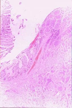
A section (on the right) of an intestinal carcinoid mass arising from the mucosa (150 X, courtesy of Professor Pantaleo Bufo, University of Foggia, Italy).
Click to see larger pictureClick to see detailView Full Size Image
Picture Type: Photo
Caption: Picture 2. Malignant carcinoid syndrome. A section of a carcinoid mass (350 X, courtesy of Professor Pantaleo Bufo, University of Foggia, Italy).
Click to see larger pictureClick to see detailView Full Size Image
Picture Type: Photo
Caption: Picture 3. Malignant carcinoid syndrome. 
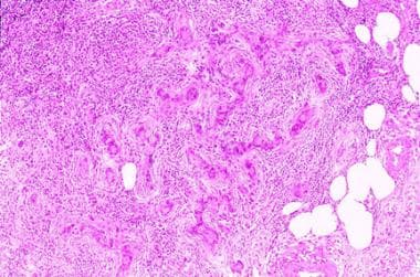
A section of a rare lymph node metastasis from adenocarcinoid tumor (250 X, courtesy of Professor Pantaleo Bufo, University of Foggia, Italy).
Click to see larger pictureClick to see detailView Full Size Image
Picture Type: Photo
  BIBLIOGRAPHY Section 11 of 11   Click here to go to the previous section in this topic Click here to go to the top of this page
Author Information Introduction Clinical Differentials Workup Treatment Medication Follow-up Miscellaneous Pictures Bibliography

  • Ahlman H: Midgut carcinoid tumors. Surg Annu 1986; 18: 65-93[Medline].
  • Anderson AS, Krauss D, Lang R: Cardiovascular complications of malignant carcinoid disease. Am Heart J 1997; 134(4): 693-702[Medline].
  • Anthony LB, Martin W, Delbeke D, Sandler M: Somatostatin receptor imaging: predictive and prognostic considerations. Digestion 1996; 57: 50-3[Medline].
  • Antonelli A, Gambuzza C, Bertoni F, Baschieri L: Treatment of the carcinoid syndrome with somatostatin, salmon calcitonin, or octreotide. Clin Ther 1992; 14(2): 178-84[Medline].
  • Caprotti R, Angelini C, Mussi C, et al: Gastrointestinal carcinoids. Prognosis and survival. Minerva Chir 2003 Aug; 58(4): 523-28,529-32[Medline].
  • Chaowalit N, Connolly HM, Schaff HV, et al: Carcinoid heart disease associated with primary ovarian carcinoid tumor. Am J Cardiol 2004 May 15; 93(10): 1314-5[Medline].
  • Codd JE, Drozda J, Merjavy J: Palliation of carcinoid heart disease. Arch Surg 1987; 122(9): 1076-7[Medline].
  • Eller R, Frazee R, Roberts J: Gastrointestinal carcinoid tumors. Am Surg 1991; 57(7): 434-7[Medline].
  • Farling PA, Durairaju AK: Remifentanil and anaesthesia for carcinoid syndrome. Br J Anaesth 2004 Jun; 92(6): 893-5[Medline].
  • Farndon JR: The carcinoid syndrome: methods of treatment and recent experience with hepatic artery ligation and infusion. Clin Oncol 1977 Dec; 3(4): 365-75[Medline].
  • Feldman JM: Carcinoid tumors and the carcinoid syndrome. Curr Probl Surg 1989 Dec; 26(12): 835-85[Medline].
  • Feldman JM: Carcinoid tumors and syndrome. Semin Oncol 1987 Sep; 14(3): 237-46[Medline].
  • Feldman JM: Histaminuria from histamine-rich foods. Arch Intern Med 1983; 143(11): 2099-102[Medline].
  • Frilling A, Rogiers X, Knofel WT, Broelsch CE: Liver transplantation for metastatic carcinoid tumors. Digestion 1994; 55 (Suppl 3): 104-6[Medline].
  • Granerus G, Lindell SE, Waldenstrom J, et al: Histamine metabolism in carcinoidosis. Lancet 1966 Jun 4; 1(7449): 1267-8[Medline].
  • Gregor M: Therapeutic principles in the management of metastasising carcinoid tumors: drugs for symptomatic treatment. Digestion 1994; 55 (Suppl 3): 60-3[Medline].
  • Hodgson HJ: Controlling the carcinoid syndrome. BMJ 1988 Nov 12; 297(6658): 1213-4[Medline].
  • Jabbour-Khoury S, Dabbous A, al-Jazzar M, et al: Anesthetic management of a patient having a carcinoid syndrome. Middle East J Anesthesiol 2003 Oct; 17(3): 435-47[Medline].
  • Joensuu H, Kumpulainen E, Grohn P: Treatment of metastatic carcinoid tumour with recombinant interferon alfa. Eur J Cancer 1992; 28A(10): 1650-3[Medline].
  • Maton PN: The carcinoid syndrome. JAMA 1988 Sep 16; 260(11): 1602-5[Medline].
  • Metcalfe DD: Differential diagnosis of the patient with unexplained flushing/anaphylaxis. Allergy Asthma Proc 2000; 21(1): 21-4[Medline].
  • Mignon M: Natural history of neuroendocrine enteropancreatic tumors. Digestion 2000; 62 Suppl 1: 51-8[Medline].
  • Mollet NR, Dymarkowski S, Bogaert J: MRI and CT revealing carcinoid heart disease. Eur Radiol 2003 Dec; 13 Suppl 4: L14-8[Medline].
  • Monsegu J, Algayres JP, Ciribilli JM, et al: Tricuspid insufficiency disclosing carcinoid syndrome. Rev Med Interne 1994; 15(9): 593-6[Medline].
  • Rorstad O: Prognostic indicators for carcinoid neuroendocrine tumors of the gastrointestinal tract. J Surg Oncol 2005 Mar 1; 89(3): 151-60[Medline].
  • Rosenberg JM, Welch JP: Ileal tumor causing carcinoid syndrome without hepatic metastases. Arch Surg 1984 Apr; 119(4): 485[Medline].
  • Shapiro RS, Shafir M, Sung M, et al: Cryotherapy of metastatic carcinoid tumors. Abdom Imaging 1998; 23(3): 314-7[Medline].
  • Sinzinger H, Renner F, Granegger S: Unsuccessful iodine-131 MIBG imaging of carcinoid tumors and apudomas. J Nucl Med 1986 Jul; 27(7): 1221-2[Medline].
  • van der Horst-Schrivers AN, Wymenga AN, Links TP, et al: Complications of midgut carcinoid tumors and carcinoid syndrome. Neuroendocrinology 2004; 80 Suppl 1: 28-32[Medline].
  • Warner RR: Diagnosis, treatment and management of carcinoid cancer. Personal communication - Carcinoid Symposium May 11, 2000; Available at: www.carcinoid.org/index.shtml.
  • Yeo CJ, Couse NF, Zinner MJ: Serotonin and substance P stimulate intestinal secretion in the isolated perfused ileum. Surgery 1989; 105(1): 86-92[Medline].
  • Zeitlin IJ: Kinin release in the carcinoid syndrome. Lancet 1971 Nov; 2(7732): 1040[Medline].

Malignant Carcinoid Syndrome excerpt