Continually Updated Clinical Reference
 
 
  All Sources     eMedicine     Medscape     Drug Reference     MEDLINE
 
You are in: eMedicine Specialties > Medicine, Ob/Gyn, Psychiatry, and Surgery > Hematology

Thrombocytosis, Essential

Last Updated: December 17, 2004
Email to a Colleague
Synonyms and related keywords: essential thrombocytosis, ET, primary thrombocythemia, idiopathic thrombocythemia, essential thrombocythemia, vascular thrombosis, acute myelogenous leukemia, AML, chronic myelogenous leukemia, CML

  AUTHOR INFORMATION Section 1 of 11    Click here to go to the next section in this topic
Author Information Introduction Clinical Differentials Workup Treatment Medication Follow-up Miscellaneous Pictures Bibliography

Author: Asheesh Lal, MBBS, Physician, Department of Internal Medicine, Lexington Medical Center

Asheesh Lal, MBBS, is a member of the following medical societies: American Society of Clinical Oncology, and American Society of Hematology

Editor(s): Wadie F Bahou, MD, Chief, Division of Hematology, Hematology/Oncology Fellowship Director, Professor, Department of Internal Medicine, State University of New York at Stony Brook; Francisco Talavera, PharmD, PhD, Senior Pharmacy Editor, eMedicine; Marcel E Conrad, MD, Distinguished Professor of Medicine, University of South Alabama; Director Cancer Center, Clinical Cancer Research Program, The Cancer Center, Mobile Infirmary Medical Center; Rajalaxmi McKenna, MD, FACP, Consulting Staff, Department of Medicine, Southwest Medical Consultants, SC, Good Samaritan Hospital, Advocate Health Systems; and Emmanuel C Besa, MD, Professor of Medicine, Department of Medicine, Division of Hematologic Malignancies, Kimmel Cancer Center, Thomas Jefferson University

Disclosure


  INTRODUCTION Section 2 of 11   Click here to go to the previous section in this topic Click here to go to the top of this page Click here to go to the next section in this topic
Author Information Introduction Clinical Differentials Workup Treatment Medication Follow-up Miscellaneous Pictures Bibliography

Background: Essential thrombocytosis (ET), first described by Epstein and Goedel in 1934, is a nonreactive, chronic myeloproliferative disorder. ET is associated with sustained megakaryocyte proliferation that increases the number of circulating platelets. Traditionally, ET was considered a clonal disorder that involved pluripotent stem cells; however, recent studies indicate that some patients may have polyclonal hematopoiesis.

ET is characterized by a platelet count greater than 600,000/mL, megakaryocytic hyperplasia, splenomegaly, and a clinical course complicated by hemorrhagic and/or thrombotic episodes.

Pathophysiology: Platelet survival is normal in ET. Megakaryocytes increase the production of platelets, causing thrombocytosis. The cause of this increase in platelet production remains unclear, though it may be a result of autonomous production, increased sensitivities to cytokines (eg, interleukin-3), decreased inhibition to platelet-inhibiting factors (eg, transforming growth factor beta), or defects in accessory cell microenvironment.

The mechanism by which thrombocythemia produces hemorrhage or thrombosis is not well defined. Several defects have been described, including a decrease in aggregation, hyperaggregation, and intracellular concentration of various chemicals. In addition, reports show a decrease in von Willebrand ristocetin cofactor activity and high molecular weight von Willebrand factor multimers. Some reports show patients with an acquired deficiency of antithrombin III, protein C, and protein S.

Frequency:

  • In the US: Clinicians diagnose approximately 6000 cases each year. Some researchers speculate that the incidence rate may be several times higher. A study from southeastern Minnesota reports an incidence of 2.38 cases per 100,000 population per year (Mesa, 1999).

Mortality/Morbidity:

  • Patients with ET have a 10-year survival rate of 64-80%, which may not be significantly different from that of the age-matched general population. Death occurs from thrombotic complications. Transformation to acute myelogenous leukemia (AML) occurs in 0.6-5% of patients and may accelerate if the patient takes chemotherapeutic agents.
  • Patients may experience symptoms relating to large vessel or microvascular thrombosis and bleeding.

Sex: In older patients, the frequency is similar in both sexes; however, the disease occurs more often in young women than in young men.

Age: ET is more frequent in older patients, although younger patients may develop the disease. The median age at diagnosis is 60 years, and perhaps up to 20% of patients are younger than 40 years. The disease is rare in children.


  CLINICAL Section 3 of 11   Click here to go to the previous section in this topic Click here to go to the top of this page Click here to go to the next section in this topic
Author Information Introduction Clinical Differentials Workup Treatment Medication Follow-up Miscellaneous Pictures Bibliography

History: Approximately 25-33% of patients are asymptomatic at diagnosis. The remainder report vasomotor symptoms or complications from thrombosis or bleeding. Most symptomatic patients present with symptoms that relate to small- or large-vessel thrombosis. Some patients present with bleeding.

  • Neurologic symptoms
    • Headache is the most common neurologic symptom.
    • Microvascular occlusion of the toes and fingers causes digital pain, gangrene, or erythromelalgia. Erythromelalgia is characterized by burning pain and dusky extremity congestion.
    • The pain increases with exposure to heat and improves with cold; a single dose of aspirin may provide relief for several days.
    • Patients also report paresthesias and episodic transient ischemic attacks. Transient neurological symptoms include the following:

      • Unsteadiness

      • Dysarthria

      • Dysphoria

      • Vertigo

      • Dizziness

      • Migraine

      • Syncope

      • Scotoma

      • Seizures
  • Thrombosis
    • Thrombosis of large veins and arteries is common and may result in occlusion of the leg, coronary, and renal arteries. Other arteries may be involved.
    • Venous thrombosis of the splenic, hepatic, or leg and pelvic veins may develop. Priapism is a rare complication. Pulmonary hypertension may result from pulmonary vasculature occlusion.
  • Bleeding
    • The gastrointestinal tract is the primary site of bleeding complications. Approximately 40% of the patients have duodenal arcade thrombosis, resulting in sloughing of the duodenal mucosa, simulating a duodenal ulcer.
    • Other sites of bleeding include the skin, eyes, gums, urinary tract, joints, and brain.
    • Bleeding is usually not severe and only rarely requires transfusion.
    • The bleeding is generally associated with a platelet count greater than 1 million/mL.
  • Constitutional symptoms (occur in 20-30% of patients). Weight loss is unusual. Other symptoms include sweating, low-grade fever, and pruritus.
  • Pregnancy complications
    • ET causes an increase in spontaneous abortions.
    • Placental infarctions may occur, resulting in intrauterine growth retardation and fetal death.

    • In most cases, fetal loss occurs during the first trimester.

    • A patient history of spontaneous abortion is the greatest risk factor for subsequent spontaneous abortions.

    • Excessive bleeding during delivery is rare.

    • Patients with successful pregnancies show a decrease in platelet count.

Physical: In most patients, physical examination findings are unremarkable. Approximately 40-50% of patients present with splenomegaly; 20% present with hepatomegaly.

Causes: The etiology and predisposing factors for ET development remain unclear. Genetic transmission of this disorder is rare, although reports show several families with multiple members affected by ET. Research suggests that a thrombopoietin production or receptor abnormality can cause familial ET.
  DIFFERENTIALS Section 4 of 11   Click here to go to the previous section in this topic Click here to go to the top of this page Click here to go to the next section in this topic
Author Information Introduction Clinical Differentials Workup Treatment Medication Follow-up Miscellaneous Pictures Bibliography

Agnogenic Myeloid Metaplasia With Myelofibrosis
Chronic Myelogenous Leukemia
Myelodysplastic Syndrome
Polycythemia Vera
Thrombocytosis, Secondary


Myeloproliferative Disorders Resource Center

View all Myeloproliferative Disorders Articles

Myeloproliferative Disorders Multimedia Library


Quick Find
Author Information
Introduction
Clinical
Differentials
Workup
Treatment
Medication
Follow-up
Miscellaneous
Pictures
Bibliography

Click for related images.

Related Articles
Agnogenic Myeloid Metaplasia With Myelofibrosis

Chronic Myelogenous Leukemia

Myelodysplastic Syndrome

Polycythemia Vera

Thrombocytosis, Secondary


Patient Education
Click here for patient education.


  WORKUP Section 5 of 11   Click here to go to the previous section in this topic Click here to go to the top of this page Click here to go to the next section in this topic
Author Information Introduction Clinical Differentials Workup Treatment Medication Follow-up Miscellaneous Pictures Bibliography

Lab Studies:

  • CBC count
    • This panel is essential for ET diagnosis.
    • The hallmark of ET is a sustained, unexplained thrombocytosis.
    • Leukocytosis, erythrocytosis, and mild anemia may be found.
    • The peripheral blood may show occasional immature precursor cells (eg, myelocytes, metamyelocytes). Large platelets (thrombocytes) are typically identifiable on routine peripheral blood smear.
    • Mild basophilia and eosinophilia may be found.
  • Bone marrow
    • Approximately 90% of patients show an increase in bone marrow cellularity.
    • Megakaryocytic hyperplasia is common.
    • Giant megakaryocytes are often observed. Clusters of megakaryocytes may be present; significant dysplasia of the megakaryocytes is unusual.
    • Hyperplasia of granulocyte and reticulocyte precursors is common.
    • Bone marrow reticulin is usually increased, but collagen fibrosis is uncommon.
    • Iron stores may be absent in the bone marrow. This may be due to gastrointestinal tract bleeding or menorrhagia. However, in ET, as in other myeloproliferative disorders, bone marrow iron stain results may be negative even when other studies do not support the presence of iron deficiency.
  • Platelet aggregation studies
    • The results of the prothrombin time and activated partial thromboplastin time studies are usually within reference ranges. The bleeding time may or may not be prolonged.
    • Platelet aggregation study findings are abnormal and show impaired platelet aggregation to epinephrine, adenosine diphosphate, and collagen but not to ristocetin and arachidonic acid.
    • Some patients may present with spontaneous platelet aggregation.
  • Chemistries
    • Chemistries reveal elevated uric acid levels in 25% of patients at diagnosis.

    • Pseudohyperkalemia may occur, and falsely elevated phosphorous and acid phosphatase levels may be noted.

    • Pseudohypoxemia may develop from extreme thrombocytosis.

    • Vitamin B-12 levels are increased in 25% of patients.
  • Cytogenetic study results are usually normal. Molecular studies (eg, polymerase chain reaction, Southern [genomic] blotting) may be used as sensitive means of excluding chronic myelogenous leukemia.

Imaging Studies:

  • CT scans or ultrasonograms of the spleen may reveal splenomegaly in patients even when this condition is not physically detectable.

Other Tests:

  • Elevation of C-reactive protein, fibrinogen, and interleukin-6 levels suggests the presence of secondary thrombocytosis, because these are acute-phase reactants.
  • Conduct an RBC mass study to exclude polycythemia vera. In ET, the RBC mass is without abnormality.
  • Conduct a sensitivity test to interleukin-3, which shows an increase in the formation of endogenous erythroid and/or megakaryocytic colonies and indicates the presence of abnormal hematopoietic progenitor cells (primarily a research tool).

Procedures:

  • Bone marrow aspirate and biopsy
    • A bone marrow aspirate and biopsy are useful. Use specialized needles to obtain the aspirate and biopsy material over the posterior iliac crest.
    • Obtaining an aspirate over the sternum may also be helpful, although most physicians prefer the posterior iliac crest.
    • Do not attempt to obtain a biopsy from the sternum.
Histologic Findings: Approximately 90% of patients show an increase in bone marrow cellularity. Megakaryocytic hyperplasia is present. Giant megakaryocytes are frequently observed, and clusters of megakaryocytes may be present. Significant dysplasia of the megakaryocytes is unusual. Hyperplasia of granulocyte and reticulocyte precursors is common. Bone marrow reticulin is usually increased, but collagen fibrosis is uncommon.

In ET, as in other myeloproliferative disorders, bone marrow iron stain results may be negative when other studies do not support the presence of iron deficiency. For practical purposes, a ferritin level that is within the reference range or increased, along with an RBC mean corpuscular volume that is within the reference range, is sufficient to exclude reactive thrombocytosis secondary to iron deficiency and the possibility of polycythemia vera masked by iron deficiency.

  TREATMENT Section 6 of 11   Click here to go to the previous section in this topic Click here to go to the top of this page Click here to go to the next section in this topic
Author Information Introduction Clinical Differentials Workup Treatment Medication Follow-up Miscellaneous Pictures Bibliography

Medical Care:

Surgical Care:

Consultations:


  MEDICATION Section 7 of 11   Click here to go to the previous section in this topic Click here to go to the top of this page Click here to go to the next section in this topic
Author Information Introduction Clinical Differentials Workup Treatment Medication Follow-up Miscellaneous Pictures Bibliography

ET treatment commonly includes the use of hydroxyurea, which is an antimetabolite similar in structure to naturally occurring compounds required for normal cell function. This structural similarity allows many of the antimetabolites to serve as substrates for important cellular enzymes. These substrates inhibit cell replication by direct inhibition of the enzymes needed for DNA replication or DNA repair or by incorporating directly into DNA. Tumors and healthy cells with high growth fractions (eg, bone marrow) are sensitive to inhibition by the antimetabolites. Anagrelide is an imidazoquinazoline drug that inhibits platelet aggregation. Anagrelide appears to decrease platelet counts by decreasing platelet production. Interferon alfa is a biologic response modifier, and phosphorous-32 is a radionuclide with direct myelosuppressive properties. Interferon alfa is not known to be teratogenic and does not cross the placenta, perhaps making it safe for use during pregnancy. Platelet counts rebound in most patientsafter stopping interferon. Platelet counts are reduced to less than 600,000/mL in 90% of cases after 3 months. Adjust drug dosing to achieve a platelet count within the reference range (target range, <450,000/mL).

Low-dose aspirin may be used to control microvascular symptoms.

Consider the patient's age, status, and adverse effect profile, in addition to the drug's cost, when choosing the treatment agent.

Drug Category: Antimetabolites -- Are similar in structure to the naturally occurring compounds required for the normal function of a cell. This structural similarity allows many of the antimetabolites to serve as substrates for important cellular enzymes, and they inhibit cell replication by direct inhibition of the enzymes needed for DNA replication or repair or by incorporating directly into DNA. Tumors and normal cells with high growth fractions (eg, bone marrow) are sensitive to inhibition by the antimetabolites.
Drug Name
Hydroxyurea (Hydrea) -- Inhibitor of deoxynucleotide synthesis and one of the drugs of choice for inducing hematologic remission in chronic myelogenous leukemia. Less leukemogenic than alkylating agents (eg, busulfan, melphalan, chlorambucil). Myelosuppressive effects last a few days to a week and are easier to control than those of alkylating agents; busulfan has prolonged bone marrow suppression and can cause pulmonary fibrosis. Dose can be administered as a single daily dose or divided into 2-3 doses at higher dose ranges. Changes in blood cell counts may take 3-4 d to be apparent after a change in the drug dose.
Adult DoseNo clear dosing guidelines exist
Suggested initial dose: 500 mg/d PO; adjust dose based on response; not to exceed 800 mg/m2 PO q4h
Pediatric DoseNo clear dosing guidelines exist
Suggested initial dose: 15 mg/kg/d PO; adjust dose based on response
ContraindicationsDocumented hypersensitivity; severe anemia or bone marrow suppression (ie, leukopenia [WBC count, <2500/mL]) count); thrombocytopenia (platelet count, <100,000/mL)
InteractionsIncreases cytotoxicity of 5-fluorouracil (5-FU) when administered after 5-FU; patients who have received irradiation therapy may have an exacerbation of postirradiation erythema
Pregnancy C - Safety for use during pregnancy has not been established.
PrecautionsTherapy requires close supervision; determine the complete status of the blood prior to and repeatedly during treatment, including bone marrow examination (if indicated), kidney function, and liver function; determine the hemoglobin level, total leukocyte counts, and platelet counts at least weekly throughout the course of therapy; if the WBC count decreases to <2500/mL or the platelet count decreases to <100,000/mL, interrupt therapy until the values rise significantly toward reference range levels; administration of hydroxyurea is associated with significant increases in the MCV of the RBCs
Drug Category: Imidazoquinazolines -- Cause suppression of megakaryocytes and decrease in platelet counts without affecting other hematopoietic cell lines.
Drug Name
Anagrelide (Agrylin) -- Mechanism by which anagrelide reduces blood platelet count remains under investigation. Inhibits cyclic nucleotide phosphodiesterase and the release of arachidonic acid from phospholipase, possibly by inhibiting phospholipase A2. Effective in polycythemia vera with elevated platelet counts. Studies in patients support a hypothesis of dose-related reduction in platelet production, resulting from a decrease in megakaryocyte hypermaturation.
Adult Dose0.5 mg PO qid or 1 mg PO bid; after 1 wk, adjust to lowest effective dose; dose must not be increased by more than 0.5 mg/d in any 1 wk; not to exceed 10 mg/d or 2.5 mg/dose
Pediatric DoseNot established
ContraindicationsDocumented hypersensitivity; congestive heart failure; child-bearing potential
InteractionsSucralfate may decrease absorption
Pregnancy C - Safety for use during pregnancy has not been established.
PrecautionsCaution in suspected heart disease, reduced renal function, or hepatic dysfunction; thrombocytopenia appears to be the main dose-limiting adverse effect; headache can occur upon initiation of drug, which can be minimized if started at low doses (1 mg bid)
Drug Category: Biologic response modifiers -- Exact mechanism of action is undetermined. May be beneficial because of myelosuppressive and antiproliferative effects.
Drug Name
Interferon alfa (Roferon-A, Intron A) -- Myelosuppressive protein product manufactured by recombinant DNA technology. Mechanism of antitumor activity is not clearly understood; however, direct antiproliferative effects against malignant cells and modulation of host immune response may play important roles.
Adult Dose3-5 million U SC 3-5 times/wk for 30 d; may take for 3-26 wk to obtain remission
Pediatric DoseNot established
ContraindicationsDocumented hypersensitivity
InteractionsTheophylline, zidovudine, and vinblastine may increase toxicity; cimetidine may increase antitumor effects
Pregnancy C - Safety for use during pregnancy has not been established.
PrecautionsVariations in dosage and adverse reactions exist among different brands of interferon; do not use different brands of interferon in a single treatment regimen; depression and suicidal ideation, suicidal attempts, and completed suicides have been reported in association with therapy; monitor blood cell counts, liver function, electrolytes, and TSH; obtain periodic ECG monitoring for patients with cardiac problems
Drug Category: Radionuclides -- Have myelosuppressive properties.
Drug Name
Phosphorous-32 -- A beta particle emitter, which is therefore myelosuppressive. Affects all cell types.
Adult Dose2.3 mCi/m2 IV, repeated in 3-6 mo; onset of action occurs in 4-8 wk
Pediatric DoseNot established; best avoided in pediatric patients
ContraindicationsBreastfeeding
InteractionsConcomitant use with myelosuppressive agents may cause cumulative marrow toxicity and severe cytopenias
Pregnancy X - Contraindicated in pregnancy
PrecautionsAssociated with transient, mild pancytopenia; prolonged pancytopenia may occur, particularly in elderly patients
  FOLLOW-UP Section 8 of 11   Click here to go to the previous section in this topic Click here to go to the top of this page Click here to go to the next section in this topic
Author Information Introduction Clinical Differentials Workup Treatment Medication Follow-up Miscellaneous Pictures Bibliography

Complications:

Prognosis:

  MISCELLANEOUS Section 9 of 11   Click here to go to the previous section in this topic Click here to go to the top of this page Click here to go to the next section in this topic
Author Information Introduction Clinical Differentials Workup Treatment Medication Follow-up Miscellaneous Pictures Bibliography

Medical/Legal Pitfalls:

  • Failure to establish the diagnosis in patients with either hemorrhage or thrombosis
  • Failure to administer cytoreductive therapy in patients older than 60 years or in those with a history of thrombosis: In high-risk patients, initiate platelet-reducing measures to prevent serious complications

Special Concerns:

  • Pregnancy
    • ET may increase the rate of pregnancy loss in women, primarily because of spontaneous abortions during the first trimester.
    • Intrauterine death and intrauterine growth retardation may occur.
    • Patients with a history of spontaneous abortion are clearly at increased risk of pregnancy loss.
    • Spontaneous decreases in the platelet count may occur with a successful pregnancy, with the nadir in the second trimester. The cause of the decline is unclear and is only partially explained as owing to hemodilution.
    • Treatment during pregnancy remains controversial; weigh potential teratogenic risks against risks to the mother.
    • Consider treatment in patients at increased risk of fetal loss.

    • Risk of teratogenicity may be lower with interferon than with other agents, and the drug appears to be well tolerated during pregnancy. Other therapies have included aspirin, hydroxyurea, and plateletpheresis.

    • Women with histories of thrombosis and ET should receive cytoreductive therapy during pregnancy to minimize the risk of complications. Interferon is the preferred drug for this purpose. Therapy with interferon may be started before conception.
  • Surgery
    • Thrombohemorrhagic complications are possible in patients undergoing surgery.

    • Before surgery, normalize platelet counts.
  • Splenectomy has been associated with serious thrombohemorrhagic complications and should be avoided.
  PICTURES Section 10 of 11   Click here to go to the previous section in this topic Click here to go to the top of this page Click here to go to the next section in this topic
Author Information Introduction Clinical Differentials Workup Treatment Medication Follow-up Miscellaneous Pictures Bibliography

Caption: Picture 1. 
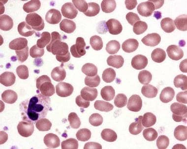
Peripheral blood smear in essential thrombocytosis showing increased platelet numbers. Courtesy Wei Wang, MD, and John Lazarchick, MD; Department of Pathology, Medical University of South Carolina.
Click to see larger pictureClick to see detailView Full Size Image
Picture Type: Photo
Caption: Picture 2. 
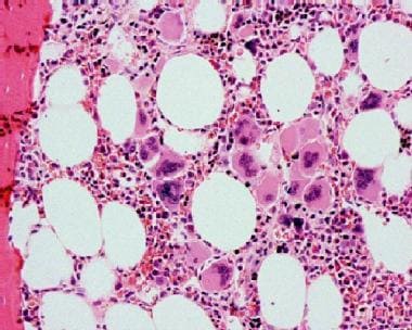
Bone marrow biopsy in essential thrombocytosis showing increased megakaryocytes. Courtesy Wei Wang, MD, and John Lazarchick, MD; Department of Pathology, Medical University of South Carolina.
Click to see larger pictureClick to see detailView Full Size Image
Picture Type: Photo
  BIBLIOGRAPHY Section 11 of 11   Click here to go to the previous section in this topic Click here to go to the top of this page
Author Information Introduction Clinical Differentials Workup Treatment Medication Follow-up Miscellaneous Pictures Bibliography

  • Barbui T, Finazzi G: Treatment indications and choice of a platelet-lowering agent in essential thrombocythemia. Curr Hematol Rep 2003 May; 2(3): 248-56[Medline].
  • Bellucci S, Janvier M, Tobelem G: Essential thrombocythemias. Clinical evolutionary and biological data. Cancer 1986 Dec 1; 58(11): 2440-7[Medline].
  • Besses C, Cervantes F, Pereira A, et al: Major vascular complications in essential thrombocythemia: a study of the predictive factors in a series of 148 patients. Leukemia 1999 Feb; 13(2): 150-4[Medline].
  • Bucalossi A, Marotta G, Bigazzi C: Reduction of antithrombin III, protein C, and protein S levels and activated protein C resistance in polycythemia vera and essential thrombocythemia patients with thrombosis. Am J Hematol 1996 May; 52(1): 14-20[Medline].
  • Budde U, Schaefer G, Mueller N: Acquired von Willebrand's disease in the myeloproliferative syndrome. Blood 1984 Nov; 64(5): 981-5[Medline].
  • Cervantes F, Tassies D, Salgado C: Acute transformation in nonleukemic chronic myeloproliferative disorders: actuarial probability and main characteristics in a series of 218 patients. Acta Haematol 1991; 85(3): 124-7[Medline].
  • Chistolini A, Filoni V, Dragoni F: Hepatitis C virus antibody in coagulopathic patients: ELISA and RIBA methods. Haematologica 1993 Jul-Aug; 78(4): 252-4[Medline].
  • Chistolini A, Mazzucconi MG, Ferrari A: Essential thrombocythemia: a retrospective study on the clinical course of 100 patients. Haematologica 1990 Nov-Dec; 75(6): 537-40[Medline].
  • Colombi M, Radaelli F, Zocchi L: Thrombotic and hemorrhagic complications in essential thrombocythemia. A retrospective study of 103 patients. Cancer 1991 Jun 1; 67(11): 2926-30[Medline].
  • Cortelazzo S, Finazzi G, Ruggeri M: Hydroxyurea for patients with essential thrombocythemia and a high risk of thrombosis. N Engl J Med 1995 Apr 27; 332(17): 1132-6[Medline].
  • el-Kassar N, Hetet G, Briere J: Clonality analysis of hematopoiesis in essential thrombocythemia: advantages of studying T lymphocytes and platelets. Blood 1997 Jan 1; 89(1): 128-34[Medline].
  • Elliott MA, Tefferi A: Interferon-alpha therapy in polycythemia vera and essential thrombocythemia. Semin Thromb Hemost 1997; 23(5): 463-72[Medline].
  • Emilia G, Sacchi S, Temperani P: Progression of essential thrombocythemia to blastic crisis via idiopathic myelofibrosis. Leuk Lymphoma 1993 Mar; 9(4-5): 423-6[Medline].
  • Epstein E, Goedel A: Hammorhagische thrombocythamie bei vascularer schrumpfmilz. Virch Arch (Pathol Anat) 1934; 292: 233.
  • Fabris F, Casonato A, Grazia del Ben M: Abnormalities of von Willebrand factor in myeloproliferative disease: a relationship with bleeding diathesis. Br J Haematol 1986 May; 63(1): 75-83[Medline].
  • Fenaux P, Simon M, Caulier MT: Clinical course of essential thrombocythemia in 147 cases. Cancer 1990 Aug 1; 66(3): 549-56[Medline].
  • Gugliotta L, Marchioli R, Fiacchini M, et al: Epidemiological, diagnostic, therapeutic and prognostic aspects of essential thrombocythemia in a retrospective study of the GIMMC group in two thousand patients [abstract]. Blood. 1997;90(suppl 1):348a. Blood 1997; 90(suppl 1): 348a.
  • Gugliotta L, Marchioli R, Fiacchini M, et al: Epidemiological, diagnostic, therapeutic and prognostic aspects of essential thrombocythemia in a retrospective study of the GIMMC group in two thousand patients [abstract]. Blood. 1997;90(suppl 1):348a. Blood 1997; 90(suppl 1): 348a.
  • Harrison CN, Gale RE, Machin SJ: A large proportion of patients with a diagnosis of essential thrombocythemia do not have a clonal disorder and may be at lower risk of thrombotic complications. Blood 1999 Jan 15; 93(2): 417-24[Medline].
  • Harrison CN, Donohoe S, Carr P, et al: Patients with essential thrombocythaemia have an increased prevalence of antiphospholipid antibodies which may be associated with thrombosis. Thromb Haemost 2002 May; 87(5): 802-7[Medline].
  • Hehlmann R, Jahn M, Baumann B: Essential thrombocythemia. Clinical characteristics and course of 61 cases. Cancer 1988 Jun 15; 61(12): 2487-96[Medline].
  • Jantunen R, Juvonen E, Ikkala E: The predictive value of vascular risk factors and gender for the development of thrombotic complications in essential thrombocythemia. Ann Hematol 2001 Feb; 80(2): 74-8[Medline].
  • Kobayashi S, Teramura M, Hoshino S: Circulating megakaryocyte progenitors in myeloproliferative disorders are hypersensitive to interleukin-3. Br J Haematol 1993 Apr; 83(4): 539-44[Medline].
  • Mesa RA, Silverstein MN, Jacobsen SJ: Population-based incidence and survival figures in essential thrombocythemia and agnogenic myeloid metaplasia: an Olmsted County Study, 1976-1995. Am J Hematol 1999 May; 61(1): 10-5[Medline].
  • Randi ML, Barbone E, Zerbinati P: Essential thrombocythemia following polycythemia vera: an unusual sequence. J Med 1996; 27(5-6): 363-8[Medline].
  • Ruggeri M, Gisslinger H, Tosetto A, et al: Factor V Leiden mutation carriership and venous thromboembolism in polycythemia vera and essential thrombocythemia. Am J Hematol 2002 Sep; 71(1): 1-6[Medline].
  • Shabbad E, Cassel A, Froom P: Effect of adherent cells on the regulation of BFU-E in patients with myeloproliferative disease. Am J Hematol 1990 Apr; 33(4): 225-9[Medline].
  • Tefferi A, Fonseca R, Pereira DL, Hoagland HC: A long-term retrospective study of young women with essential thrombocythemia. Mayo Clin Proc 2001 Jan; 76(1): 22-8[Medline].
  • van Genderen PJ, Michiels JJ, van der Poel-van de Luytgaarde SC: Acquired von Willebrand disease as a cause of recurrent mucocutaneous bleeding in primary thrombocythemia: relationship with platelet count. Ann Hematol 1994 Aug; 69(2): 81-4[Medline].
  • Zauli G, Visani G, Catani L: Reduced responsiveness of bone marrow megakaryocyte progenitors to platelet-derived transforming growth factor beta 1, produced in normal amount, in patients with essential thrombocythaemia. Br J Haematol 1993 Jan; 83(1): 14-20[Medline].

Thrombocytosis, Essential excerpt