| Patient Education |
|
Click here for patient education.
|
|
You are in: eMedicine Specialties >
Hematology > Red Blood Cells and Disorders
Thalassemia, Beta
Article Last Updated: Sep 21, 2007
AUTHOR AND EDITOR INFORMATION
Section 1 of 11
Author: Kenichi Takeshita, MD, Adjunct Associate Professor, Department of Medicine, Division of Hematology, New York University School of Medicine; Medical Director, Clinical Research and Development, Celgene
Kenichi Takeshita is a member of the following medical societies: American Society of Clinical Oncology and American Society of Hematology
Editors: Wadie F Bahou, MD, Chief, Division of Hematology, Hematology/Oncology Fellowship Director, Professor, Department of Internal Medicine, State University of New York at Stony Brook; Francisco Talavera, PharmD, PhD, Senior Pharmacy Editor, eMedicine; Marcel E Conrad, MD, BS, (Retired) Distinguished Professor of Medicine, University of South Alabama; Director, Clinical Cancer Research Program, The Cancer Center, Mobile Infirmary Medical Center; Rajalaxmi McKenna, MD, FACP, Consulting Staff, Department of Medicine, Southwest Medical Consultants, SC, Good Samaritan Hospital, Advocate Health Systems; Emmanuel C Besa, MD, Professor, Department of Medicine, Division of Hematologic Malignancies, Kimmel Cancer Center, Thomas Jefferson University
Author and Editor Disclosure
Synonyms and related keywords:
beta thalassemia syndromes, Cooley anemia, Mediterranean anemia, thalassemia major, thalassemia intermedia, thalassemia minor, thalassemia trait, hemoglobin E, hereditary disorder
Background
Beta thalassemia syndromes are a group of hereditary disorders characterized by a genetic deficiency in the synthesis of beta-globin chains. In the homozygous state, beta thalassemia (ie, thalassemia major) causes severe transfusion-dependent anemia. In the heterozygous state, the beta thalassemia trait (ie, thalassemia minor) causes mild-to-moderate microcytic anemia. In addition, hemoglobin (Hb) E, a common Hb variant found in Southeast Asia, is associated with a beta thalassemia phenotype, and this variant is included in the beta thalassemia category of diseases.
Pathophysiology
Mutations in globin genes cause thalassemias. Alpha thalassemia affects the alpha-globin gene(s). Beta thalassemia affects one or both of the beta-globin genes. These mutations result in the impaired synthesis of the beta globin protein portion, a component of Hb, thus causing anemia. In beta thalassemia minor (ie, beta thalassemia trait or heterozygous carrier-type), one of the beta-globin genes is defective. The defect can be a complete absence of the beta-globin protein (ie, beta-zero thalassemia) or a reduced synthesis of the beta-globin protein (ie, beta-plus thalassemia) (see Image 1). The genetic defect usually is a missense or nonsense mutation in the beta-globin gene, although occasional defects due to gene deletions of the beta-globin gene and surrounding regions also have been reported. In beta thalassemia major (ie, homozygous beta thalassemia), the production of beta-globin chains is severely impaired, because both beta-globin genes are mutated. The severe imbalance of globin chain synthesis (alpha >> beta) results in ineffective erythropoiesis and severe microcytic hypochromic anemia (see Image 2). The excess unpaired alpha-globin chains aggregate to form precipitates that damage red cell membranes, resulting in intravascular hemolysis. Premature destruction of erythroid precursors results in intramedullary death and ineffective erythropoiesis. The profound anemia typically is associated with erythroid hyperplasia and extramedullary hematopoiesis.
Frequency
United States
The frequency of disease varies widely, depending on the ethnic population. Beta thalassemia is reported most commonly in Mediterranean, African, and Southeast Asian populations.
International
The disease is found most commonly in the Mediterranean region, Africa, and Southeast Asia, presumably as an adaptive association to endemic malaria. The incidence may be as high as 10% in these areas.
Mortality/Morbidity
The major causes of morbidity and mortality are anemia and iron overload.
- The severe anemia resulting from this disease, if untreated, can result in high-output cardiac failure; the intramedullary erythroid expansion may result in associated skeletal changes such as cortical bone thinning. The long-term increase in red-cell turnover causes hyperbilirubinemia and bilirubin-containing gallstones.
- Increased iron deposition resulting from multiple life-long transfusions and enhanced iron absorption results in secondary iron overload. This overload causes clinical problems similar to those observed with primary hemochromatosis (eg, endocrine dysfunction, liver dysfunction, cardiac dysfunction).
Race
Beta thalassemia genes are reported throughout the world, although more frequently in Mediterranean, African, and Southeast Asian populations. Patients of Mediterranean extraction are more likely to be anemic with thalassemia trait than Africans because they have beta-zero thalassemia rather than beta-plus thalassemia.
- The genetic defect in Mediterranean populations is caused most commonly by (1) a mutation creating an abnormal splicing site or (2) a mutation creating a premature translation termination codon.
- Southeast Asian populations also have a significant prevalence of Hb E and alpha thalassemia.
- African populations more commonly have genetic defects leading to alpha thalassemia.
Sex
This genetic disorder is caused by abnormalities in the beta-globin gene, located on chromosome 11. It is not a sex-linked genetic trait.
Age
The manifestations of the disease may not be apparent until a complete switch from fetal to adult Hb synthesis occurs. This switch typically is completed by the sixth month after birth.
History
Thalassemia minor usually presents as an asymptomatic mild microcytic anemia and is detected through routine blood tests. Thalassemia major is a severe anemia that presents during the first few months after birth.
- Thalassemia minor (beta thalassemia trait) usually is asymptomatic, and it typically is identified during routine blood count evaluation.
- Thalassemia major (homozygous beta thalassemia) is detected during the first few months of life, when the patient's level of fetal Hb decreases.
Physical
- Patients with the beta thalassemia trait generally have no unusual physical findings.
- Beta thalassemia major
- The physical findings are related to severe anemia, ineffective erythropoiesis, extramedullary hematopoiesis, and iron overload resulting from transfusion and increased iron absorption.
- Skin may show pallor from anemia and jaundice from hyperbilirubinemia.
- The skull and other bones may be deformed secondary to erythroid hyperplasia with intramedullary expansion and cortical bone thinning.
- Heart examination may reveal findings of cardiac failure and arrhythmia, related to either severe anemia or iron overload.
- Abdominal examination may reveal changes in the liver, gall bladder, and spleen. Hepatomegaly related to significant extramedullary hematopoiesis typically is observed. Patients who have received blood transfusions may have hepatomegaly or chronic hepatitis due to iron overload; transfusion-associated viral hepatitis resulting in cirrhosis or portal hypertension also may be seen. The gall bladder may contain bilirubin stones formed as a result of the patient's life-long hemolytic state. Splenomegaly typically is observed as part of the extramedullary hematopoiesis or as a hypertrophic response related to the extravascular hemolysis.
- Extremities may demonstrate skin ulceration.
- Iron overload also may cause endocrine dysfunction, especially affecting the pancreas, testes, and thyroid.
Causes
Beta thalassemia is caused by a genetic mutation in the beta-globin gene; however, many additional factors influence the clinical manifestations of disease. That is, the same mutations may have different clinical manifestations in different patients. The following factors are known to influence the clinical phenotype:
- Intracellular fetal Hb concentrations
- The level of expression of fetal Hb (ie, the expression level of the gamma-globin gene) determines, in part, the severity of the disease.
- Patients with high fetal Hb have milder disease.
- Co-inheritance of alpha thalassemia
- Patients with co-inheritance of alpha thalassemia have a milder clinical course because they have a less severe alpha-beta chain imbalance.
- The coexistence of sickle cell trait and beta thalassemia is a major and symptomatic hemoglobinopathy with most of the symptoms and complications of sickle cell disease. Unlike sickle cell trait, in which most Hb-on-Hb electrophoresis is Hb A (AS), S is the dominant Hb (SA) and usually constitutes about 60% of the circulating Hb.
Lead Nephropathy
Other Problems to be Considered
Additional causes of microcytic anemia:
Lead poisoning
Sideroblastic anemia
Anemia of chronic disease
Unstable Hb levels
Red cell membrane disorders (some types)
Lab Studies
- The diagnosis of beta thalassemia minor usually is suggested by the presence of an isolated, mild microcytic anemia, target cells on the peripheral blood smear, and a normal red blood cell count. An elevation of Hb A2 (2 alpha-globin chains complexed with 2 delta-globin chains) demonstrated by electrophoresis or column chromatography confirms the diagnosis of beta thalassemia trait. The Hb A2 level in these patients usually is approximately 4-6%. In rare cases of concurrent severe iron deficiency, the increased Hb A2 level may not be observed, although it becomes evident with iron repletion. The increased Hb A2 level also is not observed in patients with the rare delta-beta thalassemia trait.
- An elevated Hb F level is not specific to patients with the beta thalassemia trait.
- Free erythrocyte porphyrin (FEP) tests may be useful in situations in which the diagnosis of beta thalassemia minor is unclear. FEP level is normal in patients with the beta thalassemia trait, but it is elevated in patients with iron deficiency or lead poisoning.
- Alpha thalassemia is characterized by genetic defects in the alpha-globin gene, and this variant has features similar to beta thalassemia. Patients with this disorder have normal Hb A2 levels. Establishing the diagnosis of the alpha thalassemia trait requires measuring either the alpha-beta chain synthesis ratio or performing genetic tests of the alpha-globin cluster (by Southern blot or polymerase chain reaction tests).
- Iron studies (iron, transferrin, ferritin) are useful in excluding iron deficiency and the anemia of chronic disorders as the cause of the patient's anemia.
- Patients may require a bone marrow examination to exclude certain other causes of microcytic anemia. Physicians must perform an iron stain (Prussian blue stain) to diagnose sideroblastic anemia (ringed sideroblasts).
- The Mentzer index is defined as mean corpuscular volume per red cell count. An index of less than 13 suggests that the patient has the thalassemia trait, and an index of more than 13 suggests that the patient has iron deficiency.
- Prenatal diagnosis is possible (see Other Tests).
Imaging Studies
- The skeletal abnormalities observed in patients with thalassemia major include an expanded bone marrow space, resulting in the thinning of the bone cortex. These changes are particularly dramatic in the skull, which may show the characteristic hair-on-end appearance. Bone changes also can be observed in the long bones, vertebrae, and pelvis.
- The liver and biliary tract of patients with thalassemia major may show evidence of extramedullary hematopoiesis and damage secondary to iron overload resulting from multiple transfusion therapy. Transfusion also may result in infection with the hepatitis virus, which leads to cirrhosis and portal hypertension. Gallbladder images may show the presence of bilirubin stones.
- The heart is a major organ that is affected by iron overload and anemia. Cardiac dysfunction in patients with thalassemia major includes conduction system defects, decreased myocardial function, and fibrosis. Some patients also develop pericarditis.
Other Tests
Molecular diagnostic tests can precisely determine whether a mutation is present any time after approximately 8 weeks of gestation. The physician can establish the diagnosis in utero using DNA obtained from amniocentesis or by chorionic villus sampling. In most laboratories, the DNA is amplified using the polymerase chain reaction technique and then is analyzed for the presence of the thalassemia mutation using a panel of oligonucleotide probes corresponding to known thalassemia mutations.
Procedures
- Physicians often use splenectomy to decrease transfusion requirements. Because postsplenectomy sepsis is possible, defer this procedure until the patient is older than 6-7 years. In addition, to minimize the risk of postsplenectomy sepsis, vaccinate the patient against Pneumococcus species, Meningococcus species, and Haemophilus influenzae. Also, administer penicillin prophylaxis to children after splenectomy.
- Patients with thalassemia minor may have bilirubin stones in their gall bladder and, if symptomatic, may require treatment. Perform a cholecystectomy using a laparoscope or at the time of the splenectomy.
Histologic Findings
The peripheral blood smear shows microcytic hypochromic red cells with target cells and anisopoikilocytosis.
Medical Care
Patients with thalassemia minor usually do not require any specific treatment. Treatment for patients with thalassemia major includes chronic transfusion therapy, iron chelation, splenectomy, and allogeneic hematopoietic transplantation.
- Thalassemia minor
- Patients in the heterozygous state usually do not require treatment.
- Inform patients that their condition is hereditary and that physicians sometimes mistake the disorder for iron deficiency.
- Some pregnant patients with the beta thalassemia trait may develop concurrent iron deficiency and severe anemia; they may require transfusional support if not responsive to iron repletion modalities.
- Thalassemia major
- The goal of long-term hypertransfusional support is to maintain the patient's Hb at 9-10 g/dL, thus improving the patient's sense of well-being while simultaneously suppressing enhanced erythropoiesis. This strategy not only treats the anemia, but also suppresses endogenous erythropoiesis so that extramedullary hematopoiesis and skeletal changes are suppressed.
- Patients receiving transfusion therapy also require iron chelation with desferrioxamine.
- Blood banking considerations for these patients include completely typing their erythrocytes prior to the first transfusion. This procedure helps future crossmatching processes.
- Allogeneic hematopoietic transplantation may be curative in some patients with thalassemia major. An Italian group led by Lucarelli has the most experience with this procedure.1 This group's research documents a 90% long-term survival rate in patients with favorable characteristics (young age, HLA match, no organ dysfunction).
Surgical Care
Patients with thalassemia minor rarely require splenectomy, although the development of bilirubin stones frequently leads to cholecystectomy.
Diet
- Drinking tea may help to reduce iron absorption through the intestinal tract.
- Vitamin C may improve iron excretion in patients receiving iron chelation. Anecdotal reports suggest that large doses of vitamin C can cause fatal arrhythmias when administered without concomitant infusion of deferoxamine.
Activity
Activity may be limited secondary to severe anemia.
Medical therapy for beta thalassemia primarily involves iron chelation. Deferoxamine is the intravenously administered chelation agent currently approved for use in the United States. Deferiprone is an oral chelation agent, recently approved for use in Europe. While the results of studies on this oral agent are encouraging, complications of hepatic fibrosis may develop.2 Deferiprone currently is not approved for use in the United States. Deferasirox is an orally administered chelation agent approved for use by the US FDA in 2005. The drug was undergoing review or investigation in other countries as of 2005. Additional treatments under development are experimental protocols to manipulate globin gene expression using gene therapy or using drugs that activate gamma-globin genes. Since fetal globin gene expression is associated with a milder phenotype, approaches to enhance intracellular Hb F levels (by activating gamma-globin gene expression) are currently under investigation. The 2 most widely studied drugs in this area are butyrates and hydroxyurea. Current obstacles in gene therapy include inability to express high levels of the beta-globin gene in erythroid cells and inability to transduce hematopoietic pluripotent stem cells at high efficiency.
Drug Category: Chelating agents
These agents bind iron and promote excretion.
| Drug Name | Deferoxamine (Desferal) |
| Description | Usually administered as slow subcutaneous infusion through portable pump. Freely soluble in water. Approximately 8 mg of iron bound by 100 mg of deferoxamine. Agent is excreted in bile and urine, resulting in red discoloration. Readily chelates iron from ferritin and hemosiderin but not from transferrin. Most effective when administered as continuous infusion. |
| Adult Dose | 20-40 mg/kg/d SC infused over 8-12 h; may be administered IV/IM if necessary |
| Pediatric Dose | Administer as in adults |
| Contraindications | Documented hypersensitivity; patients who do not have acute iron poisoning; severe renal disease and anuria (consider dose reduction after the loading dose) |
| Interactions | Coadministration of vitamin C improves iron chelation (vitamin C is contraindicated in patients with heart failure because it may exacerbate cardiac dysfunction) |
| Pregnancy | C - Fetal risk revealed in studies in animals but not established or not studied in humans; may use if benefits outweigh risk to fetus
|
| Precautions | Compliance may be poor, especially in adolescent children; follow efficacy by monitoring ferritin levels; tachycardia, hypotension, and shock may occur in patients receiving long-term therapy, and could add to the cardiovascular collapse resulting from iron toxicity; GI adverse effects include abdominal discomfort, nausea, vomiting, and diarrhea, which may add to the symptoms of acute iron toxicity; flushing and fever are reported |
| Drug Name | Deferasirox (Exjade) |
| Description | Tab for oral susp. Oral iron chelation agent demonstrated to reduce liver iron concentration in adults and children who receive repeated RBC transfusions. Binds iron with high affinity in a 2:1 ratio. Approved to treat chronic iron overload due to multiple blood transfusions. Treatment initiation recommended with evidence of chronic iron overload (ie, transfusion of about 100 mL/kg packed RBCs, about 20 U for 40-kg person, and serum ferritin level is consistently >1000 mcg/L). Serum ferritin should be monitored qmo and dosage adjusted q3-6mo based on ferritin measurements in increments of 5 or 10 mg/kg. If ferritin consistently falls below 500 mcg/L, consider holding drug. Dose should not exceed 30 mg/kg/d. |
| Adult Dose | Initial: 20 mg/kg PO qd on empty stomach 30 min ac; calculate dose to nearest whole tab Maintenance: Adjust dose by 5- to 10-mg/kg/d Note: Dissolve tab completely in water, orange juice, or apple juice, then immediately drink susp; resuspend any remaining residue in small volume of liquid and swallow |
| Pediatric Dose | <2 years: Not established >2 years: Administer as in adults |
| Contraindications | Documented hypersensitivity |
| Interactions | Data limited; do not take with aluminum-containing antacids |
| Pregnancy | B - Fetal risk not confirmed in studies in humans but has been shown in some studies in animals
|
| Precautions | Common adverse effects include diarrhea, nausea, abdominal pain, headache, pyrexia, cough, and rash; may increase serum creatinine and hepatic enzyme levels; decrease dose with persistent elevation of serum creatinine level; may cause auditory and visual disturbances; slight decreases in serum copper and zinc levels may occur; dissolve tab completely in water, orange juice, or apple juice and drink resulting susp immediately (do not swallow tab whole, do not chew or crush) |
Further Outpatient Care
See Treatment.
In/Out Patient Meds
- Administer iron chelation daily as described under medication.
- Transfuse red blood cells to maintain the Hb concentration at 9-10 g/dL.
Deterrence/Prevention
- Prenatal diagnosis is possible by analyzing DNA obtained via chorionic villi sampling at 8-10 weeks of gestation or by amniocentesis at 14-20 weeks of gestation. Since the genetic defects are quite variable, family genotyping usually must be completed for diagnostic linkage (segregation) analysis. With the anticipated availability of large-scale mutation screening by DNA chip technology, extensive pedigree analyses may be obviated. Physicians can perform fetal blood sampling for Hb chain synthesis at 18-22 weeks of gestation, but this procedure is not as reliable as DNA analysis sampling methods.
- Genetic therapy strategies are currently in the early stages of development.
Complications
- Iron overload
- Extramedullary hematopoiesis
- Asplenia secondary to splenectomy
- Medical complications from long-term transfusional therapy - Iron overload or transfusion-associated infections (eg, hepatitis)
- Increased risk for infections resulting from asplenia (eg, encapsulated organisms such as pneumococcus) or from iron overload (eg, Yersinia species)
- Cholelithiasis (eg, bilirubin stones)
Prognosis
- Individuals with thalassemia minor (thalassemia trait) usually have asymptomatic mild anemia. This state does not result in mortality or significant morbidity.
- The prognosis of patients with thalassemia major is highly dependent on the patient's adherence to long-term treatment programs, namely the hypertransfusion program and life-long iron chelation. Allogeneic bone marrow transplantation may be curative.
Patient Education
- Educate patients with thalassemia minor about the genetic (hereditary) nature of their disease, and inform them that their immediate family members (ie, parents, siblings, children) may be affected. The presence of thalassemia major in both parents implies that children will likely have a form of the disease. (The presence of compound heterozygosity in the parents makes accurate phenotypic predictions for children incomplete).
- Inform patients with thalassemia minor that they do not have iron deficiency and that iron supplementation will not improve their anemia.
Medical/Legal Pitfalls
Thalassemia is an iron-overloading disorder. Therapy with iron is contraindicated in this disease. The presence of microcytic anemia is not always due to iron deficiency.
| Media file 1:
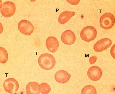
Peripheral smear in beta-zero thalassemia minor showing microcytes (M), target cells (T), and poikilocytes. |
 | View Full Size Image | |
Media type: Photo
|
| Media file 2:
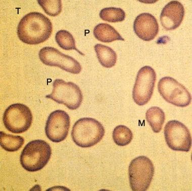
Peripheral smear from a patient with beta-zero thalassemia major showing more marked microcytosis (M) and anisopoikilocytosis (P) than in thalassemia minor. Target cells (T) and hypochromia are prominent. |
 | View Full Size Image | |
Media type: Photo
|
- Lucarelli G, Galimberti M, Polchi P. Marrow transplantation in patients with thalassemia responsive to iron chelation therapy. N Engl J Med. Sep 16 1993;329(12):840-4. [Medline]. [Full Text].
- Olivieri NF, Brittenham GM, McLaren CE, et al. Long-term safety and effectiveness of iron-chelation therapy with deferiprone for thalassemia major. N Engl J Med. Aug 13 1998;339(7):417-23. [Medline]. [Full Text].
- Cohen AR, Galanello R, Pennell DJ, et al. Thalassemia. In: Hematology (Am Soc Hematol Educ Program). 2004:14-34.
- Forget BG. Thalassemia Syndromes. In: Hematology: Basic Principles and Practice. 2000:485-509.
- Fucharoen S, Winichagoon P. Clinical and hematologic aspects of hemoglobin E beta-thalassemia. Current Opinion in Hematology. 2000;7:106-112. [Medline].
- Hoffbrand AV, AL-Refaie F, Davis B. Long-term trial of deferiprone in 51 transfusion-dependent iron overloaded patients. Blood. 1998;91:295-300. [Medline].
- Ikuta T, Atweh G, Boosalis V. Cellular and molecular effects of a pulse butyrate regimen and new inducers of globin gene expression and hematopoiesis. Ann N Y Acad Sci. 1998;850:87-99. [Medline].
- Koshy M, Dorn L, Bressler L. 2-deoxy 5-azacytidine and fetal hemoglobin induction in sickle cell anemia. Blood. 2000;96:2379-2384. [Medline].
- Malik P, Arumugam PI. Gene Therapy for {beta}-Thalassemia. In: Hematology (Am Soc Hematol Educ Program). 2005:45-50.
- May C, Rivella S, Callegari J. Therapeutic haemoglobin synthesis in beta-thalassaemic mice expressing lentivirus-encoded human beta-globin. Nature. 2000;406:82-86. [Medline].
- Perrine SP. Fetal Globin Induction--Can It Cure {beta} Thalassemia?. In: Hematology (Am Soc Hematol Educ Program). 2005:38-44.
- Rund D, Rachmilewitz E. Beta-thalassemia. N Engl J Med. Sep 15 2005;353(11):1135-46. [Medline].
- Schrier SL, Angelucci E. New strategies in the treatment of the thalassemias. Annu Rev Med. 2005;56:157-71. [Medline].
- Thein SL. Pathophysiology of {beta} Thalassemia--A Guide to Molecular Therapies. In: Hematology (Am Soc Hematol Educ Program). 2005:31-7.
- Weatherall DJ, Clegg JB. Genetic disorders of hemoglobin. Semin Hematol. Oct 1999;36(4 Suppl 7):24-37. [Medline].
Thalassemia, Beta excerpt Article Last Updated: Sep 21, 2007
|