Practice Essentials
Staphylococcal infections usually are caused by Staphylococcus aureus (S aureus). However, the incidence of infections due to Staphylococcus epidermidis (S epidermidis) and other coagulase-negative staphylococci (CoNS) also has been steadily rising. [1]
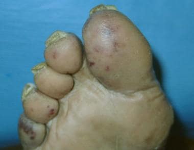
The image below depicts embolic lesions in patient with S aureus endocarditis.
Signs and symptoms
Manifestations of staphylococcal infections usually depend on the type of infection the organism causes. Common types of infections include the following [2] :
-
Skin infections (eg, folliculitis, furuncles, impetigo, wound infections, scalded skin syndrome)
-
Soft tissue infections (eg, pyomyositis, septic bursitis, septic arthritis)
-
Toxic shock syndrome
-
Purpura fulminans
-
Endocarditis
-
Osteomyelitis
-
Pneumonia
-
Food poisoning
-
Infections related to prosthetic devices (eg, prosthetic joints and heart valves; vascular shunts, grafts, catheters): Commonly associated with CoNS
-
Urinary tract infection
See Clinical Presentation for more detail.
Diagnosis
Examination in patients with staphylococcal infections may include the following findings:
-
Skin and soft tissue infections: Erythema, warmth, draining sinus tracts, superficial abscesses, bullous impetigo
-
Toxic shock syndrome: Fever higher than 38.9°C (102.02°F), diffuse erythroderma, hypotension, desquamation
-
Endocarditis: Regurgitant murmur, petechiae/cutaneous lesions, fever
Laboratory testing
-
Complete blood count: Usually shows leukocytosis with a left shift (bands); may reveal thrombocytosis with chronic staphylococcal infection
-
Erythrocyte sedimentation rate and C-reactive protein level: May be helpful in patients with subacute or chronic infections (eg, osteomyelitis)
-
Teichoic acid antibody titers: No longer routinely performed; may indicate a deep-seated (not IV line) infectious focus (eg, endocarditis, abscess, osteomyelitis)
-
Blood cultures with susceptibilities, as appropriate for site of infection
-
Peptide nucleic acid fluorescence in situ hybridization (PNA FISH): High sensitivity for S aureus (99.5%) and CoNS from positive blood cultures
-
Multiplex PCR: Also helpful and provide data regarding the presence of mecA gene typically found in methicillin-resistant S aureus (MRSA) isolates [1]
-
Screening tests for MRSA
Imaging studies
-
Transthoracic echocardiography (TTE): Should be considered in all patients with S aureus or Staphylococcus lugdunensis (S lugdunensis) bacteremia; patients with suspected endocarditis should undergo immediate transesophageal echocardiography (TEE), when possible
-
Transesophageal echocardiography (TEE): For all patients with catheter-related S aureus bacteremia (and no contraindications); for all patients with suspected S aureus endocarditis
See Workup for more detail.
Management
Promptly start antimicrobial therapy when S aureus infection is documented or strongly suspected. Appropriate choices depend on local susceptibility patterns. [3]
Temporary intravascular devices should be promptly removed if infection is suspected. [4] Long-term intravascular devices should be removed if infection with S aureus is documented.
Multiple decolonization regimens have been used in patients with recurrent staphylococcal infection. In one study, treatment with topical mupirocin, chlorhexidine gluconate washes, and oral rifampin plus doxycycline for 7 days eradicated MRSA colonization in hospitalized patients. [5]
Pharmacotherapy
Patients with serious staphylococcal infections should be initially started on agents active against MRSA until susceptibility results are available. Many CoNS are oxacillin-resistant. The duration of treatment and the use of synergistic combinations depend on the type of infection encountered. Pharmacist intervention through vancomycin dosing improved survival rates in a retrospective study of patients with MRSA bacteremia. [6]
The following antibiotics may be used in the management of staphylococcal infections (listed alphabetically, not necessarily in order of preference):
-
Cefazolin
-
Ceftaroline
-
Cefuroxime
-
Clindamycin
-
Dalbavancin
-
Daptomycin
-
Dicloxacillin
-
Doxycycline
-
Linezolid
-
Minocycline
-
Nafcillin
-
Oritavancin
-
Quinupristin/dalfopristin
-
Tedizolid
-
Telavancin
-
Tigecycline
-
Trimethoprim-sulfamethoxazole
-
Vancomycin
-
Delafloxacin
Surgery
Abscesses must be drained and/or debrided. Infections involving a prosthetic joint usually require removal of the prosthesis. Other infections involving a prosthetic device (eg, prosthetic heart valve or implanted intravascular device) may or may not require removal of the device.
See Treatment and Medication for more detail.
Background
Staphylococcal infections are usually caused by the organism S aureus. However, the incidence of infections due to S epidermidis and other CoNS has been steadily increasing in recent years. This article focuses on S aureus but also discusses infections caused by CoNS when important differences exist.
Pathophysiology
S aureus is a gram-positive coccus that is both catalase- and coagulase-positive. Colonies are golden and strongly hemolytic on blood agar. They produce a range of toxins, including alpha-toxin, beta-toxin, gamma-toxin, delta-toxin, exfoliatin, enterotoxins, Panton-Valentine leukocidin (PVL), [7] and toxic shock syndrome toxin–1 (TSST-1). [8] The enterotoxins and TSST-1 are associated with toxic shock syndrome. PVL is associated with necrotic skin [9] and lung infections and has been shown to be a major virulence factor for pneumonia [10] and osteomyelitis. [11] CoNS, particularly S epidermidis, produce an exopolysaccharide (slime) that promotes foreign-body adherence and resistance to phagocytosis.
Nienaber et al have demonstrated that methicillin-susceptible S aureus (MSSA) isolates causing endocarditis are more likely to be from a specific clonal cluster (CC30) and to possess specific virulence genes as compared to MSSA isolates from the same regions causing soft tissue infection. Isolates from patients with endocarditis were more likely to possess genes for 3 different adhesins and 5 different enterotoxins. The gene for PVL was found in the minority of both groups. [12]
S aureus has evolved to develop numerous immune evasion strategies to combat neutrophil-mediated killing, such as neutrophil activation, migration to the site of infection, bacterial opsonization, phagocytosis, and subsequent neutrophil-mediated killing. As many as 40 immune-evasion molecules of S aureus are known, and new functions are being identified for these evasion proteins. [13]
In a study of 42 S lugdunensis isolates, most isolates were able to form at least a weak biofilm, but the amount of biofilm formed by isolates was heterogeneous with poor correlation between clinical severity of disease and degree of biofilm formation. [14]
Frequency
United States
Up to 80% of people are eventually colonized with S aureus. Most are colonized only intermittently; 20-30% are persistently colonized. Colonization rates in health care workers, persons with diabetes, and patients on dialysis are higher than in the general population. The anterior nares are the predominant site of colonization in adults; carriage here has been associated with the development of bacteremia. [15] Other potential sites of colonization include the throat, [16] axilla, rectum, and perineum. The rate of MRSA hand colonization among health care workers has been shown to exceed 4% (over 8% in North America). [17]
International
S aureus infection occurs worldwide. Pyomyositis due to S aureus is more prevalent in the tropics.
Mortality/Morbidity
Mortality due to staphylococcal infections varies widely. Untreated S aureus bacteremia carries a mortality rate that exceeds 80%. The mortality rate of staphylococcal toxic shock syndrome is 3-5%. Infections due to CoNS usually carry a very low mortality rate. Because these infections are commonly associated with prosthetic devices, the most serious complication is the need to remove the involved prosthesis, although prosthetic valve endocarditis may lead to death.
Risk factors associated with increased mortality among patients with S aureus bacteremia include thrombocytopenia, an elevated score on the Charlson Comorbidity Index, MRSA infection, admission to an intensive care unit, and prior exposure to antibiotics. [18, 19]
Race
Staphylococcal infections have no reported racial predilection.
Sex
The vaginal carriage rate of staphylococcal species is approximately 10% in premenopausal women. The rate is even higher during menses.
Age
Staphylococcal species colonize many neonates on the skin, perineum, umbilical stump, and GI tract. The staphylococcal colonization rate in adults is approximately 40% at any given time.
The mortality rate of S aureus bacteremia in elderly persons is markedly increased. [20]
-
Embolic lesions in patient with Staphylococcus aureus endocarditis.
-
Close-up view of embolic lesions in patient with Staphylococcus aureus endocarditis.
-
Fifty-six-year-old man with erythema, edema, and drainage from below his right eye.
-
Gram stain in a 70-year-old woman with rheumatoid arthritis.


