Continually Updated Clinical Reference
 
 
  All Sources     eMedicine     Medscape     Drug Reference     MEDLINE
 
eMedicine - Pulmonary Fibrosis, Interstitial (Nonidiopathic) : Article by

Quick Find
Authors & Editors
Introduction
Clinical
Differentials
Workup
Treatment
Medication
Follow-up
Miscellaneous
Acknowledgments
Multimedia
References

Related Articles
Acute Respiratory Distress Syndrome

Berylliosis

Coal Worker's Pneumoconiosis

Eosinophilic Granuloma (Histiocytosis X)

Eosinophilic Pneumonia

Farmer's Lung

Hypersensitivity Pneumonitis

Lymphocytic Interstitial Pneumonia

Pneumocystis Carinii Pneumonia

Pneumonia, Aspiration

Pneumonia, Viral

Pulmonary Edema, Cardiogenic

Pulmonary Fibrosis, Idiopathic

Pulmonary Fibrosis, Interstitial (Nonidiopathic)

Sarcoidosis

Silicosis

Silo Filler's Disease


Patient Education
Click here for patient education.


Author: Eleanor M Summerhill, MD, Assistant Professor of Medicine, Division of Pulmonary and Critical Care Medicine, Warren Alpert Medical School of Brown University; Director of Internal Medicine Residency Program, Memorial Hospital of Rhode Island

Eleanor M Summerhill is a member of the following medical societies: American College of Chest Physicians, American College of Physicians, American Thoracic Society, Association of Program Directors in Internal Medicine, and Rhode Island Medical Society

Editors: Stephen P Peters, MD, PhD, Professor, Department of Medicine, Wake Forest University; Francisco Talavera, PharmD, PhD, Senior Pharmacy Editor, eMedicine; Daniel R Ouellette, MD, FCCP, Associate Professor of Medicine, Wayne State University School of Medicine; Consulting Staff, Pulmonary Disease and Critical Care Medicine Service, Henry Ford Health System; Timothy D Rice, MD, Associate Professor, Departments of Internal Medicine and Pediatrics and Adolescent Medicine, Saint Louis University School of Medicine; Zab Mosenifar, MD, Director, Division of Pulmonary and Critical Care Medicine, Director, Women's Guild Pulmonary Disease Institute, Executive Vice Chair, Department of Medicine, Cedars Sinai Medical Center; Professor of Medicine, David Geffen School of Medicine at UCLA

Author and Editor Disclosure

Synonyms and related keywords: interstitial lung disease, ILD, diffuse parenchymal lung disease, DPLD, diffuse lung disease, DLD, fibrotic lung disease, bronchiolitis obliterans-organizing pneumonia, BOOP, cryptogenic organizing pneumonia, COP, acute interstitial pneumonitis, AIP, eosinophilic pneumonia, desquamative interstitial pneumonitis, DIP, nonspecific interstitial pneumonitis, NSIP, hypersensitivity pneumonitis, HSP, lymphocytic interstitial pneumonitis, LIP, giant-cell pneumonitis, granulomatous pneumonitis, sarcoidosis, occupational interstitial lung disease, autoimmune pulmonary fibrosis

Background

Diffuse parenchymal lung disease (DPLD) is a heterogenous group of disorders. Clinical, physiologic, radiographic, and pathologic presentations of patients with these disorders are varied. However, a number of common features justify their inclusion in a single disease category.

DPLD may be idiopathic, as classically illustrated by idiopathic interstitial fibrosis (IPF), which is discussed in another section. The underlying histopathology of IPF is usual interstitial pneumonitis (UIP). Other major forms of interstitial pneumonitis include the following: desquamative interstitial pneumonia (DIP); respiratory bronchiolitis interstitial lung disease (RBILD); acute interstitial pneumonitis (AIP), also known as Hamman-Rich syndrome; nonspecific interstitial pneumonia (NSIP); cryptogenic organizing pneumonia (COP); lymphocytic interstitial pneumonia (LIP); sarcoidosis; hypersensitivity pneumonia (HSP); pulmonary Langerhans cell histiocytosis (PLCH); and lymphangioleiomyomatosis (LAM).

Some of these disorders, for example, RBILD, DIP, and PLCH, are clearly associated with smoking. Some forms of DPLD may also be related to occupational, environmental, and drug exposures as well as systemic illness such as collagen vascular disease. Although the recognized etiologies of these disorders are numerous, several groupings are clinically relevant. This article presents a broad overview, with an emphasis on those etiologies that result in pulmonary fibrosis not discussed elsewhere in this series.

Pathophysiology

A common pathophysiology has been postulated for these disorders. It is thought to begin with acute injury to the pulmonary parenchyma, leading to chronic interstitial inflammation, then fibroblast activation and proliferation, and finally progressing to the common end point of pulmonary fibrosis and tissue destruction. It now appears that inflammation is less important in IPF, which appears to be primarily a disorder of fibroblast activation and proliferation in response to some as yet unknown trigger(s).

The DPLDs typically manifest with the insidious onset of respiratory symptomatology, although onset can be acute and rapidly progressive, as in cryptogenic organizing pneumonia (COP) or acute interstitial pneumonitis (AIP).

Pathologically, all manifest histologically with disease largely within the interstitial compartment of the lung. However, alveolar and airway architecture may also be disrupted to varying degrees. The histologic patterns of usual interstitial pneumonitis (UIP), desquamative interstitial pneumonitis (DIP), nonspecific interstitial pneumonitis (NSIP), hypersensitivity pneumonitis (HSP), lymphocytic interstitial pneumonitis (LIP), cryptogenic organizing pneumonia (COP), giant cell pneumonitis, and granulomatous pneumonitis are most common and are centered in the alveolar, lobular, and lobar septa, impacting alveoli, small airways, and pulmonary vasculature.

Frequency

United States

As a group, diffuse interstitial diseases of the lung are uncommon, with overall estimates of 31.5 cases per 100,000 persons per year among men and 26.1 cases per 100,000 persons per year among women. However, the epidemiology is markedly affected by age and occupational exposures. Of patients referred to a pulmonary disease specialist, an estimated 10-15% have a DPLD.

Mortality/Morbidity

The natural history of diffuse interstitial lung diseases varies among different diagnostic entities and among individuals with the same diagnosis.

  • Some diseases are insidious in onset and gradual but unrelenting in progression (eg, similar to IPF), while other diseases are acute in onset but responsive to therapy (eg, COP).
  • Diseases that most closely approximate IPF have a similar mortality rate of approximately 50% at 5 years.
  • In the United States, mortality attributed to all types of DPLD is estimated to be 10-15% of that of chronic obstructive pulmonary disease.

Race

  • Diffuse interstitial diseases of the lung sometimes show racial predilections. Examples include sarcoidosis, which is more common in those of African ancestry in the United States. In contrast, pulmonary Langerhans cell histiocytosis (PLCH), also known as histiocytosis X, primarily affects Caucasians.

Sex

  • Several diffuse interstitial diseases of the lung show sexual predilections. IPF affects men more than women (at a ratio of 1.5:1), while LAM and pulmonary tuberous sclerosis exclusively affect women.
  • The prevalence of DPLD is 6 cases per 100,000 women and 31 cases per 100,000 men.
  • Women are much more likely to develop rheumatologic/connective-tissue disease than men and thus are more likely to experience pulmonary manifestations of those diseases. However, when affected, men with certain rheumatologic diseases (eg, rheumatoid arthritis) are more likely to develop pulmonary manifestations than women.
  • The pneumoconioses (eg, silicosis) are much more common in men than in women, probably because of higher rates of occupational exposure.

Age

  • Many of the DPLDs develop over many years and therefore are more prevalent in older adults. Others, such as sarcoidosis, LAM, connective tissue disease-associated, and inherited forms of DPLD, primarily present in younger adults.


History

The clinical history offered by patients with a DPLD is variable and related to the underlying disease process.

  • In general, all manifest primarily with respiratory symptoms that may be erroneously attributed to aging, obesity, deconditioning, or recent respiratory infection.
    • Dyspnea is the most frequent symptom, but chronic cough, wheezing, hemoptysis, and chest pain can occur.
    • Digital clubbing is common with some diagnoses (eg, IPF and asbestosis) and may first be noted by the patient. When it develops in a patient with known interstitial lung disease, it is usually indicative of advanced fibrosis. However, it may also herald an underlying bronchogenic carcinoma.
  • Clinical history may include the following:
    • Incidental diagnosis may be made from a chest radiograph or abnormal screening spirometry findings obtained for unrelated reasons.
    • Diagnosis may occur as a result of screening for high-risk occupational exposure, such as asbestos.
    • Medical attention may be sought for other manifestations of systemic illness that also affect the lungs.
  • Broadly, the manifestations of fibrotic lung disease can be grouped as follows:
    • They may be chronic, insidious, and slowly progressive.
    • They may be subacute, with a resolving, remitting, relapsing, or progressive course.
    • They may be acute, with a fulminant, progressive, remitting, or resolving course.
  • Disorders with chronic, insidious, and slowly progressive courses are those that most resemble IPF and usually share a common pathology (ie, UIP).
    • Many of the rheumatologic/connective-tissue diseases (eg, rheumatoid arthritis; calcinosis cutis, Raynaud phenomenon, esophageal motility disorder, sclerodactyly, and telangiectasia [CREST syndrome]/progressive systemic scleroderma [PSS]); systemic lupus erythematosus (SLE); mixed connective-tissue disease; the pneumoconioses (eg, asbestosis, silicosis); chronic hypersensitivity pneumonitis; and drug-related pulmonary fibrosis (eg, bleomycin) generally fit into this category.
    • Development of clinically apparent lung diseases related to occupational exposures (eg, pneumoconiosis) generally occurs many years after the exposure. Radiation fibrosis often develops months to years after radiation exposure.
    • A lag time of months or years can occur between the use of pulmonary toxic medications and the development of fibrotic disease. The effect can be dose-dependent (eg, bleomycin), although, in other cases, the relationship is less clear.
    • Pulmonary manifestations of rheumatologic/connective-tissue disease may develop in advance of, coincident with, or many years after the onset of articular disease.
    • Pulmonary sarcoidosis, although sometimes acute or subacute in onset, in some cases may present insidiously over time.
  • Subacute presentations with a variable course are typified by COP.
    • COP often develops weeks or months after the onset of a flu-like illness.
    • Patients can present to medical attention with dyspnea or exercise intolerance.
    • The course is variable and may either spontaneously remit or progress.
    • The disorder is thought to be very responsive to steroid therapy, although it may recur when steroids are withdrawn or tapered.
    • In some cases, COP may progress to end-stage fibrotic lung disease.
  • Disorders with an acute onset are typified by AIP, which is an idiopathic form of severe lung injury.
    • The histopathology is that of adult respiratory distress syndrome (ARDS) with diffuse alveolar damage (DAD).
    • Patients present either with no antecedent history of lung disease or as part of an accelerated phase of underlying interstitial disease.
    • Most patients progress rapidly to respiratory failure.
    • Some patients may improve with steroids or other immunosuppressive therapy.

Physical

  • Varied etiologies make generalization of physical examination findings difficult for patients with DPLD. However, clinical examination findings noted in patients with idiopathic pulmonary fibrosis are frequently noted in patients with other DPLDs.
    • Patients frequently are dyspneic, which may be more pronounced with activity and generally is associated with an accompanying tachypnea.
    • Central cyanosis may be present if significant hypoxemia and arterial oxygen desaturation are present.
    • Lung volumes are usually reduced. Fine end-inspiratory pulmonary rales (Velcro rales) are a common finding and may be difficult to distinguish from those auscultated in patients with congestive heart failure. Pulmonary sarcoidosis and other granulomatous disorders are often an exception.
    • Wheezes may be heard and reflect airway involvement, as in sarcoidosis.
    • A pulmonary squawk has been described with HSP.
    • A right-sided gallop (S3), an accentuated second heart sound (P2) with fixed or paradoxic splitting, and a right ventricular lift may be present. These indicate cor pulmonale.
    • Digital clubbing may accompany many of these disorders, as previously discussed.
  • Disease-specific findings include the following:
    • Generalized lymphadenopathy often occurs with sarcoidosis.
    • Cutaneous and articular findings are associated with rheumatologic disease.
    • Gastrointestinal manifestations occur with inflammatory bowel disease.

Causes

Numerous causes/diagnoses are included among the DPLDs, many of which can be grouped as follows:

  • Those that mimic interstitial lung disease in clinical presentation and chest radiographic findings, including the following:
    • Congestive heart failure
    • Atypical pneumonia (including Pneumocystis species)
    • Lymphangitic spread of cancer
  • Those associated with environmental or occupational exposures, including the following:
    • Pneumoconioses (a group of occupational lung diseases related to inhalational exposures to inorganic dusts, eg, silicosis, asbestosis, berylliosis, coal worker's pneumoconiosis [black lung])
    • HSP caused by exposure to protein antigens (eg, farmer's lung, pigeon-breeder's lung, hot-tub lung)
    • Fibrotic lung disease due to exposure to toxic gases, fumes, aerosols, and vapors (eg, silo-filler's disease)
    • Radiation exposure (ionizing radiation, frequently used in medical therapeutics)
  • Those associated with rheumatologic/connective-tissue diseases, such as the following:
    • Scleroderma (CREST/PSS)
    • Rheumatoid arthritis
    • Mixed connective-tissue disease
    • Systemic lupus erythematosus
    • The pulmonary-renal syndromes (ie, Wegner or Goodpasture disease) - Often included with this group; however, predominant manifestation is vasculitis rather than fibrosis
  • Those related to drug exposure, including the following:
    • Cytotoxic agents, eg, bleomycin, busalfan, methotrexate
    • Antibiotics, eg, nitrofurantoin, sulfasalazine
    • Antiarrhythmics, eg, amiodarone, tocainide
    • Anti-inflammatory medications, eg, gold, penicillamine
    • Illicit drugs, eg, crack cocaine, heroin
  • Sarcoidosis and other granulomatous diseases (eg, berylliosis)
  • Those related to other systemic illnesses, including the following:
    • Hepatitis C
    • Inflammatory bowel disease
    • Acquired immunodeficiency syndrome
  • Idiopathic or rare DPLDs, such as the following:
    • COP (idiopathic)
    • Pulmonary Langerhans cell histiocytosis (rare)
    • Eosinophilic pneumonia
  • Inherited DPLDs, including the following:
    • Familial IPF or sarcoidosis
    • Tuberous sclerosis
    • Neurofibromatosis
    • Niemann-Pick disease
    • Gaucher disease
    • Hermansky-Pudlak syndrome
  • Certain DPLDs, such as RBILD, DIP, and PLCH, are largely or only seen in current or former smokers.


Acute Respiratory Distress Syndrome
Berylliosis
Coal Worker's Pneumoconiosis
Eosinophilic Granuloma (Histiocytosis X)
Eosinophilic Pneumonia
Farmer's Lung
Hypersensitivity Pneumonitis
Lymphocytic Interstitial Pneumonia
Pneumocystis Carinii Pneumonia
Pneumonia, Aspiration
Pneumonia, Viral
Pulmonary Edema, Cardiogenic
Pulmonary Fibrosis, Idiopathic
Pulmonary Fibrosis, Interstitial (Nonidiopathic)
Sarcoidosis
Silicosis
Silo Filler's Disease


Lab Studies

  • Routine blood analysis and serum chemistries are of limited value, and findings generally are nonspecific.
  • Serologic testing for rheumatologic disease or vasculitis (eg, antinuclear antibodies, rheumatoid factor, erythrocyte sedimentation rate, C-reactive protein, anti-citrulline antibody, antineutrophil cytoplasmic antibodies, and antiglomerular basement membrane) may be appropriate in specific cases, as may serum precipitins for common hypersensitivity antigens. ACE testing is not very specific or sensitive but may offer a confirmatory clue to the diagnosis of sarcoidosis.

Imaging Studies

  • Chest radiography findings are frequently abnormal in patients with fibrotic lung disease.
    • Reticular and/or nodular opacities are the hallmark (see Media File 1).
    • Honeycombing is a late finding and correlates with severe histopathologic findings.
    • Findings may be normal in 10% of patients with histologically proven disease. Therefore, a complete evaluation including pulmonary function testing and in some cases, pulmonary exercise testing, should be undertaken in all patients clinically suspected to have underlying interstitial lung disease.
    • Certain patterns and distributions of abnormality are suggestive of particular diseases.
      • Sarcoid-associated interstitial disease often demonstrates symmetric hilar adenopathy.
      • IPF, asbestosis, and connective tissue disease related changes are most often basilar and peripheral in distribution.
      • Radiation fibrosis is restricted to the previous radiation port but also may have an upper lung zone predominance, as may sarcoidosis, PLCH, HSP, pneumoconioses, and drug-related DPLD due to gold or nitrofurantoin therapy.
      • Some patterns of abnormality, such as reverse congestive heart failure or a bat-wing pattern, are described with eosinophilic pneumonia.
  • High-resolution chest CT scanning is more sensitive than chest radiography and may reveal characteristic, if not diagnostic, findings (see Media File 2).
    • Linear reticular opacities are the most common findings.
    • A ground-glass pattern is less common. It is an opacification that does not obscure underlying lung markings and is thought to be a favorable prognostic finding.
    • Airspace consolidation may be present in eosinophilic pneumonia and COP.
    • The presence of nodules suggests HSP, granulomatous disease such as sarcoidosis, PLCH, and RBILD.
    • Large cystic spaces may be seen in PLCH and LAM.
    • Honeycombing is indicative of end-stage disease and carries a poor prognosis.

Other Tests

  • Pulmonary function testing may demonstrate reduced lung volumes with testing of total lung capacity (TLC), forced expiratory volume in 1 second (FEV1), and forced vital capacity (FVC).
    • The ratio of FEV1 to FVC is usually normal or increased.
    • Diffusing capacity for carbon monoxide (DLCO) is generally reduced.
    • Static pulmonary compliance is reduced.
    • The restrictive defect may be absent in patients with significant smoking history due to coexistent chronic obstructive pulmonary disease (COPD).
    • An obstructive defect may be present in patients with coexistent COPD, sarcoidosis, LAM, and tuberous sclerosis.
  • Arterial blood gas analysis often reveals an increased alveolar-arterial partial pressure of oxygen gradient and a reduced partial pressure of oxygen.
    • Oxygen desaturation is common.
    • All of these findings may be worsened with exercise.
  • Pulmonary exercise testing may demonstrate decreased exercise capacity with exercise-limiting impairments in ventilation and gas exchange.

Procedures

  • Bronchiolar lavage findings are frequently abnormal although generally not diagnostic. Bronchiolar lavage is useful in evaluating the possibility of infection or malignancy. It can also be diagnostic for eosinophilic pneumonia.
  • Transbronchial and endobronchial lung biopsies may be diagnostic, particularly for sarcoidosis or lymphangitic spread of carcinoma, but frequently are not useful for other diagnoses. This is due to the patchy distribution of the majority of these diseases.
  • Many patients require open or thoracoscopic lung biopsy to establish a definitive diagnosis.

Histologic Findings

The histopathology observed in diffuse interstitial diseases of the lung is varied. Fibrotic lung disease is associated most often with UIP, desquamative interstitial pneumonitis (DIP), or nonspecific interstitial pneumonitis (NSIP). However, other pathologic patterns may also be noted. These may be consistent with COP, granulomatous lung disease, HSP, AIP (diffuse alveolar damage), giant cell pneumonitis, eosinophilic pneumonitis, and LIP.

Interpretation of histopathologic findings may be difficult, even in experienced hands, and disagreement may occur even among expert pathologists.


Medical Care

  • Treatment is best determined by the specific diagnosis. Unfortunately, a specific etiology often is not determined. General supportive measures include the following:
    • Smoking cessation should be counseled.
    • Good pulmonary hygiene is important.
    • Supplemental oxygen therapy may be useful for any patients who demonstrate significant hypoxemia (SaO2 <89% or PaO2 <55 mm Hg while breathing room air).
    • If inhalational exposures are thought to be the etiology (occupational), removal from exposure is prudent.
    • If a toxic medication is suspected, its discontinuation is mandated.
    • Respiratory infections should be treated promptly.
  • Pharmacologic therapy with corticosteroids (eg, prednisone) and/or cytotoxic agents for their potential steroid-sparing effect (eg, cyclophosphamide, azathioprine, or methotrexate) may be indicated for specific diagnoses.
  • Other immunosuppressive or antifibrotic agents such as colchicine, cyclosporine, and D-penicillamine may have a role in specific cases. Empiric use of these medications without a specific diagnosis should be discouraged because they have significant toxicities.
    • Interferon-gamma-1b, pirfenidone, and acetylcysteine have recently been studied for treatment of IPF. Interferon-gamma-1b initially appeared to have a favorable effect. This, however, was not supported in a larger follow-up study. There is some evidence to suggest that pirfenidone and acetylcysteine may have some benefit in IPF. Further investigation is still needed, particularly in other forms of DPLD.

Surgical Care

  • Surgery in the form of either thoracoscopic (preferred) or open lung biopsy is indicated to obtain tissue specimens for definitive diagnosis.
  • More recently, lung transplantation has become a treatment option for selected patients with advanced disease.
    • Survival rates worldwide after single lung transplantation are approximately 74% at 1 year, 58% at 3 years, 47% at 5 years, and 24% at 10 years.
    • Survival rates are lower for bilateral lung transplantation.
    • Following transplantation, patients overall report improved quality of life with better physical, social, and general health functioning.

Consultations

  • Consider consultation with a pulmonary or occupational disease specialist for patients with suspected DPLD.

Diet

  • No specific dietary restrictions are warranted for affected patients.
  • Some suggest that antioxidants have a therapeutic benefit.

Activity

  • Encourage exercise and pulmonary rehabilitation because they may improve a patient's functional status. However, these activities generally have no effect on disease progression.


Medications are best used for specific diagnoses. However, corticosteroids, cytotoxic agents, and, more recently, antifibrotic, antioxidant, and other immunosuppressive agents have been used with varying success in some forms of DPLD.

Drug Category: Corticosteroids

Have anti-inflammatory properties and cause profound and varied metabolic effects. In addition, modify body's immune response to diverse stimuli.

Drug NamePrednisone (Deltasone, Meticorten, Orasone, Sterapred)
DescriptionUsed as immunosuppressant in treatment of autoimmune disorders. By reversing increased capillary permeability and suppressing PMN activity, may decrease inflammation. Oral corticosteroid with relatively less mineralocorticoid activity.
Best prescribed in consultation with a pulmonary disease specialist.
Adult Dose1-1.5 mg/kg/d PO up to 100 mg/d to start, followed by a taper after 2-4 wk to a maintenance dose of 0.25-0.5 mg/kg/d PO
Pediatric DoseNot established
ContraindicationsDocumented hypersensitivity, viral infection, peptic ulcer disease, hepatic dysfunction, connective-tissue infections, fungal or tubercular skin infections, GI disease
InteractionsCoadministration with estrogens may decrease clearance; concurrent use with digoxin may cause digitalis toxicity secondary to hypokalemia; phenobarbital, phenytoin, and rifampin may increase metabolism of glucocorticoids (consider increasing maintenance dose); monitor for hypokalemia with coadministration of diuretics
PregnancyB - Usually safe but benefits must outweigh the risks.
PrecautionsAbrupt discontinuation may cause adrenal crisis; hyperglycemia, edema, osteonecrosis, myopathy, peptic ulcer disease, hypokalemia, osteoporosis, euphoria, psychosis, myasthenia gravis, growth suppression, and infections may occur

Drug Category: Cytotoxics

Used for some of their immunosuppressant properties.

Drug NameCyclophosphamide (Cytoxan, Neosar)
DescriptionChemically related to nitrogen mustards. As an alkylating agent, mechanism of action of active metabolites may involve cross-linking of DNA, which may interfere with growth of normal and neoplastic cells of immune system. Possibly a steroid-sparing medication.
Adult Dose20-125 mg/d IV
Pediatric DoseNot established
ContraindicationsDocumented hypersensitivity, severely depressed bone marrow function
InteractionsAllopurinol may increase risk of bleeding or infection and enhance myelosuppressive effects; may potentiate doxorubicin-induced cardiotoxicity; may reduce digoxin serum levels and antimicrobial effects of quinolones
Chloramphenicol may increase half-life while decreasing metabolite concentrations; may increase effect of anticoagulants; coadministration with high doses of phenobarbital may increase rate of metabolism and leukopenic activity; thiazide diuretics may prolong cyclophosphamide-induced leukopenia and neuromuscular blockade by inhibiting cholinesterase activity
PregnancyD - Unsafe in pregnancy
PrecautionsRegularly examine hematologic profile (particularly neutrophils and platelets) to monitor for hematopoietic suppression; regularly examine urine for RBCs, which may precede hemorrhagic cystitis; opportunistic infections may occur

Drug NameAzathioprine (Imuran)
DescriptionInhibits mitosis and cellular metabolism by antagonizing purine metabolism and inhibiting synthesis of DNA, RNA, and proteins. These effects may decrease proliferation of immune cells and result in lower autoimmune activity. Possibly a steroid-sparing medication.
Adult Dose3 mg/kg/d PO; not to exceed 200 mg/d
Pediatric DoseNot established
ContraindicationsDocumented hypersensitivity
InteractionsToxicity increases with allopurinol; concurrent use with ACE inhibitors may induce severe leukopenia; may increase levels of methotrexate metabolites and decrease effects of anticoagulants, neuromuscular blockers, and cyclosporine
PregnancyD - Unsafe in pregnancy
PrecautionsIncreases risk of neoplasia; caution with liver disease and renal impairment; hematologic toxicities may occur; fever and drug-induced pulmonary fibrosis may occur; opportunistic infections may occur

Drug Category: Anti-inflammatories

May inhibit cellular division and fibrosis.

Drug NameColchicine
DescriptionDecreases leukocyte motility and phagocytosis observed in inflammatory responses.
Adult Dose0.6 mg/d PO
Pediatric DoseNot established
ContraindicationsDocumented hypersensitivity; severe renal, hepatic, GI, or cardiac disorders; blood dyscrasias
InteractionsSympathomimetic agent toxicity and effect of CNS depressants are significantly increased
PregnancyC - Safety for use during pregnancy has not been established.
PrecautionsPregnancy category D when administered parenterally; adverse effects include risk of renal failure, hepatic failure, permanent hair loss, bone marrow suppression, numbness or tingling in hands and feet, disseminated intravascular coagulopathy, and decreased sperm count


Further Inpatient Care

  • Patients with interstitial lung disease generally are treated in an outpatient setting.
  • With advancing disease, progressive respiratory failure, pneumonia, pulmonary embolism, and cor pulmonale may all occur and require hospital admission.
  • Bronchogenic cancer occurs with increased frequency.

Further Outpatient Care

  • See patients with diffuse interstitial lung disease every 3-6 months.
    • Order pulmonary function testing every 3-6 months to assess disease progression and response to therapy.
    • High-resolution CT scan is becoming an increasingly important tool in making the initial diagnosis and subsequent assessment of disease progression and/or response to therapy.

In/Out Patient Meds

  • Immunosuppressive and antifibrotic medications and supplemental oxygen may be indicated for some patients.
  • Prompt treatment is necessary for complicating pulmonary disease such as cor pulmonale (oxygen, diuretics), pulmonary embolism (anticoagulants), and infection (antibiotics).

Transfer

  • Most patients with DPLD can be treated in community settings.
  • Transfer to a tertiary care center is indicated when the diagnosis is in doubt or when treatment is ineffective.

Deterrence/Prevention

  • Prevention is predicated upon the specific diagnosis.

Complications

  • Progressive respiratory failure
  • Cor pulmonale
  • Pulmonary embolism
  • Pneumothorax
  • Pneumonia
  • Bronchogenic carcinoma

Prognosis

  • The prognosis is variable and depends on the specific diagnosis and clinical, physiologic, and pathologic severity.

Patient Education

  • Educate patients about the nature of the specific diagnosis and about potential toxicities of prescribed medications.


Medical/Legal Pitfalls

  • When a patient presents with DPLD possibly resulting from an occupational exposure, several important medicolegal questions should be considered as follows:
    • Does the diagnosis represent a sentinel health event that requires notification of the particular state's Department of Public Health?
    • Is concern sufficient enough to merit notification of the US National Institute for Occupational Safety and Health?
    • Does a workplace investigation need to be conducted?
  • Often, important compensation, disability, and product-liability issues must be addressed. These and other issues are appropriate reasons to involve an occupational disease specialist.


The authors and editors of eMedicine gratefully acknowledge the contributions of previous author, Robert S. Crausman, MD, MMS, to the development and writing of this article.


Media file 1:  
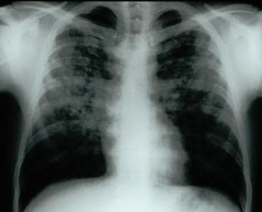
Frontal chest radiograph demonstrating bilateral reticular and nodular interstitial infiltrates with upper zone predominance.
Click to see larger pictureClick to see detailView Full Size Image
Media type:  X-RAY

Media file 2:  High-resolution chest CT scan of patient with bilateral reticular and nodular interstitial infiltrates with upper zone predominance.
Click to see larger pictureClick to see detailView Full Size Image
Media type:  CT


  • Alalawi R, Whelan T, Bajwa RS, et al. Lung transplantation and interstitial lung disease. Curr Opin Pulm Med. Sep 2005;11(5):461-6.
  • American Thoracic Society/European Respiratory Society. American Thoracic Society/European Respiratory Society International Multidisciplinary Consensus Classification of the Idiopathic Interstitial Pneumonias. This joint statement of the American Thoracic Society (ATS), and the European Respiratory Society. Am J Respir Crit Care Med. Jan 15 2002;165(2):277-304.
  • Azuma A, Nukiwa T, Tsuboi E. Double-blind, placebo-controlled trial of pirfenidone in patients with idiopathic pulmonary fibrosis. Am J Respir Crit Care Med. May 1 2005;171(9):1040-7. [Medline].
  • Demedts M, Behr J, Buhl R. High-dose acetylcysteine in idiopathic pulmonary fibrosis. N Engl J Med. Nov 24 2005;353(21):2229-42.
  • Hunninghake GW, Zimmerman MB, Schwartz DA, et al. Utility of a lung biopsy for the diagnosis of idiopathic pulmonary fibrosis. Am J Respir Crit Care Med. Jul 15 2001;164(2):193-6. [Medline].
  • Katzenstein AL, Myers JL. Idiopathic pulmonary fibrosis: clinical relevance of pathologic classification. Am J Respir Crit Care Med. Apr 1998;157(4 Pt 1):1301-15. [Medline].
  • King TE Jr, Schwarz MI, Brown K, et al. Idiopathic pulmonary fibrosis: relationship between histopathologic features and mortality. Am J Respir Crit Care Med. Sep 15 2001;164(6):1025-32. [Medline].
  • King TE Jr, Tooze JA, Schwarz MI, et al. Predicting survival in idiopathic pulmonary fibrosis: scoring system and survival model. Am J Respir Crit Care Med. Oct 1 2001;164(7):1171-81. [Medline].
  • Meier-Sydow J, Weiss S, Buhl R. Idiopathic pulmonary fibrosis: current clinical concepts and challenges in management. Semin Respir Crit Care Med. 1994;15:77-96.
  • Philit F, Etienne-MastroĂ¯anni B, Parrot A. Idiopathic acute eosinophilic pneumonia: a study of 22 patients. Am J Respir Crit Care Med. Nov 1 2002;166(9):1235-9. [Medline].
  • Raghu G, Brown KK, Bradford WZ. A placebo-controlled trial of interferon gamma-1b in patients with idiopathic pulmonary fibrosis. N Engl J Med. Jan 8 2004;350(2):125-33. [Medline].
  • Schwarz MI, King TE, Raghu G. Approach to the evaluation and diagnosis of interstitial lung disease. In: King TE, Schwarz MI, eds. Interstitial Lung Disease. Ontario, BC, Canada: BC Decker;2003:1-30.
  • Selman M, Pardo A, Barrera L. Gene expression profiles distinguish idiopathic pulmonary fibrosis from hypersensitivity pneumonitis. Am J Respir Crit Care Med. Jan 15 2006;173(2):188-98.

Pulmonary Fibrosis, Interstitial (Nonidiopathic) excerpt

Article Last Updated: Jun 16, 2006