Background
Cardiogenic pulmonary edema (CPE) is defined as pulmonary edema due to increased capillary hydrostatic pressure secondary to elevated pulmonary venous pressure. CPE reflects the accumulation of fluid with a low-protein content in the lung interstitium and alveoli as a result of cardiac dysfunction (see the image below). (See Etiology.)
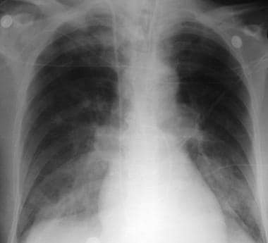 Radiograph shows acute pulmonary edema in a patient who was admitted with acute anterior myocardial infarction. Findings are vascular redistribution, indistinct hila, and alveolar infiltrates.
Pulmonary edema can be caused by the following major pathophysiologic mechanisms:
-
Imbalance of Starling forces - ie, increased pulmonary capillary pressure, decreased plasma oncotic pressure, increased negative interstitial pressure
-
Damage to the alveolar-capillary barrier
-
Lymphatic obstruction
-
Idiopathic (unknown) mechanism
Increased hydrostatic pressure leading to pulmonary edema may result from many causes, including excessive intravascular volume administration, pulmonary venous outflow obstruction (eg, mitral stenosis or left atrial [LA] myxoma), and LV failure secondary to systolic or diastolic dysfunction of the left ventricle. CPE leads to progressive deterioration of alveolar gas exchange and respiratory failure. Without prompt recognition and treatment, a patient's condition can deteriorate rapidly. (See Etiology, Prognosis, Presentation, Workup, Treatment, and Medication.)
Complications
The major complications associated with CPE are respiratory fatigue and failure. Prompt diagnosis and treatment usually prevent these complications, but the physician must be prepared to provide assisted ventilation if the patient begins to show signs of respiratory fatigue (eg, lethargy, fatigue, diaphoresis, worsening anxiety). (See Prognosis and Treatment.)
Sudden cardiac death secondary to cardiac arrhythmia is another concern, and continuous monitoring of heart rhythm is helpful in prompt diagnosis of dangerous arrhythmias.
Patient education
To help prevent recurrence of CPE, counsel and educate patients in whom pulmonary edema is due to dietary causes or medication noncompliance.
Etiology
CPE is caused by elevated pulmonary capillary hydrostatic pressure leading to transudation of fluid into the pulmonary interstitium and alveoli. Increased LA pressure increases pulmonary venous pressure and pressure in the lung microvasculature, resulting in pulmonary edema.
Mechanism of CPE
Pulmonary capillary blood and alveolar gas are separated by the alveolar-capillary membrane, which consists of three anatomically different layers: (1) the capillary endothelium; (2) the interstitial space, which may contain connective tissue, fibroblasts, and macrophages; and (3) the alveolar epithelium.
Exchange of fluid normally occurs between the vascular bed and the interstitium. Pulmonary edema occurs when the net flux of fluid from the vasculature into the interstitial space is increased. The Starling relationship determines the fluid balance between the alveoli and the vascular bed. Net flow of fluid across a membrane is determined by applying the following equation:
Q = K(Pcap - Pis) - l(Pcap - Pis),
where Q is net fluid filtration; K is a constant called the filtration coefficient; Pcap is capillary hydrostatic pressure, which tends to force fluid out of the capillary; Pis is hydrostatic pressure in the interstitial fluid, which tends to force fluid into the capillary; l is the reflection coefficient, which indicates the effectiveness of the capillary wall in preventing protein filtration; the second Pcap is the colloid osmotic pressure of plasma, which tends to pull fluid into the capillary; and the second Pis is the colloid osmotic pressure in the interstitial fluid, which pulls fluid out of the capillary.
The net filtration of fluid may increase with changes in different parameters of the Starling equation. CPE predominantly occurs secondary to LA outflow impairment or LV dysfunction. For pulmonary edema to develop secondary to increased pulmonary capillary pressure, the pulmonary capillary pressure must rise to a level higher than the plasma colloid osmotic pressure. Pulmonary capillary pressure is normally 8-12 mm Hg, and colloid osmotic pressure is 28 mm Hg. High pulmonary capillary wedge pressure (PCWP) may not always be evident in established CPE, because the capillary pressure may have returned to normal when the measurement is performed.
Lymphatics
The lymphatics play an important role in maintaining an adequate fluid balance in the lungs by removing solutes, colloid, and liquid from the interstitial space at a rate of approximately 10-20 mL/h. An acute rise in pulmonary arterial capillary pressure (ie, to >18 mm Hg) may increase filtration of fluid into the lung interstitium, but the lymphatic removal does not increase correspondingly. In contrast, in the presence of chronically elevated LA pressure, the rate of lymphatic removal can be as high as 200 mL/h, which protects the lungs from pulmonary edema.
Stages
The progression of fluid accumulation in CPE can be identified as three distinct physiologic stages.
Stage 1
In stage 1, elevated LA pressure causes distention and opening of small pulmonary vessels. At this stage, blood gas exchange does not deteriorate, or it may even be slightly improved.
Stage 2
In stage 2, fluid and colloid shift into the lung interstitium from the pulmonary capillaries, but an initial increase in lymphatic outflow efficiently removes the fluid. The continuing filtration of liquid and solutes may overpower the drainage capacity of the lymphatics. In this case, the fluid initially collects in the relatively compliant interstitial compartment, which is generally the perivascular tissue of the large vessels, especially in the dependent zones.
The accumulation of liquid in the interstitium may compromise the small airways, leading to mild hypoxemia. Hypoxemia at this stage is rarely of sufficient magnitude to stimulate tachypnea. Tachypnea at this stage is mainly the result of the stimulation of juxtapulmonary capillary (J-type) receptors, which are nonmyelinated nerve endings located near the alveoli. J-type receptors are involved in reflexes modulating respiration and heart rates.
Stage 3
In stage 3, as fluid filtration continues to increase and the filling of loose interstitial space occurs, fluid accumulates in the relatively noncompliant interstitial space. The interstitial space can contain up to 500mL of fluid. With further accumulations, the fluid crosses the alveolar epithelium in to the alveoli, leading to alveolar flooding. At this stage, abnormalities in gas exchange are noticeable, vital capacity and other respiratory volumes are substantially reduced, and hypoxemia becomes more severe.
Cardiac disorders manifesting as CPE
Atrial outflow obstruction
This can be due to mitral stenosis or, in rare cases, atrial myxoma, thrombosis of a prosthetic valve, or a congenital membrane in the left atrium (eg, cor triatriatum). Mitral stenosis is usually a result of rheumatic fever, after which it may gradually cause pulmonary edema. Other causes of CPE often accompany mitral stenosis in acute CPE; an example is decreased LV filling because of tachycardia in arrhythmia (eg, atrial fibrillation) or fever.
LV systolic dysfunction
Systolic dysfunction, a common cause of CPE, is defined as decreased myocardial contractility that reduces cardiac output. The fall in cardiac output stimulates sympathetic activity and blood volume expansion by activating the renin-angiotensin-aldosterone system, which causes deterioration by decreasing LV filling time and increasing capillary hydrostatic pressure.
Chronic LV failure is usually the result of congestive heart failure (CHF) or cardiomyopathy. Causes of acute exacerbations include the following:
-
Acute myocardial infarction (MI) or ischemia
-
Patient noncompliance with dietary restrictions (eg, dietary salt restrictions)
-
Patient noncompliance with medications (eg, diuretics)
-
Severe anemia
-
Sepsis
-
Thyrotoxicosis
-
Myocarditis
-
Myocardial toxins (eg, alcohol, cocaine, chemotherapeutic agents such as doxorubicin [Adriamycin], trastuzumab [Herceptin])
-
Chronic valvular disease, aortic stenosis, aortic regurgitation, and mitral regurgitation
LV diastolic dysfunction
Ischemia and infarction may cause LV diastolic dysfunction in addition to systolic dysfunction. With a similar mechanism, myocardial contusion induces systolic or diastolic dysfunction.
Diastolic dysfunction signals a decrease in LV diastolic distensibility (compliance). Because of this decreased compliance, a heightened diastolic pressure is required to achieve a similar stroke volume. Despite normal LV contractility, the reduced cardiac output, in conjunction with excessive end-diastolic pressure, generates hydrostatic pulmonary edema. Diastolic abnormalities can also be caused by constrictive pericarditis and tamponade.
Dysrhythmias
New-onset rapid atrial fibrillation and ventricular tachycardia can be responsible for CPE.
LV hypertrophy and cardiomyopathies
These can increase LV stiffness and end-diastolic pressure, with pulmonary edema resulting from increased capillary hydrostatic pressure.
LV volume overload
LV volume overload occurs in a variety of cardiac or noncardiac conditions. Cardiac conditions are ventricular septal rupture, acute or chronic aortic insufficiency, and acute or chronic mitral regurgitation. Endocarditis, aortic dissection, traumatic rupture, rupture of a congenital valve fenestration, and iatrogenic causes are the most important etiologies of acute aortic regurgitation that may lead to pulmonary edema.
Ventricular septal rupture, aortic insufficiency, and mitral regurgitation cause elevation of LV end-diastolic pressure and LA pressure, leading to pulmonary edema. LV outflow obstruction, such as that caused by aortic stenosis, produces increased end-diastolic filling pressure, increased LA pressure, and increased pulmonary capillary pressures.
Some sodium retention may occur in association with LV systolic dysfunction. However, in certain conditions, such as primary renal disorders, sodium retention and volume overload may play a primary role. CPE can occur in patients with hemodialysis-dependent renal failure, often as a result of noncompliance with dietary restrictions or noncompliance with hemodialysis sessions.
Myocardial infarction
One of the mechanical complications of MI can be the rupture of ventricular septum or papillary muscle. These mechanical complications substantially increase volume load in the acute setting and therefore may cause pulmonary edema.
LV outflow obstruction
Acute obstruction of the aortic valve can cause pulmonary edema. However, aortic stenosis due to a congenital disorder, calcification, prosthetic valve dysfunction, or rheumatic disease usually has a chronic course and is associated with hemodynamic adaptation of the heart. This adaptation may include concentric LV hypertrophy, which itself can cause pulmonary edema by way of LV diastolic dysfunction. Hypertrophic cardiomyopathy is a cause of dynamic LV outflow obstruction.
Elevated systemic blood pressure can be considered an etiology of LV outflow obstruction because it increases systemic resistance against the pump function of the left ventricle.
Prognosis
In-hospital mortality rates for patients with CPE are difficult to assign because the causes and severity of the disease vary considerably. In a high-acuity setting, in-hospital death rates are as high as 15-20%.
Myocardial infarction, associated hypotension, and a history of frequent hospitalizations for CPE generally increase the mortality risk.
Severe hypoxia may result in myocardial ischemia or infarction. Mechanical ventilation may be required if medical therapy is delayed or unsuccessful. Endotracheal intubation and mechanical ventilation are associated with their own risks, including aspiration (during intubation), mucosal trauma (more common with nasotracheal intubation than with orotracheal intubation), and barotrauma.
-
Radiograph shows acute pulmonary edema in a patient who was admitted with acute anterior myocardial infarction. Findings are vascular redistribution, indistinct hila, and alveolar infiltrates.
-
Radiograph shows acute pulmonary edema in a patient known to have ischemic cardiomyopathy. Findings are Kerley B lines (1mm thick and 1cm long) in the lower lobes and Kerley A lines in the upper lobes.
-
Radiograph demonstrates cardiomegaly, bilateral pleural effusions, and alveolar opacities in a patient with pulmonary edema.
-
Radiograph shows interstitial pulmonary edema, cardiomegaly, and left pleural effusion presenting at an earlier stage of pulmonary edema.
-
Lateral chest radiograph shows prominent interstitial edema and pleural effusions.


