Background
Babesiosis is a tick-borne, malaria-like illness caused by species of the intraerythrocytic protozoan Babesia. Humans are incidental hosts for Babesia when bitten by nymph or adult ticks. Babesia infection is most commonly seen in the north midwestern and northeastern United States. It can also be found throughout the world in certain parts of Europe, Asia, Africa, and South America. [1]
Human babesiosis is a zoonotic infection in which ticks transmit Babesia organisms from a vertebrate reservoir to humans [1, 2] ; humans are typically dead-end hosts. In the United States, most infections are caused by Babesia microti, a species commonly found in mice. Other species known to infect humans include B. duncani, B. divergens, B. venatorum, and B. crassa. [3]
Babesia species and organisms of the closely related genus Theileria parasitize the erythrocytes of wild and domestic animals.These parasites are members of the order Piroplasmida, named for the pear-shaped forms found within infected red blood cells (RBCs). [4]
Over 2000 cases of babesiosis were reported in the United States in 2018. [5] In healthy individuals, most infections are asymptomatic. Several groups of patients become symptomatic, and, within these subpopulations, significant morbidity and mortality occur. The disease most severely affects patients who are elderly, immunocompromised, or asplenic. [3]
Babesiosis can be difficult to diagnose. Although the index of suspicion should be high in areas endemic for Babesia infection, patients with babesiosis have few, if any, localizing signs to suggest the disease. Confirmation of the diagnosis depends on the degree of parasitemia and the expertise and experience of laboratory personnel. [4, 6]
Most patients infected by B. microti who are otherwise healthy appear to have a mild illness and typically recover without specific chemotherapy. Asymptomatic patients do not necessarily require treatment [7] ; the decision to treat should be an individualized one. For symptomatic cases, treatment is recommended. In addition, patients should be advised to take precautions against tick exposure and to refrain from donating blood until 2 years from the time of a reactive nucleic acid test result for Babesia. [6, 8]
Pathophysiology
Babesiosis is a zoonotic disease maintained by the interaction of tick vectors, transport hosts, and animal reservoirs. [9] The primary vectors of the parasite are ticks of the genus Ixodes. In the United States, the black-legged deer tick, Ixodes scapularis (also known as Ixodes dammini), is the primary vector for the parasite; in Europe, Ixodes ricinus appears to be the primary tick vector. [10] In each location, the Ixodes tick vector for Babesia is the same vector that locally transmits Borrelia burgdorferi, the agent implicated in Lyme disease.
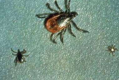 Ixodes scapularis, tick vector for babesiosis. Image courtesy of Centers for Disease Control and Prevention.
I. scapularis has 3 developmental stages—larva, nymph, and adult—each of which requires a blood meal for development into the next stage. As a larva and nymph, the tick primarily feeds on rodents (eg, the white-footed mouse, Peromyscus leucopus); however, as an adult, the tick prefers to feed on the white-tailed deer, the primary host in the United States. Female ticks are impregnated while obtaining their blood meal on the deer, with the formation of up to 20,000 eggs. In contrast, cattle constitute the primary animal reservoir in Europe.
The clinical signs and symptoms of babesiosis are related to the parasitism of RBCs by Babesia. The ticks ingest Babesia from the host during feeding; they then multiply the protozoa in their gut wall and concentrate them in their salivary glands. When they feed again on a new host, they inoculate the new host with Babesia.
Entering the host’s bloodstream during the tick bite, the parasite infects RBCs, producing differentiated and undifferentiated trophozoites. Upon infection of the host erythrocyte, mature B. microti trophozoites undergo asynchronous asexual budding and divide into 2 or 4 merozoites. As parasites leave the erythrocyte, the membrane is damaged. The precise mechanism of hemolysis is unknown.
Babesia species in the host erythrocyte range from 1 to 5 µm in length. B. microti measures 2 × 1.5 µm, B. divergens measures 4 × 1.5 µm, and B. bovis measures 2.4 × 1.5 µm. As noted, the organisms are pear-shaped, oval, or round. Their ring form and peripheral location in the erythrocyte frequently lead to their being mistaken for Plasmodium falciparum. However, they differ from P. falciparum in that the schizogony is asynchronous, and massive hemolysis does not occur.
Alterations in RBC membranes cause decreased conformability and increased RBC adherence, which can lead to development of noncardiogenic pulmonary edema and acute respiratory distress syndrome (ARDS) among those severely affected. [1, 11]
Fever, hemolytic anemia, and hemoglobinuria may result from Babesia infection. As with malaria, RBC fragments may cause capillary blockage or microvascular stasis, which could explain liver, splenic, renal, and central nervous system (CNS) involvement. Animal studies have shown that increased cytoadherence of infected RBCs could cause these vascular blockages, though further research is needed. [9] As in malaria, cells of the reticuloendothelial system (RES) in the spleen remove damaged RBC fragments from the circulation. RBC destruction results in hemolytic anemia. The amount of hemolysis does not seem to be directly related to the degree of parasitemia, though the cause is unclear. [11]
The spleen offers a critical host defense against babesiosis, as suggested by the higher incidence and greater severity of babesiosis in asplenic patients. The spleen traps the infected erythrocytes, and their ingestion by macrophages follows. Additionally, hypersplenism can lead to platelet sequestration which causes thrombocytopenia. [11]
Complement activation by Babesia may lead to the generation of tumor necrosis factor (TNF) and interleukin-1 (IL-1). Decreased complement levels, increased circulating C1q-binding activity, and decreased C4, C3, and CH50 levels are observed in patients with babesiosis. The generation of these primarily macrophage-produced mediators may explain many of the clinical features, including fever, anorexia, arthralgias, myalgias, and the fulminant shock syndrome of bovine babesiosis.
Babesiosis elicits a B-cell response and a T-cell response. Patients with acute babesiosis may have an increase in T-suppressor lymphocytes and/or T-cytotoxic lymphocytes and a decreased response to lymphocyte mitogens with a polyclonal hypergammaglobulinemia.
Etiology
Babesiosis is an infection caused by parasites of the Babesia genus. It is a zoonosis that is transmitted from vertebrates to humans through the bite of a tick from the Ixodidae family (most commonly I. scapularis in the United States, I. ricinus in Europe). Ixodes ticks are small and differ from the large Dermacentor ticks that transmit Rocky Mountain spotted fever (RMSF) and ehrlichiosis.
More than 100 species of Babesia exist, but only a small number of them are known to be responsible for the majority of symptomatic disease. The causative agent of babesiosis varies according to geographic region.
In the United States, human infection with Babesia is primarily due to the rodent strain B. microti, found mostly in northeastern and north midwestern states. A few cases have been reported in Missouri, California, and Washington. These are caused by Babesia-like agents named after their geographic location: MO-1 (Missouri, closely related to B. divergens), CA-1 (California), and WA-1 (Washington, also known as CA5 and B. duncani).
In Europe, the causative agent of babesiosis is typically the cattle strain B. divergens, though B. microti and B. microti-like agents have been identified. Another cattle strain found in Europe, B. bovis, also causes disease in humans on occasion. China and some European countries have also reported B. venatorum as a cause of babesiosis. There are multiple other species that are under investigation. [3]
Tick life cycle
The I. scapularis life cycle takes 2 years to complete, beginning with egg deposition in the spring. The white-footed mouse is the primary enzootic reservoir. After feeding on infected white-footed mice, the tick larvae become infected with B. microti. The tick then develops from the larval phase to the nymphal phase. This development takes 1 year (ie, until the next spring).
Nymphs infected with B. microti may transmit the Babesia organisms to other mice or rodents or to a human host. Nymphs feed for 3-4 days on white-footed mice or other rodents and mature into adults the following fall.
Adult Ixodes tick populations are maintained in white-tailed deer. The adults mate and feed on the deer during the spring; they then deposit their eggs and die. Although rodents are infected with Babesia, the white-tailed deer does not carry the organism. B microti is transmitted from the larval phase of I. scapularis to the nymphal phase (transstadial transmission) but not transovarially. The white-footed mouse is necessary to perpetuate the Babesia organisms, and the deer is needed to perpetuate the Ixodes tick population.
Larvae, nymphs, and adult ticks may all infect humans, but the nymph is the primary vector of B. microti infection.
Risk factors
Babesia parasites from rodents (primarily the white-footed deer mouse but also the field mouse, vole, rat, and chipmunk) are transmitted to humans through tick bites in endemic areas. As such, babesiosis is more prevalent during the periods of high tick activity, such as spring and summer. Restocking of deer populations and curtailment of hunting has increased deer herds in certain areas. The proximity of deer, mouse, and tick creates the conditions for human infection.
Several reported cases of infection via blood transfusions from donors who lived in or traveled to an endemic area have been documented. [12, 13] The incubation period in transfusion-associated disease appears to be 6-9 weeks. The rate of acquiring B. microti from a unit of packed RBCs has been estimated to be 1 in 600-1800 in endemic areas.
Case reports of transplacental or perinatal transmission have also been documented. [3]
Epidemiology
United States statistics
Human babesiosis is endemic in the northeastern coastal region of the United States, particularly Nantucket Island, Martha’s Vineyard, and Cape Cod, Massachusetts; Block Island, Rhode Island; and eastern Long Island, [14] Shelter Island, and Fire Island, New York. Disease prevalence in Cape Cod, as suggested by antibodies to B. microti, has been reported as 3.7%, whereas on Shelter Island in individuals with a high risk of exposure to ticks, it was 4.4% in June and reached 6.9% by October.
Babesiosis was a reportable condition in 40 out of 50 states in 2018. Of those, 28 states reported cases of babesiosis during that year. Most (86%) of the 2,161 cases were reported by 7 states: Connecticut, Massachusetts, New Jersey, Rhode Island, New York, Minnesota, and Wisconsin. [5]
The incidence of babesiosis has increased over the past 20 years. [15] This is thought to be the result of restocking of the deer population, curtailment of hunting, and an increase in outdoor recreational activities; however, the prevalence of this disease is unknown because most infected patients are asymptomatic.
In endemic areas, the organism may also be transmitted by blood transfusion. [3, 16, 17, 18, 19, 20, 21]
International statistics
Babesiosis is occasionally seen in areas of Europe and Asia where the tick vector and vertebrate host reside. [22] Since 1957, when the first case of human babesiosis was reported in an asplenic farmer from the former Yugoslavia, approximately 53 cases have been reported, mostly in Ireland, the United Kingdom, and France. The majority of the cases involved bovine Babesia and occurred in individuals who were splenectomized. [15]
Sporadic case reports of babesiosis in Japan, Korea, China, Mexico, Colombia, South Africa, and Egypt have also been documented.
Age-, sex-, and race-related demographics
Although persons of any age can be affected by babesiosis, clinically ill patients with intact spleens are usually aged 50 years or older, suggesting that age plays a factor in the severity of disease. Patients with babesiosis who were previously healthy individuals are generally older (mean, >60 years) than those who had previous medical problems (mean, 48 years). Vannier et al suggested that the age-associated decline in resistance to B. microti is genetically determined. [1, 2]
Babesiosis has no known predilection for sex or race.
Prognosis
Babesiosis has a spectrum of severity, which may be divided into 3 distinct parts as follows [1] :
· Asymptomatic infection
· A mild-to-moderate viral-like syndrome
· Severe disease with a fulminant course resulting in death or a persistent relapsing course
Babesiosis in otherwise healthy hosts may produce an acute infectious condition that resembles malaria. However, most cases of babesiosis are subclinical or only mildly symptomatic. In the United States, the prognosis for babesiosis is excellent; most patients recover spontaneously. About 25% of adults and 50% of children infected with Babesia are asymptomatic and improve spontaneously without treatment. Fewer than 10% of US patients with babesiosis have died; most of these have been elderly or asplenic.
In patients who are asplenic, babesiosis can be quite severe and is associated with substantial mortality. Asplenic patients tend to have a more fulminant and prolonged clinical course. [23] Highly immunocompromised patients are at a higher risk for complications such as acute respiratory distress syndrome, shock, warm autoimmune hemolytic anemia, heart failure, and death. [3] In a 1998 review by White et al, 9 of 139 (6.5%) patients who were hospitalized with babesiosis in New York State from 1982-1983 died. [24]
Immunocompromised patients are at a higher risk of relapse, especially if they have HIV with acquired immunodeficiency syndrome (AIDS), if they are transplant patients on immunosuppression, if they are receiving rituximab, or if they have malignancy and asplenia. [3]
In Europe, babesiosis often comes from B. divergens, which can cause life-threatening infection. About 83% of infected European patients are asplenic, contributing to a poor prognosis. More than 50% of patients with babesiosis in Europe become comatose and die.
Deaths have been reported from transfusion-transmitted babesiosis within the immunocompromised population in areas where Babesia infection is not endemic. [25] The mortality rate of B.microti infection from a transfusion is about 20%. [3]
Approximately 10-20% of patients with babesiosis are co-infected with Lyme disease. [3] The symptoms experienced by these patients are more severe and prolonged than symptoms experienced by patients who have either disease alone. Babesia co-infection should be considered when a patient with Lyme disease does not respond to typical therapy or when a patient with Lyme disease has atypical symptoms.
-
Peripheral smear showing babesiosis.
-
Ixodes scapularis, tick vector for babesiosis. Image courtesy of Centers for Disease Control and Prevention.
-
Blood smear showing Babesia species in erythrocytes. Image courtesy of Centers for Disease Control and Prevention.
-
Babesia species, tetrad formation. Image courtesy of Centers for Disease Control and Prevention.