Continually Updated Clinical Reference
 
 
  All Sources     eMedicine     Medscape     Drug Reference     MEDLINE
 
eMedicine - Azotemia : Article by

Quick Find
Authors & Editors
Introduction
Clinical
Differentials
Workup
Treatment
Medication
Follow-up
Miscellaneous
Multimedia
References

Related Articles
Acute Tubular Necrosis

Chronic Renal Failure

Glomerulonephritis, Acute

Glomerulonephritis, Chronic

Nephritis, Interstitial

Obstructed Megaureter

Uremia


Patient Education
Click here for patient education.


Author: Moro O Salifu, MD, MPH, Director, Nephrology Fellowship Program, Associate Professor, Department of Internal Medicine, Division of Nephrology, SUNY Downstate Medical Center

Moro O Salifu is a member of the following medical societies: American College of Physicians-American Society of Internal Medicine, American Medical Association, American Society for Artificial Internal Organs, American Society of Nephrology, and National Kidney Foundation

Coauthor(s): Onyekachi Ifudu, MD, Director of Inpatient Dialysis Services, Associate Professor, Department of Internal Medicine, State University of New York Health Science Center at Brooklyn

Editors: Frank C Brosius III, MD, Nephrology Program Director, Department of Internal Medicine, Division of Nephrology, Professor of Internal Medicine and Physiology, University of Michigan School of Medicine; Francisco Talavera, PharmD, PhD, Senior Pharmacy Editor, eMedicine; George R Aronoff, MD, Director, Professor, Departments of Internal Medicine and Pharmacology, Section of Nephrology, Kidney Disease Program, University of Louisville School of Medicine; Rebecca J Schmidt, DO, FACP, FASN, Clinical Associate Professor of Medicine, West Virginia School of Osteopathic Medicine; Professor of Medicine, Section Chief, Department of Medicine, Section of Nephrology, West Virginia University School of Medicine; Vecihi Batuman, MD, FACP, FASN, Chief, Medical Service, VA Medical Center, New Orleans, Professor of Medicine, Tulane University School of Medicine; Chief, Renal-Hypertension Section, Department of Medicine, Tulane University Medical Center, New Orleans Veterans Affairs Medical Center

Author and Editor Disclosure

Synonyms and related keywords: elevated urea nitrogen, elevated creatinine, acute renal failure, ARF, chronic renal failure, chronic kidney disease, CKD, uremia


Background

Each human kidney contains approximately 1 million functional units, called nephrons, which are primarily involved in formation. Formation ensures that the body eliminates the final products of metabolic activities and excess water in an attempt to maintain a constant internal environment (homeostasis).

Urine formation by each nephron involves 3 main processes, as follows: filtration at the glomerular level, selective reabsorption from the filtrate passing along the renal tubules, and secretion by the cells of the tubules into this filtrate. Perturbation of any of these processes impairs the kidney's excretory function, resulting in azotemia, which is elevation of blood urea nitrogen (BUN) (reference range, 8-20 mg/dL) and serum creatinine (normal value, 0.7-1.4 mg/dL) levels.

The quantity of glomerular filtrate produced each minute by all nephrons in both kidneys is referred to as the glomerular filtration rate (GFR). Average GFR is about 125 mL/min (10% less for women) or 180 L/d. About 99% (178 L/d) is reabsorbed, and the rest (2 L/d) is excreted.

Measuring renal function

Radionuclide assessment of GFR is the criterion standard for measuring kidney function. However, because it is expensive and not widely available, serum creatinine concentration and creatinine clearance (CrCl) more commonly are used.

An inverse relationship between serum creatinine and GFR exists. However, the serum creatinine and CrCl are not sensitive measures of kidney damage for two reasons. First, substantial renal damage can take place before any decrease in GFR occurs. Second, a substantial decline in GFR may lead to only slight elevation in serum creatinine, as shown in Media file 1. An elevation in serum creatinine is apparent only when the GFR falls to about 60-70 mL/min.
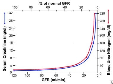
 This is due to compensatory hypertrophy and hyperfiltration of the remaining healthy nephrons.

Because creatinine normally is filtered as well as secreted into the renal tubules, the CrCl may cause the GFR to be substantially overestimated, especially as kidney failure progresses because of maximal tubular excretion. More accurate determinations of GFR require the use of inulin clearance or a radiolabeled compound, such as iothalamate. In practice, precise knowledge of the GFR is not required, and disease process usually can be monitored by the estimated GFR (eGFR) using different methods, as shown below. 

The CrCl is best calculated by obtaining a 24-hour collection for creatinine and volume and then using the following formula: CrCl (mL/min) = U/P X V where U is the 24-hour creatinine in mg/dL, P is the serum creatinine in mg/dL, and V is the 24-hour volume/1440 (number of min in 24 h). Using the 24-hour creatinine in grams and the serum creatinine in milligrams, CrCl (mL/min) = creatinine [g/d]/serum creatinine [mg/dL]) X 70. An adequate 24-hour collection usually reflects a creatinine generation of 15-20 mg/kg in women and 20-25 mg/kg in men. When 24-hour creatinine is measured, the adequacy of the collection must be established prior to calculation of the creatinine clearance.

Alternatively, a bedside formula (Cockroft and Gault) using the patient's serum creatinine, age, and lean weight (in kg) can be used to estimate the GFR, as follows: CrCl (mL/min) = (140 - age) X weight (kg) / (72 X serum creatinine) in mg/dL (X 0.85 for women).

Another formula was derived from data collected in a large study called the Modification of Diet in Renal Disease (MDRD). This formula is known as the MDRD formula or the Levey formula. It is now widely accepted as more accurate than the Cockroft and Gault formula and is an alternative to radioisotope clearance. Because serum creatinine levels alone cannot detect earlier stages of chronic kidney disease (CKD), the MDRD formula also takes into account the patient's age and race. Although more accurate, it is much more difficult to calculate manually. However, software for estimating GFR by the MDRD formula is available on most pocket digital assistants (PDA) or can be found on the Internet.

Pathophysiology

There are three pathophysiologic states in azotemia, as follows: prerenal azotemia, intrarenal azotemia, and postrenal azotemia. 

Prerenal azotemia

Prerenal azotemia refers to elevation in BUN and creatinine levels because of problems in the systemic circulation that decrease flow to the kidneys. In prerenal azotemia, decrease in renal flow stimulates salt and water retention to restore volume and pressure. When volume or pressure is decreased, the baroreceptor reflexes located in the aortic arch and carotid sinuses are activated. This leads to sympathetic nerve activation, resulting in renal afferent arteriolar vasoconstriction and renin secretion through b1-receptors. Constriction of the afferent arterioles causes a decrease in the intraglomerular pressure, reducing GFR proportionally. Renin converts angiotensin I to angiotensin II, which, in turn, stimulates aldosterone release. Increased aldosterone levels results in salt and water absorption in the distal collecting tubule.

A decrease in volume or pressure is a nonosmotic stimulus for antidiuretic hormone production in the hypothalamus, which exerts its effect in the medullary collecting duct for water reabsorption. Through unknown mechanisms, activation of the sympathetic nervous system leads to enhanced proximal tubular reabsorption of salt and water, as well as BUN, creatinine, calcium, uric acid, and bicarbonate. The net result of these 4 mechanisms of salt and water retention is decreased output and decreased urinary excretion of sodium (<20 mEq/L).

Intrarenal azotemia

Intrarenal azotemia, also known as acute renal failure (ARF), renal-renal azotemia, and acute kidney injury (AKI), refers to elevation in BUN and creatinine levels because of problems in the kidney itself. There are several definitions, including a rise in serum creatinine levels of about 30% from baseline or a sudden decline in output below 500 mL/d. If output is preserved, it is called nonoliguric ARF. If output falls below 500 mL/d, it is called oliguric ARF. Any form of ARF may be so severe to virtually stop formation, a condition called anuria (<100 mL/d).

The most common causes of nonoliguric ARF are acute tubular necrosis (ATN), aminoglycoside nephrotoxicity, lithium toxicity, or cisplatin nephrotoxicity. Tubular damage is less severe than in oliguric ARF. Normal output in nonoliguric ARF does not reflect normal GFR. Patients may still make 1440 mL/d of urine even when the GFR falls to about 1 mL/min because of decreased tubular reabsorption.

Some studies indicate that nonoliguric forms of ARF are associated with less morbidity and mortality than oliguric ARF. Uncontrolled studies also suggest that volume expansion, potent diuretic agents, and renal vasodilators can convert oliguric to nonoliguric ARF if administered early.

The pathophysiology of acute oliguric or nonoliguric ARF depends on the anatomical location of the injury. In ATN, epithelial damage leads to functional decline in the ability of the tubules to reabsorb salt, water, and other electrolytes. Excretion of acid and potassium also is impaired. In more severe ATN, the tubular lumen is filled with epithelial casts, causing intraluminal obstruction, resulting in the decline of GFR.

Acute interstitial nephritis is characterized by inflammation and edema, resulting in azotemia, hematuria, sterile pyuria, white cell casts with variable eosinophiluria, proteinuria, and hyaline casts. The net effect is a loss of urinary concentrating ability, with low osmolality (usually <500 mOsm/L), low specific gravity (<1.015), high urinary sodium (>40 mEq/L), and, occasionally, hyperkalemia and renal tubular acidosis. However, in the presence of a superimposed prerenal azotemia, the specific gravity, osmolality, and sodium may be misleading.

Glomerulonephritis or vasculitis is suggested by the presence of hematuria, red cells, white cells, granular and cellular casts, and a variable degree of proteinuria. Nephrotic syndrome usually is not associated with active inflammation and often presents as proteinuria greater than 3.5 g/24 h.

Glomerular diseases may reduce GFR due to changes in basement membrane permeability, as well as stimulation of the renin-aldosterone axis. Glomerular diseases often manifest as nephrotic or nephric syndrome. In nephrotic syndrome, the urinary sediment is inactive, and there is gross proteinuria (>3.5 g/d), hypoalbuminemia, hyperlipidemia, and edema. Azotemia and hypertension are uncommon initially, but their presence may indicate advanced disease.

In nephritic syndrome, the urinary sediment is active with white or red cell casts, granular casts, and azotemia. Proteinuria is less obvious, but increased salt and water retention in glomerulnephritis can lead to hypertension, edema formation, decreased output, low urinary excretion of sodium, and increased specific gravity.

Acute vascular diseases include vasculitis syndromes, malignant hypertension, scleroderma renal crisis, and thromboembolic disease, all of which cause renal hypoperfusion and ischemia leading to azotemia. Chronic vascular diseases are due to hypertensive benign nephrosclerosis, which has not been conclusively associated with end-stage renal disease and ischemic renal disease from bilateral renal artery stenosis.

In bilateral renal artery stenosis, maintenance of adequate intraglomerular pressure for filtration greatly depends on efferent arteriolar vasoconstriction. Azotemia sets in when angiotensin-converting enzyme (ACE) inhibitors or angiotensin type 2 receptor blockers cause efferent arteriolar dilatation, thereby decreasing intraglomerular pressure and filtration. Therefore, converting enzyme inhibitors and receptor blockers are contraindicated in bilateral renal artery stenosis.

In addition to accumulation of urea creatinine and other waste products, a substantial reduction in GFR in CKD results in decreased production of erythropoietin (causing anemia) and vitamin D-3 (causing hypocalcemia, secondary hyperparathyroidism, hyperphosphatemia, and renal osteodystrophy); reduction in acid, potassium, salt, and water excretion (causing acidosis, hyperkalemia, hypertension, and edema); and platelet dysfunction, which leads to increased bleeding tendencies.

The syndrome associated with the signs and symptoms of accumulation of toxic waste products (uremic toxins) is termed uremia and often occurs at a GFR of about 10 mL/min. Some of the uremic toxins (ie, urea, creatinine, phenols, guanidines) have been identified, but none has been found responsible for all the manifestations of uremia.

Postrenal azotemia

Postrenal azotemia refers to elevation in BUN and creatinine levels because of obstruction in the collecting system. Obstruction to flow leads to a reversal of Starling forces responsible for glomerular filtration. Progressive bilateral obstruction causes hydronephrosis with an increase in the Bowman capsular hydrostatic pressure and tubular blockage resulting in progressive decline and ultimate cessation in glomerular filtration, azotemia, acidosis, fluid overload, and hyperkalemia.

Unilateral obstruction rarely causes azotemia. With relief of complete ureteral obstruction within 48 hours of onset, there is evidence that relatively complete recovery of GFR can be achieved within a week, while little or no further recovery occurs after 12 weeks. Complete or prolonged partial obstruction can lead to tubular atrophy and irreversible renal fibrosis. Hydronephrosis may be absent if obstruction is mild or acute or if the collecting system is encased by retroperitoneal tumor or fibrosis.

Frequency

United States

Considerable variability exists in reports about the incidence of hospital or community-acquired ARF. In one report, community-acquired ARF occurred in about 1% of all hospital admissions. Overall, ARF occurs in about 5% of all hospital admissions. However, differences exist in ARF occurring in the intensive care unit (about 15%) and in the coronary care unit (about 4%). In CKD, progressive azotemia leading to end-stage renal disease requiring dialysis or kidney transplantation occurs in a number of chronic diseases with frequencies for diabetes (36%), hypertension (24%), glomerulonephritis (15%), cystic kidney disease (4%), uncertain (5%), and all other known miscellaneous renal disorders (15%).

International

A report from Madrid evaluated 748 cases of ARF at 13 tertiary hospital centers. The most frequent causes were ATN (45%); prerenal (21%); acute or chronic renal failure, mostly due to ATN and prerenal disease (13%); urinary tract obstruction (10%); glomerulonephritis or vasculitis (4%); acute interstitial nephritis (2%); and atheroemboli (1%). Etiologies of CKD differ around the world. Diabetic nephropathy as a cause of CKD is on the rise in developed and developing countries.

Mortality/Morbidity

  • Prognosis in ARF generally is poor and depends on the severity of the underlying disease and the number of failed organs. While mortality rate in simple ARF without other underlying disease is 7-23%, the mortality in the patient in the intensive care unit on mechanical ventilation is as high as 80%.
  • The prognosis of patients with CKD depends on the etiology of the failure. Patients with diabetic kidney disease, hypertensive nephrosclerosis, and ischemic nephropathy (ie, large-vessel arterial occlusive disease) tend to have progressive azotemia resulting in end-stage renal disease. Different types of glomerulonephritis have major differences in prognosis, with some being quite benign and rarely progressing to end-stage renal disease, whereas others have rapid progression to end-stage renal disease within months. About 50% of patients with polycystic kidney disease progress to end-stage renal disease by the fifth or sixth decade of life.

Race

In the 2006 annual report of the United States Renal Data System (USRDS), more than 500,000 patients with end-stage renal disease were receiving dialysis or a kidney transplant in the United States. Racial distribution was reported as Asian/Pacific Islander (4.0%), black (33.0%), white (61.0%), American Indian (1.3%), and other/unknown (1.7%).

Sex

Of the patients reported in the 2006 annual report of the USRDS, male frequency is 56.0% and female frequency is 44.0%.

Age

Of the patients reported in the 2006 annual report of the USRDS, frequencies for patients aged 0-19 years is 1%; aged 20-44 years, 17.0%; aged 45-64 years, 41.0%; aged 65-74 years, 22.0%; and older than 75 years, 18.0%.


History

It is necessary to quickly establish if azotemia is acute or chronic and whether it is due to prerenal, intrarenal, or postrenal causes. This is vital in initiating treatment and in preventing progression.

Clinical evaluation requires a thorough history, physical examination, and specific laboratory tests. Tests include serologies, urinalysis, and, if indicated, radiologic studies and kidney biopsy.

  • Prerenal azotemia: History of diarrhea, vomiting, profound heat exhaustion, excessive sweat loss, concurrent illness that impairs patient's ability to eat and drink adequately, hemorrhage, liver disease, congestive heart failure, and polyuria (eg, caused by lithium intoxication, diuretics, diabetes, diabetes insipidus)
  • Intrarenal azotemia
    • History of nocturia, polyuria, proteinuria, shock, and edema
    • Personal or family history of congenital or systemic diseases, especially diabetes, hypertension, systemic lupus erythematosus, other collagen vascular diseases, hepatitis B (HBV), hepatitis C (HCV), syphilis, multiple myeloma, and AIDS
    • Detailed medication history (looking for nephrotoxic medications, especially antibiotics, NSAIDs, ACE inhibitors, diuretics, and herbal remedies), chemical exposure, and intravenous drug abuse (exposure to HIV, HBV, and HCV infections)
  • Postrenal azotemia: Renal colic, dysuria, frequency, hesitancy, urgency incontinence, pelvic malignancy or irradiation, and benign prostatic hypertrophy

Physical

Physical examination should be detailed but focused on signs of high diagnostic yield.

  • Prerenal azotemia: Look for tachycardia; orthostatic hypotension (systolic BP drop of >20 mm Hg, diastolic BP drop of >10 mm Hg or more from the supine to standing position); hypotension; signs of dehydration, including dry mucous membranes, loss of skin turgor, and loss of axillary sweat; and signs of congestive heart failure or hepatic insufficiency.
  • Intrarenal azotemia: Look for hypertension and its end organ effects, such as hypertensive retinopathy and left ventricular hypertrophy (apical impulse displaced lateral to midclavicular line), rash, joint swelling or tenderness, needle tracks, hearing abnormality, palpable kidneys, abdominal bruits, pericardial rub, and asterixis. The latter 2 signs are suggestive of uremia. The presence of uremic pericarditis requires immediate dialysis.
  • Postrenal azotemia: A palpable bladder that is dull to percussion and a rectal or pelvic mass on digital examination is suggestive of postrenal azotemia (obstruction).

Causes

Causes are broadly classified as prerenal, intrarenal, and postrenal.

  • Prerenal azotemia
    • Prerenal azotemia occurs as a result of impaired renal blood flow or decreased perfusion resulting from decreased blood volume, decreased cardiac output (congestive heart failure), decreased systemic vascular resistance, decreased effective arterial volume from sepsis or hepatorenal syndrome, and renal artery abnormalities.
    • It may be superimposed on a background of chronic renal failure.
    • Iatrogenic causes of prerenal azotemia, such as excessive diuresis and treatment with ACE inhibitors, should be ruled out.
  • Intrarenal azotemia
    • Intrarenal azotemia occurs as a result of injury to the glomeruli, tubules, interstitium, or small vessels.
    • It may be acute oliguric, acute nonoliguric, or chronic.
    • The presence of systemic disease, nocturia, proteinuria, loss of urinary concentrating ability (low urine specific gravity), anemia, and hypocalcemia are suggestive of chronic intrarenal azotemia.
  • Postrenal azotemia
    • Postrenal azotemia occurs when an obstruction to urine flow is present.
    • It is observed in bilateral ureteral obstruction from tumors or stones, retroperitoneal fibrosis, neurogenic bladder, and bladder neck obstruction from prostatic hypertrophy or carcinoma and posterior urethral valves.
    • It may be superimposed on a background of chronic renal failure.


Acute Tubular Necrosis
Chronic Renal Failure
Glomerulonephritis, Acute
Glomerulonephritis, Chronic
Nephritis, Interstitial
Obstructed Megaureter
Uremia

Other Problems to be Considered

Causes of elevated BUN or creatinine levels that are unrelated to kidney function are GI hemorrhage, large protein meal, total parenteral nutrition (TPN), steroids, and ketoacidosis.

Therapy with medications, such as trimethoprim, cimetidine, cefoxitin, and flucytosine, should be considered, as these medications impair creatinine excretion.


Lab Studies

  • Obtain CBC count, biochemical profile, urinalysis, and urine electrolytes for the initial evaluation. In addition to establishing the presence of systemic disease, clues for the origin of azotemia may emerge from these tests. Diagnostic indices commonly used to differentiate prerenal from intrarenal or postrenal azotemia are summarized in Media file 2.
  • Prerenal azotemia
    • In prerenal azotemia, hemoconcentration results in elevation of hematocrit, total protein/albumin, calcium, bicarbonate, and uric acid from their baselines.
    • Oliguria (urine volume is <500 mL/d), anuria (<100 mL/d), high urine specific gravity (>1.015), normal urinary sediment, and low urinary sodium (<20 fractional excretion of sodium <1.0%) are seen.
    • When volume depletion is predominant, exaggerated proximal tubular reabsorption results in azotemia, hypernatremia, elevated levels of calcium, uric acid, and bicarbonate, while hemoconcentration results in the elevation of total protein, albumin, and hematocrit levels from their baselines. When hypoperfusion due to decreased cardiac output or effective arterial volume is present, patients exhibit edema, hyponatremia, and hypoalbuminemia. The hematocrit, calcium, uric acid, and bicarbonate levels vary widely in this category. These patients often are critically ill.
  • Intrarenal azotemia
    • Anemia, thrombocytopenia, hypocalcemia, and high anion gap metabolic acidosis may suggest intrarenal azotemia.
    • Low urine specific gravity (<1.015), active urinary sediment (see Pathophysiology), high urinary sodium (>40 mEq/L), fractional excretion of sodium (FENa) (>5%), plasma BUN–creatinine ratio (<20), and low urine osmolality may suggest intrarenal azotemia.
    • For patients with long-standing CKD, a renal sonogram usually shows small, contracted kidneys. Some causes of CKD can be associated with normal or large-sized kidneys, such as HIV nephropathy, diabetes, and renal amyloidosis. The renal sonogram usually is diagnostic for patients with polycystic kidney disease. In patients with active urinary sediment, progressive azotemia, proteinuria, and/or normal-sized kidneys on sonogram, a renal biopsy should be considered. Consultation with a nephrologist is imperative in all such patients.
  • Postrenal azotemia
    • Urinary indices in postrenal azotemia due to complete bilateral obstruction are usually nondiagnostic. The prima facie finding here is anuria and, occasionally, hypertension. Urine output still may be present if overflow (in bladder outlet obstruction) or partial ureteral obstruction is present.
    • A Foley catheter should be inserted as part of the initial evaluation to rule out obstruction below the bladder outlet. Unilateral ureteral obstruction rarely leads to azotemia; it occurs acutely (due to obstruction from calculi, papillary necrosis, or hematoma), producing renal colic, or may be chronic and asymptomatic, producing hydronephrosis.
    • Bilateral partial obstruction may be associated with azotemia in the presence of normal urine output. When patients are subjected to maneuvers that increase urinary flow (eg, diuretic renogram, perfusion pressure flow studies), they may exhibit an increase in size or pressure of the collecting system or experience pain.
    • In addition to azotemia, polyuria due to loss of concentrating ability and type 1 renal tubular acidosis, with hyperkalemia, hypercalcemia from metastatic pelvic tumor, and elevated prostate-specific antigen (PSA), may be clues to postrenal azotemia. Hydronephrosis in the absence of hydroureter may be seen in early (<3 d) obstruction, retroperitoneal process, or partial obstruction. Renal sonogram is the test of choice to rule out obstructive uropathy. If renal sonogram is equivocal, a lasix washout scan, as discussed below in Imaging Studies, should be performed.

Imaging Studies

  • Plain film of abdomen
    • If symptoms suggest nephrolithiasis, a plain film of the abdomen is performed to screen for presence of a radiopaque stone.
    • Calcium-containing, struvite, and cystine stones can be identified, but radiolucent ones, such as uric acid stones, will be missed.
  • Renal sonogram is the most commonly used renal imaging study because of its ease of use and broad application for the following:
    • Renal sonogram can determine renal size, which is important when considering renal biopsy. Small kidneys (<9 cm) may be suggestive of scarring from advanced renal disease, whereas normal or large kidneys with smooth contours may indicate a potentially reversible process.
    • It can differentiate cystic lesions from solid lesions.
    • It is a test of choice for diagnosing urinary tract obstruction.
    • Renal sonogram can detect kidney stones.
  • Doppler renal ultrasonography can be used to evaluate renal vascular flow (eg, renal vein thrombosis, renal infarction, renal artery stenosis).
  • Intravenous pyelogram
    • The risk of contrast nephrotoxicity should be weighed against the benefits of making a diagnosis that will not change management.
    • Intravenous pyelogram can provide detailed information concerning calyceal anatomy and the size and shape of the kidney.
    • It is extremely useful for detecting renal stones.
    • It is the preferred technique in the evaluation and diagnosis of certain structural disorders, such as chronic pyelonephritis, medullary sponge kidney, and papillary necrosis.
    • It can provide data on the degree of obstruction.
  • CT scan is complementary to ultrasonography, especially when the diagnosis is uncertain. Contrast nephrotoxicity should be weighed against the benefits. CT scan does the following:
    • It can be used to distinguish (in most cases) neoplastic lesions from simple cyst and is the criterion standard for radiologic diagnosis of renal stone disease, including radiolucent stones.
    • It can be used to evaluate and stage renal cell carcinoma and to diagnose renal vein thrombosis. It can diagnose polycystic kidney disease with higher sensitivity than ultrasonography, particularly in younger patients.
  • Magnetic resonance imaging or magnetic resonance angiography
    • MRI or magnetic resonance angiography (MRA) is used only when CT scanning and ultrasonography are nondiagnostic.
    • They are the criterion standard for diagnosis of renal vein thrombosis.
    • They are used in the evaluation of renal cell carcinoma and renal artery stenosis or vasculitis.
  • Renal arteriography
    • The availability of procedures not requiring contrast material (ie, sonography, MRI, MRA) and the risk of contracting contrast nephrotoxicity have reduced use of this test.
    • Renal arteriography is used in polyarteritis nodosa and renal artery stenosis to demonstrate multiple aneurysms and/or stenoses.
  • Renal venography
    • Risk of contrast nephrotoxicity is present.
    • Renal venography is the criterion standard for diagnosis of renal vein thrombosis.
  • Radionuclide studies
    • Technetium dimercaptosuccinic acid (Tc 99m DMSA) is used to detect renal scarring.
    • Technetium diethylenetriamine pentaacetic acid (Tc 99m DTPA) is used for quantitative assessment of renal function (perfusion, filtration, and excretion).
  • Voiding cystourethrogram can be performed with a radionuclide study to detect vesicoureteral reflux.
  • Retrograde or anterograde pyelography
    • This test has limited use because of the availability of sonography.
    • It may be used in patients with a high index of suspicion for hydronephrosis for whom sonogram results are normal, such as in retroperitoneal fibrosis.
  • Lasix washout scan
    • The two agents used in performing lasix washout renal scans are technetium diethylenetriamine-pentaacetic acid (DTPA) and mercaptoacetyltriglycine (MAG3).
    • Usually, the renal scan is performed first. Then, if needed, the lasix washout is performed after the radionuclide has accumulated in the collecting system. Lasix is used as a part of the renogram to separate nonobstructive hydronephrosis from obstructive hydronephrosis. If there is no obstruction, flow induced by lasix and containing little or no radionuclide will fill the collecting system, washing out urine that contains radionuclide from the system. However, in the presence of obstruction, the radionuclide is not washed out as quickly.
    • The half life or clearance of the radioisotope is plotted on a curve. A half life of less than 10 minutes is considered normal, of greater than 20 minutes is considered obstruction, and of between 10-20 minutes is subject to further interpretation.
    • Conditions that can make it difficult to interpret the lasix washout curve include a megaureter or pelvis that accepts a large bolus of urine and poor renal function. In patients with a megaureter, it can be difficult to determine when the renal pelvis is full, and, in patients with renal disease, lasix onset of action may be prolonged. To overcome the problem of poor renal function or relative hypovolemia if a patient has been fasting, the patient should be well hydrated with IV fluids prior to the study.
    • The test also is operator dependent, as the lasix should be administered when it is thought that the renal pelvis is full. A full bladder also delays washout of isotope. Therefore, the patient's bladder needs to be catheterized before the study can be performed.

Procedures

  • Renal biopsy is indicated when glomerulonephritis, vasculitis, and, occasionally, interstitial nephritis are suspected to establish correct diagnosis and to guide therapy. The following are common indications for renal biopsy:
    • Isolated glomerular proteinuria or hematuria
    • Nephrotic syndrome
    • Acute nephritic syndrome
    • Unexplained acute or subacute renal failure
  • Percutaneous renal biopsy complications
    • Severe bleeding causing hypotension occurs in 1-2% of patients.
    • Bleeding requiring transfusion occurs in about 0.1-0.3% of patients. Bleeding complications can be minimized by data obtained from tests for bleeding time, prothrombin time, partial thromboplastin time, and platelet count. NSAIDs should be stopped at least 1 week prior to a scheduled elective biopsy. Patients on coumadin should be started on heparin at least 3 days prior to renal biopsy. Patients on heparin for other reasons should be stopped for at least 1 day.
  • Percutaneous renal biopsy contraindications
    • Contraindications include uncorrectable bleeding diathesis, small kidneys, severe hypertension, multiple bilateral cysts or renal tumor, hydronephrosis, active renal or perirenal infection, and uncooperative patient.
    • Percutaneous biopsy may be performed in selected patients with a solitary kidney because of the generally low risk of bleeding.
  • Open renal biopsy may be performed if percutaneous attempt either is unsuccessful or contraindicated and the benefits of diagnosis outweigh the risks.


Medical Care

  • Prerenal azotemia
    • If volume depletion is due to free water loss, the serum sodium is often greater than 10 mEq/L. The amount of fluid replacement in liters (free water deficit) can be estimated from serum sodium (patients Na-140/140 X 0.5 X weight in kg). The free water deficit should be administered intravenously over 2-8 hours and should consist of hypotonic solutions, such as 0.5% NaCl or D5W. Alert patients should be encouraged to drink free water as much as tolerated; otherwise, free water can be administered via a nasogastric tube. Serum sodium should be measured every 6-8 hours, and fluid replacement should be adjusted to avoid a precipitous decline. The rate of decrease in serum sodium should be no more than 0.7 mEq/h (17 mEq/24 h) to avoid brain edema. Volume depletion due to blood loss requires IV saline and transfusion to maintain pressure (as well as interventions to halt further loss).
    • Diarrhea often causes isotonic volume loss requiring replacement with normal saline. Normal anion gap metabolic acidosis occurring with diarrhea requires bicarbonate in 0.5% normal saline infusion.
    • Diuretic induced volume depletion, especially in the elderly, manifests as dehydration, hyponatremia, and, occasionally, hypokalemia. The treatment of choice is normal saline infusion and correction of hypokalemia.
    • Decreased cardiac output requires optimizing cardiac performance by careful use of diuretics, an ACE inhibitor, beta-blockers, nitrates, positive inotropic agents (including dobutamine), and, when indicated, specific therapy for the cause of impaired cardiac function. When ACE inhibitors are contraindicated because of hyperkalemia, the combination of nitrates and hydralazine offers an alternative. As these patients tend to have risk factors for macrovascular disease, the diagnosis of ischemic nephropathy or atheroembolic disease should be entertained when renal function continues to worsen despite optimization of cardiac function.
    • Decreased effective arterial volume due to systemic shunting can result from sepsis or liver failure (hepatorenal syndrome [HRS]). These patients often pose a management problem because of severe edema, hyponatremia, and hypoalbuminemia. Decreased oncotic pressure and increased vascular permeability, as well as exaggerated salt and water retention, shift the Starling forces toward formation of interstitial fluid. Effective treatment of sepsis with the appropriate antibiotics and hypotension with dopamine and norepinephrine is mandated. Crystalloid replacement can be tried, but it often leads to more edema.
    • For the severely hypoalbuminemic patient, salt-poor albumin infusion can be undertaken, but there is no conclusive evidence of benefit. Adequate nutrition and effective treatment of sepsis may improve oncotic pressure and normalize vascular permeability, thereby decreasing the systemic shunting. The net result is improved renal perfusion, decreased salt and water retention, improved output, and edema. In the HRS, the average survival is 1-2 weeks; however, there is evidence that the kidneys will recover with early liver transplantation. Occasionally, renal function is advanced, requiring replacement therapy.
  • Intrarenal azotemia
    • Acute renal failure
      • Ischemic or nephrotoxic ATN
        • The initial approach is to restore pressure (with fluid replacement) and to withdraw nephrotoxic drugs. If oliguria persists, albumin in combination with high-dose furosemide should be tried. The use of albumin in this context allows more lasix to be bound to albumin for delivery to the organic anion transporter in the kidney, thereby allowing more lasix to enter the tubule than would otherwise. 
        • Other approaches that have no conclusive benefit include renal dose dopamine and synthetic atrial natriuretic peptide. The renal failure phase usually lasts 7-21 days if the primary insult can be corrected. Postischemic polyuria can be seen in the recovery phase and represents an attempt to excrete excess water and solute. Saline may be replaced (75% of output) as maintenance fluid because of salt wasting during this phase. Hypokalemia may result from the saline diuresis. However, matching the hourly output with intravenous replacement is not recommended. Recovery is marked by the return of BUN and creatinine levels to near baseline values.
      • Acute interstitial nephritis: Management is by withdrawal of the offending nephrotoxin, avoidance of further nephrotoxic exposure, and dehydration. The creatinine level begins to improve within 3-5 days. Renal biopsy may be indicated if renal failure is severe or azotemia is not improving. Once the diagnosis is confirmed, a trial of oral prednisone (starting at 1 mg/kg/d and tapering over 6 wk) or intravenous pulse methylprednisolone (1 g for 3 d) in severe cases can be considered. If the patient is a poor candidate for biopsy but the diagnosis is strongly suspected, therapy should be started.
      • Radiocontrast-induced azotemia: This becomes evident 3-5 days after exposure. It is best prevented with adequate hydration with half-normal saline at 1 mL/kg/h 12 hours prior to administration of contrast and the use of smaller amounts of contrast. Clearly explain the risks of such procedures to the patient. The benefits of N-acetylcysteine and sodium bicarbonate are still being debated. Until further evidence is derived from clinical trials, there are no contraindications for using these agents to help prevent contrast-induced nephropathy.
    • Chronic kidney disease
      • It is important that patients with CKD be referred early to a nephrologist for the management of complications and for the transition to renal replacement therapy (ie, hemodialysis, peritoneal dialysis, renal transplantation). There is some evidence that early referral of patients with CKD improves short-term outcome.
      • Disease progression can be slowed by various maneuvers, such as aggressive control of diabetes, hypertension, and proteinuria, and dietary protein and phosphate restriction, as well as specific therapies for some of the glomerular diseases, such as lupus.
      • Anemia, hyperphosphatemia, acidosis, and hypocalcemia should be aggressively managed prior to renal replacement therapy.
  • Postrenal azotemia
    • Relief of the obstruction is the mainstay of therapy.
    • In anuria, bladder catheterization is mandatory to rule out bladder neck obstruction, whereas in progressive azotemia, catheterization should be done after the patient has voided to determine the postvoid residual volume. A postvoid residual volume of 100 mL or more is suggestive of obstructive uropathy, and the cause should be further investigated.

Surgical Care

  • Unilateral or bilateral percutaneous nephrostomy
    • If hydronephrosis is due to ureteral obstruction, unilateral or bilateral stents or percutaneous nephrostomy is performed. Recovery of renal function takes 7-10 days, but renal function may be severely impaired, requiring dialysis until such time that partial recovery is adequate for withdrawal of dialysis.
    • Up to 500-1000 mL/min of postobstructive polyuria can be seen with relief of obstruction, which is appropriate and represents an attempt to excrete the excess fluid during the period of obstruction.
    • Because of salt wasting during this phase, dehydration and hypokalemia are likely. Thus, two thirds of the urine output should be replaced with half-normal saline and potassium chloride if hypokalemic. Close monitoring is indicated to prevent hypotension and prerenal azotemia.
    • Matching the hourly urine output with intravenous fluids is not recommended since excess water retention during the period of obstruction cannot be lost if hourly urine output is matched.


The goals of therapy are to increase renal perfusion and to maintain urine output. Specific therapy for various systemic conditions affecting the kidney is discussed in other articles.

Drug Category: Diuretics

To induce urine output in ATN, treat edema and hypertension. These drugs increase urine excretion by inhibiting sodium and chloride reabsorption at different sites in the nephron.

Drug NameFurosemide (Lasix)
DescriptionDrug of choice as a diuretic. Inhibits sodium chloride reabsorption in the thick ascending limb of the loop of Henle.
Adult Dose1-2 mg/kg/dose, 1 or 2 doses PO/IV qd; maximum 600 mg/d is rarely used due to risk of ototoxicity; continuous IV infusion can be given at 0.1 mg/kg/h
Pediatric DoseNot established
ContraindicationsDocumented hypersensitivity, sulfonamides, hepatic coma, anuria, and state of severe electrolyte depletion
InteractionsMetformin decreases furosemide concentrations; furosemide interferes with hypoglycemic effect of antidiabetic agents and antagonizes muscle-relaxing effect of tubocurarine; auditory toxicity appears to be increased with coadministration of aminoglycosides and furosemide; hearing loss of varying degrees may occur; anticoagulant activity of warfarin may be enhanced when taken concurrently with this medication; increased plasma lithium levels and toxicity are possible when taken concurrently with this medication
PregnancyC - Fetal risk revealed in studies in animals but not established or not studied in humans; may use if benefits outweigh risk to fetus
PrecautionsCaution for hypokalemia, hyponatremia, hypomagnesemia, and excessive diuresis with volume depletion or hypotension; perform frequent serum electrolyte, carbon dioxide, glucose, creatinine, uric acid, calcium, and BUN determinations during first few months of therapy and periodically thereafter

Drug NameMetolazone (Mykrox, Zaroxolyn)
DescriptionAdjunct to furosemide in severe edematous states or when furosemide alone does not achieve adequate diuresis. Increases excretion of sodium, water, potassium, and hydrogen ions by inhibiting reabsorption of sodium in distal tubules. Metolazone may be more effective in impaired renal function.
Adult Dose5-20 mg PO qd
Pediatric DoseNot established
ContraindicationsDocumented hypersensitivity; hepatic coma or anuria
InteractionsThiazides may decrease effect of anticoagulants, sulfonylureas, and gout treatments; anticholinergics and amphotericin B may increase toxicity of thiazides; effects of thiazides may decrease when used concurrently with bile acid sequestrants, NSAIDs, or methenamine; when administered concurrently, thiazides increase toxicity of anesthetics, diazoxide, digitoxin, lithium, loop diuretics, antineoplastics, allopurinol, calcium salts, vitamin D, and nondepolarizing muscle relaxants
PregnancyB - Fetal risk not confirmed in studies in humans but has been shown in some studies in animals
PrecautionsCaution in hepatic or renal disease, diabetes mellitus, gout, or lupus erythematosus, electrolyte abnormalities, especially hyponatremia and hypokalemia

Drug Category: Adrenergic agents

These agents stimulate vasodilation of the renal vasculature and enhance perfusion.

Drug NameDopamine (Intropin)
DescriptionAbove a critical dose (renal dose), this drug becomes a potent vasoconstrictor. Renal dose dopamine is used widely, but benefit has not been established clearly.
Adult Dose1-5 mcg/kg/min continuous IV infusion for renal vasodilation; 5-20 mcg/kg/min IV for treatment of shock
Pediatric DoseNot established
ContraindicationsDocumented hypersensitivity; pheochromocytoma or ventricular fibrillation
InteractionsPhenytoin, alpha- and beta-adrenergic blockers, general anesthesia, and MAOIs increase and prolong effects of dopamine
PregnancyC - Fetal risk revealed in studies in animals but not established or not studied in humans; may use if benefits outweigh risk to fetus
PrecautionsMonitor closely for urine flow, cardiac output, pulmonary wedge pressure, and blood pressure during the infusion; prior to infusion, correct hypovolemia with either whole blood or plasma, as indicated; monitoring central venous pressure or left ventricular filling pressure may be helpful in detecting and treating hypovolemia

Drug Category: Plasma volume expanders

Increase plasma oncotic pressure and mobilize fluid from the interstitial space into the intravascular space in hypoalbuminemic patients. Enhance delivery of furosemide to distal tubule.

Drug NameAlbumin (Albuminar, Albumisol, Albunex, Albutein)
DescriptionSupplied as a 5% solution in 250 mL or 25% in 50 mL. Preference is based on whether patient requires additional fluid replacement. Not used for nutritional supplementation; thus, attempts should be made to improve patient's nutrition.
Adult Dose0.5-1 g/kg/dose IV not to exceed 6 g/kg/d in severe depletion in intravascular volume when hypoalbuminemia is present
Pediatric DoseNot established
ContraindicationsDocumented hypersensitivity to albumin, severe anemia, or congestive heart failure
InteractionsNone reported
PregnancyC - Fetal risk revealed in studies in animals but not established or not studied in humans; may use if benefits outweigh risk to fetus
PrecautionsCaution for volume overload and pulmonary edema; capillary leak syndrome

Drug Category: Corticosteroids

Potent anti-inflammatory agent and immunosuppressant. Suppresses humoral and cellular response to tissue injury, thereby reducing inflammation.

Drug NamePrednisone (Deltasone, Orasone, Meticorten, Sterapred)
DescriptionUsed commonly for many forms of glomerulonephritis and interstitial nephritis
Adult Dose0.5-2 mg/kg/d, PO at least 60 mg/d for many forms of glomerulonephritis; for interstitial nephritis, dose is continued for 2 wk and tapered over 6 wk
Pediatric DoseNot established
ContraindicationsDocumented hypersensitivity; serious infections except in septic shock or tuberculous meningitis; systemic fungal and varicella infections
InteractionsNone reported
PregnancyB - Fetal risk not confirmed in studies in humans but has been shown in some studies in animals
PrecautionsCaution in patients with hypertension, diabetes, heart failure, peptic ulcer disease, and thromboembolic disorders; taper dose to discontinue


Patient Education


Medical/Legal Pitfalls

  • Failure to adequately increase renal perfusion
  • Failure to maintain urine output


Media file 1:  The graph shows the relationship of glomerular filtration rate (GFR) to steady-state serum creatinine and BUN levels. As shown in this figure, in early renal disease, substantial decline in GFR may lead to only slight elevation in serum creatinine. Elevation in serum creatinine is apparent only when the GFR falls to about 70 mL/min.
Click to see larger pictureClick to see detailView Full Size Image
Media type:  Graph

Media file 2:  Diagnostic indices in prerenal, intrarenal, and postrenal azotemia.
Click to see larger pictureClick to see detailView Full Size Image
Media type:  Graph


  • Bellomo R. Defining, quantifying, and classifying acute renal failure. Crit Care Clin. Apr 2005;21(2):223-37. [Medline].
  • Burgess E. Conservative treatment to slow deterioration of renal function: evidence-based recommendations. Kidney Int Suppl. Jun 1999;70:S17-25. [Medline].
  • Espinel CH, Gregory AW. Differential diagnosis of acute renal failure. Clin Nephrol. Feb 1980;13(2):73-7. [Medline].
  • Johnson CA, Levey AS, Coresh J, Levin A, Lau J, Eknoyan G. Clinical practice guidelines for chronic kidney disease in adults: Part I. Definition, disease stages, evaluation, treatment, and risk factors. Am Fam Physician. Sep 1 2004;70(5):869-76. [Medline].
  • Kassirer JP. Clinical evaluation of kidney function--glomerular function. N Engl J Med. Aug 12 1971;285(7):385-9. [Medline].
  • Kimmel PL. Management of the patient with chronic renal disease. In: Greenberg A, ed. Primer on Kidney Diseases. San Diego, Calif: Academic Press; 1994:289.
  • Levey AS. Measurement of renal function in chronic renal disease [clinical conference]. Kidney Int. Jul 1990;38(1):167-84. [Medline].
  • Levey AS. Measurement of renal function in chronic renal disease. Kidney Int. Jul 1990;38(1):167-84. [Medline].
  • Liu KD, Matthay MA, Chertow GM. Evolving practices in critical care and potential implications for management of acute kidney injury. Clin J Am Soc Nephrol. Jul 2006;1(4):869-73. [Medline].
  • Marenzi G, Assanelli E, Marana I, Lauri G, Campodonico J, Grazi M, et al. N-acetylcysteine and contrast-induced nephropathy in primary angioplasty. N Engl J Med. Jun 29 2006;354(26):2773-82. [Medline].
  • Martinez FD, Solomon S, Holberg CJ, Graves PE, Baldini M, Erickson RP. Linkage of circulating eosinophils to markers on chromosome 5q. Am J Respir Crit Care Med. Dec 1998;158(6):1739-44. [Medline].
  • Memoli B, Libetta C, Conte G, Andreucci VE. Loop diuretics and renal vasodilators in acute renal failure. Nephrol Dial Transplant. 1994;9 Suppl 4:168-71. [Medline].
  • Michel DM, Kelly CJ. Acute interstitial nephritis. J Am Soc Nephrol. Mar 1998;9(3):506-15. [Medline].
  • Nissenson AR. Acute renal failure: definition and pathogenesis. Kidney Int Suppl. May 1998;66:S7-10. [Medline].
  • Pilote L, Eisenberg MJ. Prevention of radiocontrast-induced renal insufficiency. N Engl J Med. Apr 13 1995;332(15):1035-6. [Medline].
  • Recio-Mayoral A, Chaparro M, Prado B, Cózar R, Méndez I, Banerjee D, et al. The reno-protective effect of hydration with sodium bicarbonate plus N-acetylcysteine in patients undergoing emergency percutaneous coronary intervention: the RENO Study. J Am Coll Cardiol. Mar 27 2007;49(12):1283-8. [Medline].
  • Schrier RW, Abraham WT. Hormones and hemodynamics in heart failure. N Engl J Med. Aug 19 1999;341(8):577-85. [Medline].
  • Tepel M, van der Giet M, Schwarzfeld C, Laufer U, Liermann D, Zidek W. Prevention of radiographic-contrast-agent-induced reductions in renal function by acetylcysteine. N Engl J Med. Jul 20 2000;343(3):180-4. [Medline].
  • Uchino S, Kellum JA, Bellomo R, Doig GS, Morimatsu H, Morgera S, et al. Acute renal failure in critically ill patients: a multinational, multicenter study. JAMA. Aug 17 2005;294(7):813-8. [Medline].
  • Venkataraman R. Prevention of acute renal failure. Crit Care Clin. 2005;21:281-9. [Medline].

Azotemia excerpt

Article Last Updated: Sep 20, 2007