Continually Updated Clinical Reference
 
 
  All Sources     eMedicine     Medscape     Drug Reference     MEDLINE
 
You are in: eMedicine Specialties > Medicine, Ob/Gyn, Psychiatry, and Surgery > Hematology

Polycythemia Vera

Last Updated: May 17, 2006
Email to a Colleague
Synonyms and related keywords: polycythemia vera, PV, plethora vera, primary polycythemia, stem cell disorders, bone marrow disorder, red cell hyperproliferation, increased red blood cells, blood hyperviscosity, impaired microcirculation, leukemia, red blood cell hyperproliferation, bone marrow cancer, bone marrow neoplasm, marrow neoplasm, bone marrow malignancy, neoplastic marrow disorder, panhyperplastic marrow disorder, pan-hyperplastic marrow disorder, malignant marrow disorder, unregulated neoplastic proliferation, Budd-Chiari syndrome, hepatic portal vein thrombosis, mesenteric vein thrombosis, uncontrolled red blood cell production, panmyelosis, myeloproliferative disease, MPD, hyperhomocystinemia, acquired von Willebrand syndrome, von Willebrand factor, headache, dizziness, vertigo, tinnitus, angina pectoris, intermittent claudications, epistaxis, gum bleeding, ecchymoses, GI bleeding, venous thrombosis, thromboembolism, stroke, arterial thromboses, splenomegaly, splenic infarction, hepatomegaly, plethora, ruddy complexion, hypertension, deletion of 20q, deletion of 13q, trisomy 8 , trisomy 9, trisomy of 1q, deletion of 5q, monosomy 5, deletion of 7q, monosomy 7, Janus kinase-2, JAK2

  AUTHOR INFORMATION Section 1 of 11    Click here to go to the next section in this topic
Author Information Introduction Clinical Differentials Workup Treatment Medication Follow-up Miscellaneous Pictures Bibliography

Author: Emmanuel C Besa, MD, Professor of Medicine, Department of Medicine, Division of Hematologic Malignancies, Kimmel Cancer Center, Thomas Jefferson University

Coauthor(s): Ulrich Woermann, MD, Consulting Staff, Division of Instructional Media, Institute for Medical Education, University of Bern, Switzerland

Emmanuel C Besa, MD, is a member of the following medical societies: American Association for Cancer Education, American Association for the Advancement of Science, American College of Clinical Pharmacology, American Federation for Medical Research, American Society of Clinical Oncology, American Society of Hematology, and New York Academy of Sciences

Editor(s): Karen Seiter, MD, Professor, Department of Internal Medicine, Division of Oncology/Hematology, New York Medical College; Francisco Talavera, PharmD, PhD, Senior Pharmacy Editor, eMedicine; Troy H Guthrie, Jr, MD, Director of Cancer Institute, Baptist Medical Center; Rajalaxmi McKenna, MD, FACP, Consulting Staff, Department of Medicine, Southwest Medical Consultants, SC, Good Samaritan Hospital, Advocate Health Systems; and Michael E Zevitz, MD, Assistant Professor of Medicine, Finch University of the Health Sciences, The Chicago Medical School; Consulting Staff, Private Practice

Disclosure


  INTRODUCTION Section 2 of 11   Click here to go to the previous section in this topic Click here to go to the top of this page Click here to go to the next section in this topic
Author Information Introduction Clinical Differentials Workup Treatment Medication Follow-up Miscellaneous Pictures Bibliography

Background: Polycythemia vera (PV) is a stem cell disorder characterized as a panhyperplastic, malignant, and neoplastic marrow disorder. The most prominent feature of this disease is an elevated absolute red blood cell mass because of uncontrolled red blood cell production. This is accompanied by increased white blood cell (myeloid) and platelet (megakaryocytic) production, which is due to an abnormal clone of the hematopoietic stem cells with increased sensitivity to the different growth factors for maturation.

Pathophysiology: Normal stem cells are present in the bone marrow of patients with PV. Also present are abnormal clonal stem cells that interfere with or suppress normal stem cell growth and maturation. Evidence indicates that the etiology of panmyelosis is unregulated neoplastic proliferation. The origin of the stem cell transformation remains unknown (see Image 1).

Progenitors of the blood cells in these patients display abnormal responses to growth factors, suggesting the presence of a defect in a signaling pathway common to different growth factors. The observation that in vitro erythroid colonies grow when no endogenous erythropoietin (Epo) is added to the culture and the presence of a truncated Epo receptor in familial erythrocytosis indicate that the defect is in the transmission of the signal. The sensitivity of PV progenitors to multiple cytokines suggests that the defect may lie in a common pathway downstream from multiple receptors. Increased expression of BCLX suggests an additional decrease in cellular apoptosis.

Several reasons suggest that a mutation on the Janus kinase-2 gene (JAK2) is the most likely candidate gene involved in PV pathogenesis since JAK2 is directly involved in the intracellular signaling following exposure to cytokines to which PV progenitor cells display hypersensitivity. A recurrent unique acquired clonal mutation in JAK2 was recently found in most patients with PV and other myeloproliferative diseases (MPDs) including essential thrombocythemia and idiopathic myelofibrosis. A unique valine to phenylalanine substitution at position 617 (V617F) in the pseudokinase JAK2 domain has been identified called JAK2 V617F leading to a permanently turned on signaling at the affected cytokine receptors. How these mutations interact with the wild type kinase genes and how they manifest into different forms of MPDs need to be elucidated.

Thromboses and bleeding are frequent in persons with PV and MPD, and they result from the disruption of hemostatic mechanisms because of (1) an increased level of red blood cells and (2) an elevation of the platelet count. Recent findings indicate the additional roles of tissue factor and polymorphonuclear leukocytes in clotting, the platelet surface as a contributor to phospholipid-dependent coagulation reactions, and the entity of microparticles. Tissue factor is also synthesized by blood leukocytes, the level of which is increased in persons with MPD, which can contribute to thrombosis.

Hyperhomocystinemia is a risk factor for thrombosis and is also widely prevalent in patients with MPD (35% in controls, 56% in persons with PV).

Acquired von Willebrand syndrome is an established cause of bleeding in persons with MPD, accounting for approximately 12-15% of all patients with this syndrome. This condition is largely related to the absorption of von Willebrand factor onto the platelets; reducing the platelet count should alleviate the bleeding and the syndrome.

Frequency:

  • In the US: PV is relatively rare, occurring in 0.6-1.6 persons per million population.

Mortality/Morbidity: See Prognosis.

Race: Originally, Jews were thought to have a higher predilection for PV than persons of other ethnic groups; however, many studies show that PV occurs in persons of all ethnic groups.

Sex: PV has no sex predilection, although the Polycythemia Vera Study Group (PVSG) found that slightly more males are affected than females.

Age: The peak incidence of PV is age 50-70 years. However, it occurs in persons of all age groups, including those in early adulthood and childhood, albeit rarely.


  CLINICAL Section 3 of 11   Click here to go to the previous section in this topic Click here to go to the top of this page Click here to go to the next section in this topic
Author Information Introduction Clinical Differentials Workup Treatment Medication Follow-up Miscellaneous Pictures Bibliography

History: Symptoms are often insidious in onset. They are often related to blood hyperviscosity secondary to a marked increase in the cellular elements of blood, which impairs microcirculation.

  • Symptoms are related to hyperviscosity, sludging of blood flow, and thromboses, which lead to poor oxygen delivery and symptoms that include headache, dizziness, vertigo, tinnitus, visual disturbances, angina pectoris, or intermittent claudications.
  • Bleeding complications (1%) include epistaxis, gum bleeding, ecchymoses, and GI bleeding.
  • Thrombotic complications (1%) include venous thrombosis or thromboembolism and an increased prevalence of stroke and other arterial thromboses.
  • Abdominal pain due to peptic ulcer disease is present because PV is associated with increased histamine levels and gastric acidity or possible Budd-Chiari syndrome (hepatic portal vein thrombosis) or mesenteric vein thrombosis.
  • Splenomegaly, when present, can cause early satiety because of (1) gastric filling being impaired by the enlarged spleen or, rarely, (2) symptoms of splenic infarction. Weight loss may result from early satiety or from the increased myeloproliferative activity of the abnormal clone.
  • Pruritus results from increased histamine levels released from increased basophils and mast cells and can be exacerbated by a warm bath or shower. This occurs in up to 40% of patients.

Physical: Physical findings are due to manifestations of the myeloproliferative process and excess of the cellular elements of blood.

  • The following symptoms are due to the manifestations of myeloproliferative disorders with extramedullary hematopoiesis:
    • Splenomegaly - Present in 75% of patients at the time of diagnosis
    • Hepatomegaly - Present in approximately 30% of patients with PV
  • Plethora or a ruddy complexion is characteristic of PV and results from the marked increase in total red blood cell mass. This manifests in the face, palms, nailbeds, mucosa, and conjunctiva.
  • Hypertension is common in patients with PV. The red blood cell mass should differentiate PV from Gaisbock syndrome, which is hypertension and pseudopolycythemia (ie, high hemoglobin levels due to low plasma volume).
  • Diagnostic laboratory tests have been developed to increase the ability to diagnose primary myeloproliferative disorders and to differentiate them from reactive conditions associated with increased blood cell levels, which can mimic MPDs.
  • Polycythemia is characterized by increased cell counts in all cell lines in the myeloid series (ie, red blood cells, white blood cells [preferentially granulocytes], and platelets). Thus, if red blood cell levels are increased, several conditions must be excluded, including (1) conditions that increase red blood cells secondary to systemic hypoxic conditions or an artificial condition stimulating Epo secretion in the kidneys; (2) granulocytosis from infections or mobilization by secondary causes, as in leukemoid reactions; and (3) thrombocytosis from bleeding and iron deficiency. Then, once an MPD (Philadelphia chromosome–negative) is documented, it must be differentiated from essential thrombocytosis (ET), chronic myelogenous leukemia (CML), and agnogenic myeloid metaplasia (AMM), which have manifestations that overlap with PV.
  • The Polycythemia Vera Study Group (PVSG) was the first to set rigorous criteria for the diagnosis of PV in the 1970s. With the establishment of PCR-based methods for detecting JAK2 V617F mutation, this may become the first molecular diagnostic marker for PV similar to BCR/ABL for CML. However, because of a paucity of centers doing red cell mass measurements, demonstrating an elevated red cell mass continues to become more difficult to obtain. Diagnostic criteria set by the PVSG are as follows:
    • Category A

      1. Total red blood cell mass - In males, greater than or equal to 36 mL/kg; in females, greater than or equal to 32 mL/kg

      2. Arterial oxygen saturation greater than or equal to 92%

      3. Splenomegaly
  • Diagnosis is established with A1 plus A2 plus A3 or A1 plus A2 plus any 2 criteria from category B
  • Among the PVSG criteria above, the red blood cell mass measurement (if elevated in addition to the other criteria) is confirmatory of PV and specifically eliminates pseudoerythrocytosis. The red blood cell mass is becoming difficult to obtain because the chromium Cr 51 isotope needed to perform the test is no longer readily unavailable and institutions willing to perform the test are few as a result of small demand and lack of profit in performing the test.
  • The major diagnostic issue related to PV is distinguishing it from other forms of erythrocytosis, which are more common than PV. Several tests have been proposed and used to help diagnose PV, as follows:
    • Serum Epo assay: Epo levels in patients with PV are often below the lower limit of normal compared with patients with secondary erythrocytosis and pseudoerythrocytosis, but the levels for PV and secondary erythrocytosis or pseudoerythrocytosis overlap and are nonspecific for differentiating these conditions.
    • Cytogenetics: Karyotyping the PV bone marrow cells can detect less than 30% of patients with PV. Cytogenetic studies show the presence of an abnormal karyotype in the hematopoietic progenitor cells in approximately 34% of patients with PV, depending on which stage of the disease the study was performed. This test is not useful unless the result is abnormal.
    • Clonal assays: The formation of endogenous erythroid colonies in vitro (colony-forming unit, erythroid; burst-forming unit, erythroid) is a feature that was believed to be unique to PV, but tests for this are not widely available and are not standardized. Additionally, results have recently been found to be insensitive because (1) identical results can occur with other MPDs; (2) results can be similar, but at a lower level, in healthy individuals; and (3) positive results can be absent in patients with PV.
    • Bone marrow morphology and histology: Overall hypercellularity with expansion of all cell lines with megakaryocytic proliferation and the presence of myelofibrosis can help diagnose PV and MPD, but PV patients may have normal bone marrow findings. Again, these results are nonspecific and may be observed in the other Philadelphia chromosome–negative MPDs.
    • Clonal assays using G6PD markers: Clonality assays using the 2 markers for the G6PD gene expressed on females' X chromosomes of can definitively demonstrate hematopoietic cells derived from a clone with a single G6PD marker associated with PV. This assay is not generally available clinically and is limited to female patients.

    • CT scan or ultrasound: CT and ultrasound can detect nonpalpable splenomegaly, but the results for diagnosis of PV have not been standardized.
    • Research markers proposed to diagnose PV and MPD

      • Thrombopoietin receptor MPL expression: In contrast to patients with secondary erythrocytosis, patients with PV, ET, or AMM frequently have impaired expression of MPL, showing low expression in megakaryocytes and in CD34-positive bone marrow cells.

      • Expression of PRV1 mRNA in granulocytes: PRV1 transcription is increased in circulating neutrophils in PV patients and in some patients with ET or AMM, but it is not increased in patients with CML or secondary erythrocytosis. PRV1 transcription is not differentially increased in the bone marrow hematopoietic progenitor cells of MPD patients. Presently, if granulocytic PRV1 overexpression is present, then an MPD is highly likely. On the other hand, if PRV1 is not overexpressed, then an MPD cannot be excluded.

Causes: The causes of PV are unknown, but a number of approaches are now being studied to define the molecular lesion or lesions. The JAK2 V617F mutation can give rise to a turned on cytokine receptor leading to pancytosis similar to the PV phenotype. This is similar to the biologic properties of the BCR/ABL abnormality in that they both mimic cytokine signaling.

  • Clonality studies using a rare polymorphism in the G6PD gene demonstrate predominant expression of a single allele in all blood cell lines. X-chromosome inactivation studies have played a pivotal role in establishing current concepts of many hematologic malignancies. Approximately 90% of patients with PV show a skewed pattern of X inactivation in all their blood cell lines, indicating support for the concept of a transformed multipotential stem cell.
  • Cytogenetic studies show the presence of an abnormal karyotype in the hematopoietic progenitor cells in approximately 34% of patients with PV, depending on at which stage of the disease the study was performed. Approximately 20% of patients have cytogenetic abnormalities at diagnosis, increasing to more than 80% for those with more than 10 years of follow-up care.
  • The following abnormalities have been observed in patients with PV. These are similar to the abnormal karyotypes observed in patients with myelodysplastic syndromes and other MPDs.
    • Deletion of 20q (8.4%)
    • Deletion of 13q (3%)
    • Trisomy 8 (7%)
    • Trisomy 9 (7%)
    • Trisomy of 1q (4%)
    • Deletion of 5q or monosomy 5 (3%)
    • Deletion of 7q or monosomy 7 (1%)
  DIFFERENTIALS Section 4 of 11   Click here to go to the previous section in this topic Click here to go to the top of this page Click here to go to the next section in this topic
Author Information Introduction Clinical Differentials Workup Treatment Medication Follow-up Miscellaneous Pictures Bibliography

Chronic Myelogenous Leukemia
Polycythemia, Secondary


Other Problems to be Considered:

Chronic myelomonocytic leukemia
Essential thrombocythemia
AMM with myelofibrosis


Myeloproliferative Disorders Resource Center

View all Myeloproliferative Disorders Articles

Myeloproliferative Disorders Multimedia Library


Quick Find
Author Information
Introduction
Clinical
Differentials
Workup
Treatment
Medication
Follow-up
Miscellaneous
Pictures
Bibliography

Click for related images.

Related Articles
Chronic Myelogenous Leukemia

Polycythemia, Secondary


Patient Education
Click here for patient education.


  WORKUP Section 5 of 11   Click here to go to the previous section in this topic Click here to go to the top of this page Click here to go to the next section in this topic
Author Information Introduction Clinical Differentials Workup Treatment Medication Follow-up Miscellaneous Pictures Bibliography

Lab Studies:

  • Automated red blood cell counts and hematocrit values (including hemoglobin levels) may be deceptive with regard to the total red blood cell mass. Direct measurement of the red blood cell mass should show an increase with a normal or slightly decreased plasma volume. This is a nuclear medicine test that uses radiochromium-labeled red blood cells to measure actual red blood cell and plasma volume. However, patients with hemoglobin concentrations of at least 20 g/dL or hematocrit values of at least 60% in males and 56% in females always have an elevated red blood cell mass.
  • The red blood cells in patients with PV are usually normochromic normocytic unless the patient has been bleeding from underlying peptic ulcer disease or phlebotomy treatment (wherein the cells may be hypochromic and microcytic, reflecting low iron stores).
  • An elevated white blood cell count (>12,000/µL) occurs in approximately 60% of patients. It is mainly composed of neutrophils with a left shift and a few immature cells.
    • Mild basophilia occurs in 60% of patients.
    • The leukocyte alkaline phosphatase score is elevated (>100 U/L) in 70% of patients. This technique is only semiquantitative and is susceptible to interobserver and laboratory errors unless it can be performed by flow cytometry, which is not routinely available.
  • The platelet count is elevated to 400,000-800,000/mL in approximately 50% of patients.
    • The release of potassium into the serum caused by the increased number of platelets during in vitro coagulation may cause a pseudohyperkalemia in the serum, while the true plasma potassium level in vivo is actually within the reference range, as shown by measuring plasma levels and the lack of ECG changes.
    • Morphologic abnormalities in platelets include macrothrombocytes and granule-deficient platelets.
    • Abnormal platelet function (as measured by platelet aggregation tests with epinephrine, adenosine diphosphate, or collagen) may be demonstrated, but bleeding time may be normal. Some patients' platelet-rich plasma aggregates spontaneously without the addition of any of the above substances. This indicates a propensity for thromboses.
  • Routine coagulation test results are normal, with a high turnover rate for fibrinogen. These test results may be reported as abnormal in patients with increased hematocrit because of an alteration of the ratio of plasma to anticoagulant in the test tube, and the results do not reflect a true coagulopathy. Thus, the volume of the ratio of anticoagulant to blood must be modified when drawing blood for coagulation tests in patients who are polycythemic.
    • The prothrombin time and activated partial thromboplastin time may be artifactually prolonged because of the erythrocytosis.
    • The amount of plasma collected is low in relation to the anticoagulant in the tube.
  • Bone marrow studies are not necessary to establish the diagnosis, but the finding of hypercellularity and hyperplasia of the erythroid, granulocytic, and megakaryocytic cell lines or myelofibrosis supports the diagnosis of a myeloproliferative process.
  • Iron stores are decreased or absent because of the increased red blood cell mass, and macrophages may be masked in the myeloid hyperplasia that is present.
  • Fibrosis is increased and detected early by silver stains for reticulin.
  • Cytogenetics of the bone marrow cells show a clonal abnormality in 30% of patients who are not treated and in 50% of patients who are treated with alkylating or myelosuppressive agents.
    • These chromosomal abnormalities include deletions of the long arm of chromosome 5 or 20 (5q-, 20q-) and trisomy 8 (+8) or 9 (+9).
    • Leukemic transformation is usually associated with multiple or complex abnormalities.
  • Vitamin B-12 levels are elevated to more than 900 pg/mL in approximately 30% of patients, and 75% of patients show an elevation in the unbound vitamin B-12 binding capacity greater than 2200 pg/mL. This is because of increased transcobalamin-III, a binding protein found in white blood cells, and reflects the total white blood cell counts in the peripheral blood and bone marrow.
  • Hyperuricemia occurs in 40% of patients and reflects the high turnover rate of bone marrow cells releasing DNA metabolites.

Imaging Studies:

  • An enlarged spleen is often palpable and does not require any imaging studies. In some patients with posteriorly enlarged spleens or in those who are obese, ultrasonography or CT scanning may be able to detect an enlargement missed during the physical examination.

Other Tests:

  • Measuring arterial oxygen saturation (SaO2) and carboxyhemoglobin (COHB) levels is important to rule out hypoxia as a secondary cause for erythrocytosis.
    • Pulse oximetry is the most convenient method for measuring SaO2; however, in people who smoke cigarettes, the COHB must be determined directly and subtracted to give an accurate value for SaO2. A value below 92% indicates a causal relationship with erythrocytosis. If the fall is due to increased COHB, this is less likely to cause erythrocytosis.
    • Nocturnal oxygen desaturation due to sleep apnea is observed in 20% of patients.
  • Measuring spontaneous growth of erythroid progenitors in cultures (burst-forming unit, erythroid) in the absence of Epo is a very sensitive test for PV or familial erythrocytosis but is not routinely available for clinical use.
  • The hemoglobin-oxygen dissociation curve may be useful in a rare condition to detect a congenital hemoglobinopathy with increased oxygen affinity. This can occur in families.
  TREATMENT Section 6 of 11   Click here to go to the previous section in this topic Click here to go to the top of this page Click here to go to the next section in this topic
Author Information Introduction Clinical Differentials Workup Treatment Medication Follow-up Miscellaneous Pictures Bibliography

Medical Care: Phlebotomy or bloodletting has been the mainstay of therapy for this disease process for a long time. The object is to remove excess cellular elements, mainly red blood cells, to improve the circulation of blood by lowering the blood viscosity.

Patients with hematocrit values of less than 70% may be bled twice a week to reduce the hematocrit to the range of less than 45%. Patients with severe plethora who have altered mentation or associated vascular compromise can be bled more vigorously, with daily removal of 500 mL of whole blood.

  • Elderly patients with some cardiovascular compromise or cerebral vascular complications should have the volume replaced with saline solution after each procedure to avoid postural hypotension.
    • Because phlebotomy is the most efficient method of lowering the hemoglobin and hematocrit levels to the reference range, all new patients are initially phlebotomized to decrease the risk of complications. The presence of elevated platelet counts that may be exacerbated by the phlebotomy is an indication to use myelosuppressive agents to avoid thrombotic or hemorrhagic complications.

    • Once the patient's hemoglobin and hematocrit values are reduced to within the reference range (ie, <45%), implement a maintenance program either by inducing iron deficiency by continuous phlebotomies (frequency of the procedure depends on the rate of reaccumulation of red blood cells) or using a myelosuppressive agent. The choice depends on the risks of secondary leukemias and the rate of thrombosis or bleeding. Patients must be cautioned to not take iron supplements.
  • The risks for secondary leukemia depend on the type of therapy (eg, phlebotomy, radioactive phosphorus P 32, chlorambucil) or type of myelosuppressive agents (eg, hydroxyurea [HU], anagrelide, interferon alfa) and duration of therapy.

    • The PVSG demonstrated a decreased survival rate and increased mortality rate from acute leukemia in the first 5 years, and a total of 17% of patients had leukemia after 15 years with chlorambucil and with 32P. An increased incidence of thrombotic complications occurred in the phlebotomy arm.

    • This indicates that phlebotomy is not ideal for patients with elevated platelet counts and previous thrombosis, as are observed in patients who are older. In this situation, using HU has decreased these complications.

  • Hydroxyurea has been the mainstay therapy for PV after the PVSG results indicated it is an effective agent for myelosuppression; however, concerns have been raised regarding long-term risks for leukemic transformation. In the PVSG trial, HU therapy reduced the risk of thrombosis compared with phlebotomy alone and should be the drug of choice for patients older than 40 years.

    • The role of HU in leukemic transformation is not clear, but several nonrandomized studies have supported or refuted a significant rise in leukemic conversion with the long-term use of HU in persons with ET (from 0% to 5.5%) and in persons with PV (from 2.1% to 10%).

    • The PVSG closed the chlorambucil arm because of increased rates of acute leukemia after 7 years. However, in the 15-year follow-up of the HU arm compared with the phlebotomy-alone arm, the trend for leukemic transformation was greater in the HU arm but the differences did not meet statistical significance. PVSG data after a follow-up examination of a median of 8.6 years and a maximum of 795 weeks showed that 5.4% of patients developed leukemia in the HU arm compared with 1.5% of patients treated with phlebotomy alone.

    • Other series have reported secondary leukemia in 3-4% of patients, which is relatively low compared with the benefits of preventing thrombotic complications.

  • Anagrelide (Agrylin) is a cyclic adenosine monophosphate phosphodiesterase inhibitor that prevents platelet aggregation and inhibits megakaryocyte maturation, thereby decreasing platelet counts. The total response rate for controlling platelet counts with anagrelide is greater than 70% in patients with MPDs (ie, PV, ET). Acute side effects include headaches, palpitations, and fluid retention.

    • The most frequent adverse effect was headache (44%), followed by palpitation (26%) and diarrhea (26%) due to lactose deficiency and intolerance. Approximately 17% of patients withdrew from therapy because of intolerance or adverse effects.

    • Long-term treatment with anagrelide in 3660 patients with PV or ET, with a maximum follow-up of 7 years, was efficacious and safe with respect to leukemic transformation. To date, this agent does not appear to increase the risk of acute leukemia in patients with ET and PV over time.
  • Interferon alfa has been demonstrated in small anecdotal studies to possibly be useful in patients who have relapsed or progressed into AMM or large hepatosplenomegaly. However, only low doses are tolerated and significant adverse effects from long-term use may limit its usefulness.

  • Do not administer alkylating agents to younger patients (<40 y) who need long-term treatment. Alternative nonleukemogenic agents are needed for these patients.
    • Although HU has been considered safe for long-term maintenance, a recent study assessed the use of low doses of aspirin (40 mg/d). Therapy with low-dose aspirin in patients with thrombocytosis suppresses thromboxane biosynthesis by platelets, which is increased in PV and ET.
    • The Italian study, European Collaboration on Low-dose Aspirin in Polycythemia Vera (ECLAP), found aspirin efficacious for preventing thrombosis and controlling microvascular painful symptoms (erythromelalgia), which result from spontaneous platelet aggregation, in patients with PV and ET without a bleeding diathesis.

Surgical Care: Consider splenectomy in patients with painful splenomegaly or repeated episodes of thrombosis causing splenic infarction.

  • Budd-Chiari syndrome (BCS) occurs in MPD patients and most frequently in young women. Surgical approaches to the management of BCS are, therefore, relevant to patients with MPD.
    • BCS is a liver-related condition associated with large vessel thromboses and outflow obstruction with inferior vena cava or portal vein thrombosis. This is associated with the development of ascites, hepatosplenomegaly, abdominal pain, and GI bleeding, but 20% of patients are asymptomatic.
    • Diagnosis is made by using ultrasound to identify portal vein patency. In addition to the standard CT scan and MRI, BCS patients may need invasive angiographic imaging to determine the hemodynamics of the liver and the intrahepatic and vena caval gradients to determine the best surgical procedure. The histology of the liver helps determine the acuteness of the problem, the presence of chronic changes, and the degree of cirrhosis. This determines if a patient requires a shunt or a liver transplant.
    • The following procedures have been used in patients with BCS:

      • Transjugular intrahepatic portosystemic shunt

      • Side-to-side portocaval shunt or mesocaval shunt, portocaval/cavoatrial shunt, or mesoatrial shunt.
    • These procedures have been reported to be successful in 38-100% of patients, with follow-up ranging from 9-98 months.

Consultations: A consultation with a hematologist is recommended because experience in long-term follow-up care of these patients and managing complications of the disease and their treatment can be difficult.
  MEDICATION Section 7 of 11   Click here to go to the previous section in this topic Click here to go to the top of this page Click here to go to the next section in this topic
Author Information Introduction Clinical Differentials Workup Treatment Medication Follow-up Miscellaneous Pictures Bibliography

Another objective of therapy is to control the myeloproliferative activity of PV. Evidence of an increase in white blood cells and/or platelets and organomegaly indicate uncontrolled myeloproliferative activity that requires a myelosuppressive agent. Studies by the PVSG have led to the abandonment of long-term therapy with 32P and most alkylating agents (eg, busulfan, chlorambucil), and the use HU instead. However, long-term data seem to indicate a possible slight late increase in cases of acute leukemia in patients with PV who are treated with HU for more than 15 years.

Drug Category: Antimetabolites -- HU is a nonalkylating agent that inhibits DNA synthesis and cell replication by blocking the enzyme ribonucleoside diphosphate reductase.
Drug Name
Hydroxyurea (Droxia, Hydrea) -- Inhibitor of deoxynucleotide synthesis and DOC for inducing hematologic remission in CML. Less leukemogenic than alkylating agents such as busulfan, melphalan, or chlorambucil. Myelosuppressive effects last a few days to a week and are easier to control than those of alkylating agents; busulfan has prolonged marrow suppression and can cause pulmonary fibrosis. Can be administered at higher doses in patients with extremely high WBC counts (>300,000/mL) and adjusted accordingly as counts fall and platelet counts drop. Dose can be administered as a single daily dose or divided into 2-3 doses at higher dose ranges. Droxia, available in smaller tabs of 200, 300, and 400 mg, is for patients with sickle cell disease.
Adult DoseInitial: 1-2 g/d (500-mg tab), CBC count q2wk, adjust accordingly to maintain at normal
Maintenance: 20-30 mg/kg PO qd (usual total maintenance dose in adults is 500-1000 mg/d)
Pediatric DoseNot established
ContraindicationsDocumented hypersensitivity; severe anemia or bone marrow suppression
InteractionsCoadministration with fluorouracil can increase neurotoxicity
Pregnancy D - Unsafe in pregnancy
PrecautionsContinuous surveillance is necessary in long-term management; experienced hematologists should monitor patients on maintenance therapy because fine dose adjustments are often necessary; after maintenance dose is determined, decrease follow-up care to q2-3mo depending on efficacy of control; late dose adjustments often necessary as disease changes into spent phase; most common adverse reaction is myelosuppression (dose related); effects last approximately 1-2 wk; unusual reactions include febrile reaction; long-term effects include nail color changes and skin ulcers
Drug Category: Imidazole quinazolines -- Demonstrated to have powerful antiaggregating effects on platelets and to cause thrombocytopenia.
Drug Name
Anagrelide hydrochloride (Agrylin) -- Primary activity is to lower platelet levels but shows slight decrease in mean hemoglobin and hematocrit while WBC counts maintained. Effective in PV with elevated platelet counts. Adjust dosage to lowest effective dose to reduce and maintain platelet counts, WBC count, and hemoglobin levels within reference range.
Adult Dose0.5 mg PO qid or 1 mg PO bid for 7 d
Pediatric DoseNot established
ContraindicationsDocumented hypersensitivity
InteractionsSucralfate may decrease absorption
Pregnancy C - Safety for use during pregnancy has not been established.
PrecautionsCaution in possible heart disease, reduced renal function, or hepatic dysfunction; thrombocytopenia appears to be main dose-limiting adverse effect; adverse effects include headaches (27%), fluid retention (20%), diarrhea (lactose intolerance) (20%), nausea (19%), cardiac arrhythmias (generally atrial) (approximately 15-20%), abdominal pain (18%), and dizziness (11%); rare life-threatening adverse effects are CHF and pulmonary effusion
Drug Category: Interferons -- Recombinant interferon alfa is a biologic response modifier with myelosuppressive activity.
Drug Name
Recombinant alfa-2a (Roferon) or alfa-2b (Intron) interferon -- Protein product manufactured by recombinant DNA technology. Can lower counts and shrink enlarged spleens.
Adult Dose1.5-3 million U SC 3 times/wk
Pediatric DoseNot established
ContraindicationsDocumented hypersensitivity; low platelet and WBC counts
InteractionsTheophylline may increase toxicity; cimetidine may increase antitumor effects; zidovudine and vinblastine may increase toxicity
Pregnancy C - Safety for use during pregnancy has not been established.
PrecautionsCaution in brain metastases, severe hepatic or renal insufficiencies, seizure disorders, multiple sclerosis, or compromised CNS; adverse effects include acute phase with flulike symptoms; long-term effects include chronic fatigue, liver function abnormalities, and occasional neurologic or psychotic reactions
  FOLLOW-UP Section 8 of 11   Click here to go to the previous section in this topic Click here to go to the top of this page Click here to go to the next section in this topic
Author Information Introduction Clinical Differentials Workup Treatment Medication Follow-up Miscellaneous Pictures Bibliography

Further Inpatient Care:

Further Outpatient Care:

  • Thrombosis in PV is substantially more frequent in patients treated with phlebotomy alone without myelosuppression. This risk is believed to be related to thrombocytosis, which was not observed in the study. Platelet numbers alone are not likely to be the primary factor responsible for the increased risk of thrombosis; the presence of abnormal platelets is more likely.
  • The initial PVSG study using antiplatelet drugs also used aspirin at 300 mg 3 times a day plus dipyridamole at 75 mg 3 times a day. This showed an increase in the incidence of hemorrhage. Lower doses of aspirin have been suggested to be more effective without increasing bleeding complications, although this has not yet been demonstrated in a prospective randomized trial.
  • A syndrome specific to PV and other myeloproliferative syndromes is termed erythromelalgia, and it is associated with an increased risk of thrombosis. The symptoms are burning pain in the feet, hands, and digits, sometimes associated with pallor, erythema, or cyanosis of the distal portions of the extremities. Occasionally, it may progress to frank gangrene. In some instances, this is treated with aspirin (50-300 mg/d) and dipyridamole (75 mg orally 3 times a day). Myelosuppressive therapy plus phlebotomies, with the intent of normalizing the erythrocyte and platelet counts, also decreases or eliminates these symptoms. Proven thrombotic complications warrant the use of long-term anticoagulation with warfarin (see Image 4).

Complications:

  • Bleeding complications (1%) include epistaxis, gum bleeding, ecchymoses, and GI bleeding.
  • Thrombotic complications (1%) include venous thrombosis or thromboembolism and an increased prevalence of stroke and other arterial thromboses.
  • In young patients and during pregnancy, alpha interferon may be useful when hydroxyurea is unsuitable. Anagrelide may be useful when alpha interferon is not tolerated.
  • In the very elderly patients for whom regular clinic attendance is impractical, 32P or intermittent busulfan may still be used.

Prognosis:

  MISCELLANEOUS Section 9 of 11   Click here to go to the previous section in this topic Click here to go to the top of this page Click here to go to the next section in this topic
Author Information Introduction Clinical Differentials Workup Treatment Medication Follow-up Miscellaneous Pictures Bibliography

Medical/Legal Pitfalls:

  • Make the proper diagnosis, distinguishing PV from secondary erythrocytosis. Myelosuppressive therapy is not indicated and may be dangerous in patients with secondary causes. Pure erythrocytosis that occurs in families may be secondary to an altered oxygen-hemoglobin dissociation curve (high oxygen affinity in hemoglobin M). Do not administer myelosuppressive agents to these patients.
  • Failure to diagnose or to treat PV to maintain the erythrocyte and platelet counts in the reference range subjects a patient to a high risk of thrombotic problems, which may be lethal or disabling, and a moderate risk of significant bleeding problems.
  • Inappropriate and unjustified use of alkylating agents or 32P in patients for whom better alternatives are available (eg, HU, anagrelide, interferon alfa) may subject a patient to an inordinately high risk of developing acute leukemia over the next 5-15 years.
  • Elective surgery in patients with uncontrolled PV results in an extremely high risk of thrombotic complications.

Special Concerns:

  • The finding that HU may have a late effect in increasing the rate of transformation to acute leukemia after 15 years raises concern for patients aged 50-70 years or younger because these patients may easily receive more than 15 years of therapy. However, this remains controversial because most studies show no significantly increased risk of transformation to acute leukemia with HU therapy and because the major study that showed a slightly increased risk included patients who also had received alkylating agents in the past.
  • Investigate other agents such as anagrelide and interferon alfa for efficacy and long-term safety.
  PICTURES Section 10 of 11   Click here to go to the previous section in this topic Click here to go to the top of this page Click here to go to the next section in this topic
Author Information Introduction Clinical Differentials Workup Treatment Medication Follow-up Miscellaneous Pictures Bibliography

Caption: Picture 1. Bone marrow film at 100X magnification demonstrating hypercellularity and increased number of megakaryocytes. Courtesy of U. Woermann, MD, Division of Instructional Media, Institute for Medical Education, University of Bern, Switzerland.
Click to see larger pictureClick to see detailView Full Size Image
Picture Type: Photo
Caption: Picture 2. 
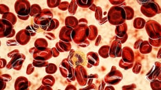
Blood film at 400X magnification demonstrating polyglobulia and thrombocytosis. Courtesy of U. Woermann, MD, Division of Instructional Media, Institute for Medical Education, University of Bern, Switzerland.
Click to see larger pictureClick to see detailView Full Size Image
Picture Type: Photo
Caption: Picture 3. Bone marrow film at 400X magnification demonstrating dominance of erythropoiesis. Courtesy of U. Woermann, MD, Division of Instructional Media, Institute for Medical Education, University of Bern, Switzerland.
Click to see larger pictureClick to see detailView Full Size Image
Picture Type: Photo
Caption: Picture 4. This blood film at 10,000X magnification shows a giant platelet and an eosinophil. Erythrocytes show signs of hypochromia as a result of repeated phlebotomies. Courtesy of U. Woermann, MD, Division of Instructional Media, Institute for Medical Education, University of Bern, Switzerland.
Click to see larger pictureClick to see detailView Full Size Image
Picture Type: Photo
  BIBLIOGRAPHY Section 11 of 11   Click here to go to the previous section in this topic Click here to go to the top of this page
Author Information Introduction Clinical Differentials Workup Treatment Medication Follow-up Miscellaneous Pictures Bibliography

  • Anagrelide Study Group: Anagrelide, a therapy for thrombocythemic states: experience in 577 patients. Am J Med 1992 Jan; 92(1): 69-76[Medline].
  • Berk PD, Goldberg JD, Donovan PB, et al: Therapeutic recommendations in polycythemia vera based on Polycythemia Vera Study Group protocols. Semin Hematol 1986 Apr; 23(2): 132-43[Medline].
  • Berlin NI: Diagnosis and classification of the polycythemias. Semin Hematol 1975 Oct; 12(4): 339-51[Medline].
  • Cimino R, Rametta V, Matera C, et al: Recombinant interferon alpha-2b in the treatment of polycythemia vera. Am J Hematol 1993 Nov; 44(3): 155-7[Medline].
  • Fruchtman SM, Mack K, Kaplan ME, et al: From efficacy to safety: a Polycythemia Vera Study group report on hydroxyurea in patients with polycythemia vera. Semin Hematol 1997 Jan; 34(1): 17-23[Medline].
  • Fruchtman SM, Pettit RM, Gilbert HS, et al: Anagrelide. Analysis of long-term safety and leukemogenic potential in myeloproliferative diseases (MPDs) [abstract]. Blood 2002; 100: 70.
  • James C, Ugo V, Le Couedic JP, et al: A unique clonal JAK2 mutation leading to constitutive signalling causes polycythaemia vera. Nature 2005 Apr 28; 434(7037): 1144-8[Medline].
  • Klein AS, Sitzmann JV, Coleman J, et al: Current management of the Budd-Chiari syndrome. Ann Surg 1990 Aug; 212(2): 144-9[Medline].
  • Kralovics R, Teo SS, Buser AS, et al: Altered gene expression in myeloproliferative disorders correlates with activation of signaling by the V617F mutation of Jak2. Blood 2005 Nov 15; 106(10): 3374-6[Medline].
  • Landolfi R: Bleeding and thrombosis in myeloproliferative disorders. Curr Opin Hematol 1998 Sep; 5(5): 327-31[Medline].
  • Landolfi R, Marchioli R, Kutti J, et al: Efficacy and safety of low-dose aspirin in polycythemia vera. N Engl J Med 2004 Jan 8; 350(2): 114-24[Medline].
  • Levine RL, Wadleigh M, Cools J, et al: Activating mutation in the tyrosine kinase JAK2 in polycythemia vera, essential thrombocythemia, and myeloid metaplasia with myelofibrosis. Cancer Cell 2005 Apr; 7(4): 387-97[Medline].
  • Streiff MB, Smith B, Spivak JL: The diagnosis and management of polycythemia vera in the era since the Polycythemia Vera Study Group: a survey of American Society of Hematology members' practice patterns. Blood 2002 Feb 15; 99(4): 1144-9[Medline].
  • Weinfeld A, Swolin B, Westin J: Acute leukaemia after hydroxyurea therapy in polycythaemia vera and allied disorders: prospective study of efficacy and leukaemogenicity with therapeutic implications. Eur J Haematol 1994 Mar; 52(3): 134-9[Medline].

Polycythemia Vera excerpt