Continually Updated Clinical Reference
 
 
  All Sources     eMedicine     Medscape     Drug Reference     MEDLINE
 
eMedicine - Aortic Stenosis : Article by

Quick Find
Authors & Editors
Introduction
Clinical
Differentials
Workup
Treatment
Medication
Follow-up
Miscellaneous
Multimedia
References

Related Articles
Mitral Regurgitation

Mitral Valve Prolapse


Patient Education
Circulatory Problems Center

Angina Pectoris Overview

Angina Pectoris Causes

Angina Pectoris Symptoms

Angina Pectoris Treatment


Author: James V Talano, MD, MBA, MM, FACC, FAHA, Director of Cardiovascular Medicine, SWICFT Institute

James V Talano is a member of the following medical societies: American College of Cardiology, American College of Chest Physicians, American College of Physician Executives, American College of Physicians, American Heart Association, American Society of Echocardiography, American Society of Nuclear Cardiology, Heart Failure Society of America, and Society of Geriatric Cardiology

Coauthor(s): Bekir Hasan Melek, MD, Assistant Professor of Clinical Medicine, Department of Medicine, Section of Cardiology, Tulane University School of Medicine

Editors: Alan D Forker, MD, Professor of Medicine, Program Director of Cardiovascular Fellowship, MidAmerica Heart Institute, University of Missouri at Kansas City School of Medicine; Director, Outpatient Lipid Diabetes Research Center, MidAmerica Heart Institute of Saint Luke's Hospital; Francisco Talavera, PharmD, PhD, Senior Pharmacy Editor, eMedicine; Steven J Compton, MD, FACC, FACP, Director of Cardiac Electrophysiology, Alaska Heart Institute, Providence and Alaska Regional Hospitals; Amer Suleman, MD, Consultant in Electrophysiology and Cardiovascular Medicine, Department of Internal Medicine, Division of Cardiology, Medical City Dallas Hospital; Richard A Lange, MD, E Cowles Andrus Professor of Cardiology, Professor of Medicine, Johns Hopkins University School of Medicine

Author and Editor Disclosure

Synonyms and related keywords: aortic stenosis, AS, valvular aortic stenosis, valvular AS, aorta stenosis, aortic valve surgery, heart failure, syncope, angina pectoris, pulsus parvus et tardus, heart valve obstruction, aortic obstruction, aortic valve obstruction, aortic valve replacement, AVR, sudden cardiac death, SCD, calcific embolization, infective endocarditis

Background

With the aging of the United States population, diseases in the elderly are a major interest among health care professionals. Valvular aortic stenosis (AS) is no exception; senile degenerative AS is now the leading indication for aortic valve replacement (AVR). The favorable long-term outcome following aortic valve (AV) surgery and the relatively low operative risk emphasize the importance of an accurate and timely diagnosis.

Pathophysiology

The pathophysiologic mechanisms responsible for symptoms in patients with AS include an increase in afterload, progressive hypertrophy of the left ventricle, and a decrease in systemic and coronary flow as consequences of valve obstruction.

In infants and children with congenital AS, the increase in obstruction occurs gradually because the valve orifice changes little as the child grows. Similarly, in adults with AS, left ventricular (LV) obstruction usually develops and increases gradually over a long period of time. The progressive LV outflow obstruction results in increased LV mass by parallel replication of sarcomeres producing concentric hypertrophy at the expense of cavity size. This increase in wall thickness is a compensatory mechanism to normalize LV wall stress. Indeed, wall thickness appears to be a critical determinant of ventricular performance in patients with AS; an inverse relationship exists between LV wall stress and ejection fraction (EF). Inadequate development of hypertrophy, depression of myocardial contractility, or a combination of these factors usually leads to impairment of ventricular performance (so-called afterload mismatch).

LV systolic function is usually well preserved, and cardiac output (CO) is maintained for many years despite a large pressure gradient across the AV. During this time, the patient does not experience a reduction in resting CO, LV dilatation, or development of symptoms. Although the CO at rest is normal in most patients with severe AS, it often fails to rise significantly during exercise. If afterload mismatch occurs, the CO, stroke volume, and the left ventricle and aorta pressure gradient all decline.

Another consequence to the pathophysiologic response to AS, LV diastolic function, is commonly abnormal, resulting in elevated LV filling pressures, which is reflected onto the pulmonary circulation. Diastolic dysfunction occurs as a consequence of both impaired LV relaxation and decreased LV compliance that is caused by increased afterload, a thick noncompliant LV, and relative myocardial ischemia. Chamber stiffness can revert toward normal as LV hypertrophy regresses following relief of valvular obstruction, and in some patients, muscle stiffness also may revert to normal. Extensive myocardial fibrosis develops with long-standing hypertrophy, which may not disappear despite regression of hypertrophy.

In patients with severe AS, the left atrial (LA) pressure waveform usually demonstrates a large a wave because of a combination of vigorous contraction of a hypertrophic left atrium and reduced LV compliance. Atrial contraction plays a particularly important role in mitral valve conductance and filling of the left ventricle in AS. It raises LVEDP while preventing a concomitant elevation of mean LA pressure. This prevents pulmonary venous and capillary pressures from rising to levels that would normally produce pulmonary congestion, while at the same time elevating LVEDP sufficiently to ensure effective LV contraction. Therefore, development of atrial fibrillation in AS is often catastrophic to the maintenance of normal forward stroke volume.

AS intensifies the severity of existing mitral regurgitation (MR) by increasing the ventricular pressure gradient responsible for driving blood from the left ventricle to the left atrium. Additionally, functional MR as a consequence of LV dilatation in late stages of AS may superimpose the hemodynamic changes associated with this lesion on those produced by AS.

Coronary blood flow at rest is increased in absolute terms but is normal when corrected for LV mass. However, myocardial blood flow reserve is often reduced. Increased LV mass, increased LV systolic pressure, and prolongation of the systolic ejection phase all elevate the myocardial oxygen requirement, especially in the subendocardial region. Myocardial perfusion is also compromised by the relative decline in myocardial capillary density and by a reduced diastolic transmyocardial (coronary) perfusion gradient due to elevated LVEDP. Therefore, the subendocardium is susceptible to low nutrient flow, and this underperfusion results in myocardial ischemia.

Frequency

United States

Aortic sclerosis (considered a precursor of calcific degenerative AS) increases with age and is present in 29% of individuals older than 65 years and in 37% of individuals older than 75 years. In elderly persons, the prevalence of AS is between 2% and 9%.

Mortality/Morbidity

The natural history of AS is well known. Patients with severe AS may be asymptomatic for many years despite the presence of severe left ventricular outflow tract (LVOT) obstruction. The peak AV systolic pressure gradient can exceed 150 mm Hg, and peak LV pressure can reach 300 mm Hg with normal end-diastolic volume (EDV) and end-systolic volume (ESV). In one series, 40% of patients with severe AS treated medically survived 5 years, whereas the 10-year survival rate was 20%. In another series of patients with significant AS treated medically, the 5-year survival rate was 64%. With the appearance of symptoms, a rapidly progressive downhill course is observed. Onset of angina is associated with an average survival of 5 years, the onset of syncope is associated with an average survival of 2-3 years, whereas the onset of congestive heart failure (CHF) is associated with an average survival of 1.5-2 years.

  • Among symptomatic patients with moderate-to-severe AS treated medically, mortality rates from the onset of symptoms were approximately 25% at 1 year and 50% at 2 years. More than 50% of deaths were sudden. Death in general, including sudden death, occurs primarily in symptomatic patients.
  • Asymptomatic patients, even with critical AS, have an excellent prognosis regarding survival, with an expected death rate of less than 1% per year; only 4% of sudden cardiac deaths in severe AS occur in asymptomatic patients.
  • Although the obstruction tends to progress more rapidly in patients with degenerative calcific AV disease than in those with congenital or rheumatic disease, predicting the rate of progression in individual patients is not possible. Therefore, careful clinical follow-up is mandatory in all patients with moderate-to-severe AS. Catheterization and echocardiographic studies suggest that the valve area may decline 0.1-0.3 cm2 per year; the systolic pressure gradient across the valve can increase by as much as 10-15 mm Hg per year. A higher rate of progression is observed in elderly patients with coronary artery disease (CAD) and chronic renal insufficiency.

Race

No racial predilection is associated with congenital or acquired AS.

Age

See Causes.


History

In AS of adults, a long latent period exists during which the LV outflow obstruction and the pressure load on the myocardium gradually increase while patients remain asymptomatic.

  • The classic symptom triad of AS includes angina pectoris, syncope, and heart failure, which most commonly manifest after the sixth decade of life.
    • In patients in whom the AV obstruction remains unrelieved, the onset of these symptoms predicts a poor outcome. The approximate time interval from the onset of symptoms to death is 2 years for heart failure, 3 years for syncope, and 5 years for angina.
    • Exertional dyspnea is the most common initial complaint, even with normal LV systolic function, and it relates to abnormal LV diastolic function.
    • Angina pectoris occurs in approximately two thirds of patients with critical AS, of which 50% have significant CAD. Because angina commonly is precipitated by exertion and relieved by rest, it often simulates symptoms of CAD. Angina results from a concomitant increased oxygen requirement by the hypertrophic myocardium and diminished oxygen delivery secondary to the excessive compression of coronary vessels and relative subendocardial myocardial ischemia. Of course, angina also can result from coexistent CAD.
    • The cause of syncope is multifactorial. It often occurs upon exertion when the arterial systolic blood pressure drops because of systemic vasodilatation in the presence of a fixed forward stroke volume. Exertional hypotension also may manifest as blackout spells, lightheadedness, or dizziness upon effort. It also may be caused by atrial or ventricular tachyarrhythmias, commonly with premonitory symptoms.
    • Syncope at rest may be due to transient ventricular tachycardia from which the patient recovers spontaneously. Episodes of atrial fibrillation with precipitous decline in CO or transient AV block due to extension of the calcification of the valve into the conduction system also can be culprits. Another cause of syncope is abnormal vasodepressor reflexes caused by increased LV intracavitary pressure (vasodepressor syncope), which is probably a common mechanism in patients with severe AS.
  • Paroxysmal nocturnal dyspnea, orthopnea, and pulmonary edema usually are late-occurring symptoms of heart failure.
  • Gastrointestinal bleeding, idiopathic or due to small bowel angiodysplasia or other vascular malformations, is present at a higher than expected frequency in patients with calcific AS; it usually resolves following AV surgery.
  • Risk of infective endocarditis is higher in younger patients with mild valvular deformity than in older patients with degenerated calcified AVs, but it can occur in both. It can occur frequently at any age with hospital-acquired Staphylococcus aureus bacteremia, which frequently results in aortic valve replacement.
  • Calcific AS may cause emboli of calcium in various organs, including the heart, kidney, and brain.
  • Because resting CO is usually well maintained for many years in patients with severe AS, marked fatigability, debilitation, peripheral hypoperfusion, and other manifestations of a low CO are usually not prominent until quite late in the natural history of the disease.
  • Atrial fibrillation, pulmonary hypertension, and systemic venous hypertension in patients with isolated AS are preterminal findings.
  • Sudden cardiac death is rare and usually occurs in symptomatic patients.

Physical

In severe AS, the carotid arterial pulse is small and rises slowly (pulsus parvus et tardus); however, in elderly individuals, this may not be present despite severe stenosis because of more rigid aorta and carotid vessels. A lag time may be present between the apical impulse and the carotid impulse. Systolic hypertension can coexist with AS, but a systolic blood pressure higher than 200 mm Hg is rare in patients with critical AS. In advanced-stage AS, both systolic blood pressure and pulse pressure are decreased.

  • Pulsus alternans can occur with the onset of LV dysfunction. The jugular venous pulse may show prominent a waves reflecting reduced RV compliance consequent to hypertrophy of the interventricular septum.
  • At the apex, a precordial a wave often is visible and palpable. A hyperdynamic LV is unusual and suggests concomitant aortic regurgitation (AR) or MR. A systolic "thrill" may be present at the second right intercostal space or at the suprasternal notch and usually indicates a mean AV gradient higher than 50 mm Hg. The thrill is best felt while the patient is leaning forward. On occasion, it can be transmitted to the carotids.
  • Rarely, RV failure with systemic venous congestion, hepatomegaly, and edema precede LV failure. This probably is due to the bulging of the interventricular septum into the right ventricle, with impedance in filling, elevated jugular venous pressure, and a prominent a wave (Bernheim effect).
  • S1 is usually normal or soft. The aortic component of the second heart sound, A2, is usually diminished or absent because the AV is calcified and immobile and/or aortic ejection is prolonged or buried in the prolonged systolic ejection murmur. The presence of a normal or A2 speaks against the presence of severe AS. Paradoxical splitting of the S2 also occurs because of late closure of A2. P2 may also be accentuated when LV failure leads to secondary pulmonary hypertension.
  • The presence of an ejection sound (eg, ejection click) is dependent on the mobility of the valve cusps and disappears when they become immobile and severely calcified. Thus, it is common in children and young adults with congenital AS but rare in elderly individuals with acquired calcific AS with rigid valves. This sound occurs approximately 40-60 milliseconds after the onset of S1 and is frequently heard best with the diaphragm of the stethoscope along the mid-lower left sternal border; it is often well transmitted to the apex and may be confused with a split S1. In contrast to a pulmonic ejection sound, the aortic ejection sound usually does not vary with respiration.
  • A prominent S4 is usually present due to forceful atrial contraction into a hypertrophied left ventricle. The presence of an S4 in a young patient with AS indicates significant AS, but with AS in an elderly person, this is not necessarily true.
  • The classic crescendo-decrescendo systolic murmur of AS is best heard at the second intercostal space in the right upper sternal border; it is harsh at the base and radiates to both carotid arteries. However, it may be more prominent at the apex in elderly persons with calcific AS due to radiation of the high-frequency components of the murmur to the apex (Gallavardin phenomenon) leading to its misinterpretation as a murmur of MR. Accentuation of the AS murmur following a long R-R interval (as in atrial fibrillation or following a premature beat) distinguishes it from the MR murmur, which usually does not change. Nevertheless, coexistent MR can be present, due to progressive LV dilatation, CAD, or mitral annular calcification. A high-pitched decrescendo diastolic murmur secondary to aortic regurgitation is common in many patients with dominant AS.
  • The intensity of the systolic murmur does not correspond to the severity of AS, rather, the timing of the peak and the length or duration of the murmur corresponds to the severity of AS. The more severe the stenosis, the longer the duration of the murmur and the more likely it peaks at mid-to-late systole.
  • The murmur of valvular AS is augmented by the inhalation of amyl nitrite upon squatting or in a postpremature beat; the murmur intensity is reduced during Valsalva strain, which is contrary to what occurs with hypertrophic obstructive cardiomyopathy where a Valsalva maneuver increases the intensity of the murmur.
  • When the left ventricle fails and cardiac output falls, the AS murmur becomes softer and may be barely perceptible. Atrial fibrillation with short R-R intervals can also decrease the murmur intensity or make it appear absent. In an elderly person with symptoms of CHF and a soft systolic ejection murmur, noninvasive evaluation for AS is needed because occult AS may be a cause of intractable heart failure. Embolization from a calcified or infected AV that results in unilateral vision loss, focal neurologic signs, and myocardial infarction can be the first signs of AV pathology.

Causes

Most cases of AS are due to the obstruction at the valvular level. Common causes are summarized in Table 1. Valvular AS can be either congenital or acquired.

  • Congenital valvular aortic stenosis
    • Congenitally unicuspid, bicuspid, tricuspid, or even quadricuspid valves may be the cause of AS. In neonates and infants younger than 1 year, a unicuspid valve can produce severe obstruction and is the most common anomaly in patients with fatal valvular AS.
    • In general, unicuspid valves are most frequent in cases of symptomatic AS in patients younger than 15 years. In adults, congenital AS is usually due to a bicuspid valve but usually does not cause significant narrowing of the aortic orifice during childhood because most of the bicuspid valves are not stenotic at birth. Their altered architecture induces turbulent flow with continuous trauma to the leaflets, ultimately resulting in fibrosis, increased rigidity and calcification of the leaflets, and narrowing of the aortic orifice.
    • These anatomical/pathological changes are similar to those observed in senile degenerative calcific stenosis of a tricuspid AV, except that in congenital bicuspid valve, these changes occur several decades earlier. Congenitally malformed tricuspid AVs with unequally sized cusps and some commissural fusion can also cause turbulent flow leading to fibrosis and, ultimately, to calcification and stenosis. Clinical manifestations of congenital AS in adults usually occur after the fourth decade of life.
  • Acquired valvular aortic stenosis
    • The main causes of acquired AS include rheumatic heart disease and senile degenerative calcification.
    • In rheumatic AS, the underlying process includes progressive fibrosis of the valve leaflets with varying degrees of commissural fusion, often with retraction of the leaflet edges and, in certain cases, calcification. As a consequence, the rheumatic valve often is regurgitant and stenotic. Coexistent mitral valve disease is common. This form of AS is uncommon in the United States.
    • Degenerative (senile) calcific AS involves progressive calcification of the leaflet bodies resulting in limitation of the normal cusp opening during systole. This represents a consequence of long-standing hemodynamic stress on the valve and is currently the most frequent cause of AS requiring AV surgery. It usually occurs in individuals older than 75 years. Cellular aging and degeneration have been implicated. Diabetes mellitus and hypercholesterolemia are risk factors for the development of this lesion. The calcification may also involve the mitral annulus or extend into the conduction system, resulting in atrioventricular or intraventricular conduction defects.
      • The pathophysiologic changes are preceded by structural changes in the valvular apparatus. Recent histopathologic studies have provided important insight into the pathogenesis of calcific AS. The available data suggest that the development and progression of calcific AS are due to an active disease process at the cellular and molecular level that shows many similarities with atherosclerosis, ranging from endothelial dysfunction to, ultimately, calcification.
      • These similarities between calcific AV disease and atherosclerosis at tissue level imply that they may share similar association with clinical risk factors. Indeed, the Cardiovascular Health Study has suggested that calcific AV disease is associated with older age, male sex, serum LDL and Lp(a) levels, systemic arterial hypertension, diabetes mellitus, and smoking.
      • The early lesion in the pathogenesis of degenerative calcific AV disease is focal endothelial thickening on the aortic side of the cusps that is initiated by endothelial disruption due to increased mechanical stress and decreased shear stress. These lesions consist of intracellular and extracellular lipids (mainly LDL and Lp[a] with evidence of lipoprotein oxidation), inflammatory cells (mainly macrophages with some foam cells and few T lymphocytes), extracellular matrix, and microscopic calcification associated with active production of osteopontin protein by a subset of macrophages. The upregulated matrix metalloproteinase expression induces remodeling of the extracellular matrix via cytokine stimulation. Phenotypically altered fibroblasts and HLA-DR also accumulate in the abnormal region of the AV.
      • Another important finding is the presence of angiotensin-converting enzyme and angiotensin II, which suggests a potential role of the renin-angiotensin system in the lesion pathogenesis. After initial inflammatory changes, calcification predominates later in the process. The extension of the lesion into adjacent fibrosa of the cusps leads to disruption of the normal collagen fiber architecture that provides tensile strength to valve leaflets, resulting in dysfunction.
    • Coexistent coronary artery calcification is also common and has been found to be correlated with aortic-valve calcium based on electron beam computed tomography (EBCT) findings, also called Ultrafast CT. Calcific AS is also observed in end-stage renal disease. Rheumatoid involvement of the valve (ie, systemic lupus erythematosus, rheumatoid arthritis) can result in nodular thickening of the valve cusps and involvement of the proximal part of the aorta but is a rare cause of AS.
  • Other infrequent causes of AS include obstructive vegetations, homozygous type II hypercholesteroemia, Paget disease, Fabry disease, ochronosis, and irradiation.
  • The likely cause of AS depends on the age of the patient at presentation. In patients who develop symptoms in their teens and early twenties, the cause usually is a congenitally unicuspid or fused bicuspid AV. If symptoms arise between the fifth and seventh decade, the culprit is either a calcified bicuspid AV or it is degenerative in nature. Nowadays, the most common presentation is an elderly patient with senile degenerative AV with calcific deposits at the base of the cusps in the absence of commissural fusion.

    Table 1. Common Reasons of Aortic Stenosis Requiring Surgery

    Age <70 years (n=324)Age >70 years (n=322)
    Bicuspid AV (50%)
    Postinflammatory (25%)
    Degenerative (18%)
    Unicommissural (3%)
    Hypoplastic (2%)
    Indeterminate (2%)
    Degenerative (48%)
    Bicuspid (27%)
    Postinflammatory (23%)
    Hypoplastic (2%)


Mitral Regurgitation
Mitral Valve Prolapse

Other Problems to be Considered

Supravalvular aortic stenosis
Congenital subvalvular aortic stenosis
Hypertrophic obstructive cardiomyopathy


Imaging Studies

  • Echocardiography
    • Two-dimensional Doppler echocardiography is the imaging modality of choice to help diagnose and estimate the severity of AS and localize the level of obstruction. American College of Cardiology/American Heart Association (ACC/AHA) recommendations for echocardiography in AS are summarized in Table 2.
    • In valvular AS, the etiology (bicuspid, rheumatic, or senile degenerative) may be assessed from the parasternal short-axis view. LV size, mass, and function should be evaluated in each patient. Although the presence of AS is readily diagnosed on 2D echocardiography (doming), severity of AS cannot be judged based on the 2D-echocardiographic images alone. Doppler echocardiography is an excellent tool for assessing the severity of AS. Using the modified Bernoulli equation, a maximum instantaneous and mean AV gradient can be derived from the continuous-wave Doppler velocity across the AV. To avoid underestimating the AV gradient, the Doppler beam should be kept as parallel as possible to the stenotic jet.
    • In a laboratory with experienced personnel, Doppler-derived AV gradients are accurate and reproducible and correlate well with those obtained with hemodynamically measured pressures during cardiac catheterization.
    • AV gradient depends not only on the severity of obstruction but also on aortic flow. In patients with low CO, the stenosis still may be severe even though the transvalvular gradient is low. To overcome this problem, 2-dimensional Doppler echocardiography can also provide a reliable estimation of aortic valve area (AVA) by the continuity equation, where LVOT indicates LV outflow tract and TVI indicates time velocity integral. The continuity equation is AVA = LVOT area multiplied by LVOT TVI/AV TVI. The echocardiographic criteria for assessment of AS severity are outlined in Table 3.
    • The major limitation of Doppler echocardiography in estimating the severity of AS is underestimation of the gradient if the beam is not parallel to the AS velocity jet. Thus, in a patient with clinical features of severe AS but echo/Doppler findings of mild-to-moderate AS, further evaluation with repeat Doppler or catheterization is required. Very rarely, Doppler may overestimate the mean gradient in cases of severe anemia (hemoglobin <8 g/dL), a small aortic root, or sequential stenoses in parallel (coexistent LVOT and valvular obstruction). Furthermore, echocardiographic calculation of AVA with the continuity equation is highly dependent on accurate measurement of the diameter of the LVOT. In cases with poor transthoracic images, transesophageal echocardiography (TEE) may be used to measure the mean and peak gradient and a planimeter may be used to measure the AVA.

      Table 2. ACC/AHA Recommendations for Echocardiography in Aortic Stenosis

      Indication Class
      Diagnosis and assessment of severity of ASI
      Assessment of LV size, function, and/or hemodynamicsI
      Reevaluation of patients with known AS with changing symptoms or signsI
      Assessment of changes in hemodynamic severity and ventricular function in patients with known AS during pregnancyI
      Reevaluation of asymptomatic patients with severe ASI
      Reevaluation of asymptomatic patients with mild-to-moderate AS and evidence of LV dysfunction or hypertrophyIIa
      Routine reevaluation of asymptomatic adult patients with mild AS who have stable physical signs and normal LV size and functionIII

      Table 3. Criteria for Determining Severity of Aortic Stenosis

      SeverityMean gradient (mm Hg)Aortic valve area (cm2)
      Mild<25>1.5
      Moderate25-501-1.5
      Severe>50<1
      (or <0.5 cm2/m2 body surface area)
      Critical>80<0.5
  • Radionuclide ventriculography may provide information on LV function, including LV ejection fraction, ESV, and EDV.
  • Multislice computed tomography: Three-dimensional volume quantification of AV calcification using this method demonstrated a close nonlinear relationship to echocardiographic parameters of severity of AS. This method is not yet clinically validated.
  • Cardiac magnetic resonance imaging: Comparison of the AV TVI and AVA measurements via echocardiography/Doppler and cardiac magnetic resonance imaging has shown excellent correlation. This method is not yet clinically validated.

Other Tests

  • Electrocardiogram
    • Although the ECG findings may be entirely normal, the principal finding is left ventricular hypertrophy (LVH), which is found in 85% of patients with severe AS; however, its absence does not preclude critical AS. The correlation between absolute voltages in precordial leads and the severity of obstruction, unlike in children with congenital AS, is poor in adults.
    • T-wave inversion and ST-segment depression in leads with predominantly positive QRS complexes are common. ST depression exceeding 0.3 mV in patients with AS indicates LV strain and suggests that severe LVH is present. Occasionally, a septal pseudoinfarct pattern can be seen. LA enlargement with preterminal negative p wave in V1 is noted in 80% of severe isolated cases of AS. The QRS vector may be directed posteriorly and superiorly in the left sagittal plane, and counterclockwise rotation with major forces being in the left posterior quadrant may be present in the transverse plane.
    • The rhythm usually is normal sinus. Atrial fibrillation is uncommon and can be seen at late stages or as a consequence of coexistent MV disease or hyperthyroidism.
    • Extension of calcification into the conduction system can cause atrioventricular or intraventricular block in 5% of cases. Approximately 10% of all cases of left anterior fascicular block are secondary to calcified AV disease. Ambulatory ECG monitoring frequently shows complex ventricular arrhythmias, particularly in cases with myocardial dysfunction.
  • Chest roentgenogram
    • The cardiac size often is normal, with rounding of the LV border and apex despite significant AS. Poststenotic dilatation of the ascending aorta is common.
    • On lateral view, AV calcification is found in almost all adults with hemodynamically significant AS. Although its absence on fluoroscopy in individuals older than 35 years rules out severe valvular AS, its presence does not prove severe obstruction in individuals older than 60 years.
    • Cardiomegaly is a late feature of severe isolated AS. The left atrium may be slightly enhanced, and pulmonary venous hypertension may be seen.
  • Exercise stress testing is usually not needed in patients with severe AS. It may precipitate ventricular tachyarrhythmias, including ventricular fibrillation. However, closely monitored exercise stress testing may be of value to assess exercise capacity in asymptomatic patients. Abnormal results may prove greater disability than the patient could admit.
  • Ambulatory ECG recordings may be necessary in the occasional patients in whom tachyarrhythmia or silent ischemia is suspected.
  • Provocative testing is used in cases when the severity of the AS is uncertain or in the presence of heart failure because of a small stroke volume (SV) and a small mean AV gradient (low-gradient AS).
    • The AVA is determined to be small even using Doppler echocardiography.
    • Infusion of an inotropic agent such as dobutamine, which results in an increase in SV and heart rate, usually is helpful in establishing the correct diagnosis.
    • Measuring CO and LV and aortic pressures simultaneously both before and during dobutamine infusion is important.
    • The measured AVA does not change and the mean pressure gradient increases significantly with an intravenous dobutamine infusion in patients with an initially low-pressure gradient but severe AS. In contrast, a marked increase in both the SV and the measured AVA usually occurs in patients who have a low CO due to concomitant myocardial dysfunction rather than due to severe AS alone.

Procedures

  • Cardiac catheterization
    • In general, if clinical findings are not consistent with Doppler echocardiogram results, cardiac catheterization is recommended for further hemodynamic assessment. ACC/AHA recommendations for cardiac catheterization in AS are summarized in Table 4.
    • Measurement of simultaneous LV and aortic pressures or pullback techniques may be used to estimate the peak pressure gradient. The pullback method is inaccurate in patients with irregular rhythm or low CO states. Using femoral artery pressure during a simultaneous measurement technique provides inaccurate results because the difference between central aortic pressure and femoral artery pressure may be significant. At the time of catheterization, CO should be assessed for the calculation of AVA, preferably by the Fick principle. The thermodilution technique has limited accuracy in assessing cardiac output in patients with irregular rhythms or low output states or those with pulmonic or tricuspid valve regurgitation.
    • Exclusion of CAD by coronary angiography is needed in all patients older than 35 years who are being considered for valve surgery. It also should be performed in patients younger than 35 years if they have LV systolic dysfunction, symptoms/signs suggestive of CAD, or 2 or more risk factors for premature CAD, excluding sex. Generally, the incidence of associated CAD has been reported to be 50% in individuals older than 50 years.
    • Measuring the left ventricular end-diastolic volume (LVEDV) and ESV and calculating the EF also can quantitate the status of LV systolic pump function. However, EF may underestimate LV performance in the presence of the increased afterload associated with severe AS. Aortic root angiogram may show thickening and doming of the AV as well as a poststenotic dilatation of the aortic root.

      Table 4. Recommendations for Cardiac Catheterization in Aortic Stenosis

      Indication Class
      Coronary angiography before AVR in patients at risk for CADI
      Assessment of severity of AS in symptomatic patients when AVR is planned or when noninvasive tests are inconclusive or a discrepancy exists in the clinical findings regarding severity of AS or the need for surgeryI
      Assessment of severity of AS before AVR when noninvasive tests are adequate and concordant with clinical findings and coronary angiography is not neededIIb
      Assessment of LV function and severity of AS in asymptomatic patients when noninvasive tests are adequateIII


Medical Care

The primary management of symptomatic patients with valvular AS is interventional. Medical treatment essentially is reserved for patients who have complications of AS such as heart failure, infective endocarditis, or arrhythmias.

  • Digitalis can be used as an inotropic agent and also to control the ventricular rate in cases with atrial fibrillation. Diuretics may be used for pulmonary congestive symptoms, and vasodilators may be used for heart failure and for hypertension. Both classes of agents should be used with caution to avoid critically reducing preload in a patient with significant AS and a hypertrophic noncompliant LV. The same precaution also is valid for beta-blockers and calcium channel blockers.
  • Endocarditis prophylaxis is recommended in all patients regardless of the etiology or the patient's age and surgical status. The risk of valve-ring abscess is highest of all of the valve lesions.
  • Recently, several small observational studies suggested that HMG-CoA reductase inhibitor use can reduce AV leaflet calcification and delay the progression of AS severity. However, prospective, randomized, placebo-controlled trials have not shown statins to be beneficial. Some believe that statin therapy was not initiated early enough in the process to delay the progression of AS in these studies, so ongoing studies are attempting to address this issue.
  • In similar observational studies, use of ACE inhibitors was associated with slower calcium accumulation in AV; however, no hemodynamic benefit was seen during the same period. A recent randomized clinical trial of atorvastatin versus placebo showed no difference in AVA or pressure gradients.

Surgical Care

The primary management of symptomatic patients with valvular AS is interventional. The timing of intervention is determined by the severity of the stenosis, the age of the patient, and the presence of symptoms. In asymptomatic patients with severe AS, follow-up is needed every 6 months. The natural history suggests that patients with a mean gradient of less than 25 mm Hg have a 20% chance of requiring intervention in 15 years. Overall, 40% of those in the medically treated group require intervention in 15 years, although in certain cases, progression to severe AS may be faster.

The age of patients undergoing AVR is rising steadily because the incidence of calcific AS increases as the age of the population increases. Early detection and close follow-up of patients with AS improves the duration and quality of life. Once symptoms develop, intervention is needed.

  • Percutaneous balloon valvuloplasty was introduced approximately 10 years ago. Generally, it is used as a palliative measure in critically ill patients who are not surgical candidates or as a bridge in critically ill patients before they undergo AVR.
  • In cases of congenital AS without calcified unicuspid or bicuspid AV in children, adolescents, and young adults, percutaneous balloon valvuloplasty is an accepted alternative to surgical valvotomy and carries a risk of 1%, but its value is limited in adults with calcific AS. The high rate of restenosis and the absence of a mortality benefit preclude its use as a definitive treatment method in these cases.
    • The best results from valvuloplasty are obtained in patients with congenital commissural bicuspid AVs, where a 60-70% reduction in gradient and a 60% increase in AVA can be expected. It is recommended for patients with gradients higher than 50-60 mm Hg and/or a valve area of less than 0.5 cm2/m2, even in asymptomatic patients, because of the low risk associated with balloon valvuloplasty, the high desire for unrestricted or minimally limited lifestyle in younger populations, and the incidence of certain rare cases of sudden cardiac death. The risk rate of causing significant AR is 10%.
    • Restenosis is common, particularly in patients with unicuspid valves or with valves affected by severe dysplasia (>60% at 6 mo, virtually 100% at 2 y). More than mild AR poses a contraindication for this procedure.
    • Valvuloplasty also can be performed in patients with severe CHF or cardiogenic shock (1) as a bridge to valve replacement as a palliative measure, (2) for patients with other comorbid conditions with a very short life expectancy, (3) for those who refuse surgery, (4) for those with heart failure who need an urgent major noncardiac surgical procedure, or (5) in pregnant patients with critical AS.
    • In critically ill patients, the mortality rate associated with the procedure is 3-7%. Another 6% develop serious complications including perforation, myocardial infarction, and severe AR.
  • Aortic valve replacement
    • ACC/AHA recommendations for AV replacement in patients with valvular AS are summarized in Table 5. In most adults with calcific AS and in those patients with calcified, bicuspid, severely stenotic AVs, AVR is the surgical treatment of choice. It is recommended if hemodynamic evidence of severe obstruction and symptoms due to AS are present. It also is recommended in asymptomatic patients with severe AS and LV dysfunction. AVR should be performed in all symptomatic patients with severe AS regardless of LV function. Even in the presence of LV dysfunction, survival is better with surgical treatment than with medical treatment.
    • Prior to AV surgery, complete hemodynamic assessment of AS with either Doppler or catheterization is required. Assessment of LV function and MV disease also is required. If significant MR is present, the degree of regurgitation should be evaluated intraoperatively after replacement of the AV to determine the need for MV repair or replacement, unless intrinsic disease of the MV apparatus is present. Coronary angiography is needed in all patients aged 35-40 years and in those patients with risk factors for CAD.
    • Successful AVR produces substantial clinical and hemodynamic improvement in patients with AS, including octogenarians. The choice of prosthesis is determined by the anticipated longevity of the patients and their ability to tolerate anticoagulation.
    • Postoperatively, end-diastolic pressure and ESV drop significantly, and symptoms of elevated LA pressure and ischemia improve. Improvement in EF invariably occurs over the following 6 months, and increased LV mass tends to decrease within 18 months postoperatively.
    • The surgical mortality risk in patients with normal LV systolic function and no other comorbid conditions is less than 5%. Risk factors for increased operative mortality include (1) high New York Heart Association (NYHA) class (25-30% mortality in patients with class IV), (2) preoperative LV systolic dysfunction (strongest predictor of postoperative LV dysfunction), (3) age (up to 30% mortality in patients >80 y but is not a contraindication for AVR), and (4) the presence of associated AR.
    • The skill level of the surgical team cannot be ignored.
    • If concomitant coronary disease is present, AVR and coronary artery bypass graft (CABG) should be performed at the same time. Risk factors for late death include preoperative NYHA class and LV systolic function, preoperative ventricular arrhythmias, concomitant AR, atrial fibrillation, and CAD, particularly a history of myocardial infarction. Overall, the 5-year survival rate in all adults after AVR is 80-94%, whereas 10-year survival rate is 68-89%.

      Table 5. Recommendations for Aortic Valve Replacement in Aortic Stenosis

      IndicationClass
      Symptomatic patients with severe ASI
      Patients with severe AS undergoing
      coronary artery bypass surgery
      I
      Patients with severe AS undergoing surgery on the aorta or other heart valvesI
      Patients with moderate AS undergoing coronary artery bypass surgery or surgery on the aorta or other heart valvesIIa
      Asymptomatic patients with severe AS and the following:
      LV systolic dysfunctionIIa
      Abnormal response to exercise (eg, hypotension)IIa
      Ventricular tachycardiaIIb
      Marked or excessive LVH (>15 mm)IIb
      Valve area <0.6 cm2 IIb
      Prevention of sudden death in asymptomatic patients with none of the findings listed under asymptomatic patients with severe ASIII
    • The Ross procedure is another option in young patients as an initial procedure or for reoperation after prior valvotomy. In this procedure, the patient's own pulmonary valve and main pulmonary artery are transplanted to the aortic position, with reimplantation of coronary arteries. A homograft is placed in the pulmonary position. Its durability is better than with tissue valves, and anticoagulation is not required. This procedure is technically demanding.
    • Despite the proven efficacy of surgical valve replacement, it still carries high operative mortality and morbidity rates in the growing population of elderly patients with significant comorbidities. Recently, percutaneous AV replacement using the antegrade transseptal and retrograde arterial approaches has been introduced as an alternative to surgical AV replacement in a subset of patients with AS who are at high surgical risk. Although percutaneous AV replacement may be an exciting alternative to the standard surgical approach, its use and long-term durability are unknown. At this time, it should only be performed as part of an investigational study.

Activity

Patients with mild AS can lead a normal life. In cases of moderate AS, moderate-to-severe physical exertion and competitive sports should be avoided.


Treatment of valvular AS is interventional. When intervention is not an option, signs of heart failure must be treated with inotropic therapy, diuretics, and nitrates. Drug therapy essentially is reserved for bacterial endocarditis prophylaxis.

Drug Category: Antibiotics

For endocarditis prophylaxis during dental/oral/respiratory tract and gastrointestinal/gastrourinary (GI/GU) procedures.

Drug NameAmoxicillin (Amoxil, Trimox)
DescriptionInterferes with synthesis of cell wall mucopeptides during active multiplication, resulting in bactericidal activity against susceptible bacteria.
Used as prophylaxis in minor procedures.
Adult Dose2 g PO 1 h before the procedure; alternatively, 3 g PO 1 h before procedure followed by 1.5 g 6 h after initial dose
Pediatric Dose50 mg/kg PO 1 h before procedure
ContraindicationsDocumented hypersensitivity
InteractionsReduces efficacy of oral contraceptives
PregnancyB - Fetal risk not confirmed in studies in humans but has been shown in some studies in animals
PrecautionsAdjust dose in renal impairment; may enhance chance of candidiasis

Drug NameAmpicillin (Marcillin, Omnipen)
DescriptionFor prophylaxis in patients undergoing surgical procedures. Coadministered with gentamicin for prophylaxis.
Adult Dose2 g PO/IV/IM 30 min prior to procedure and 1 g 6 h after first dose
Pediatric Dose50 mg/kg IV/IM 30 min prior to procedure
ContraindicationsDocumented hypersensitivity
InteractionsProbenecid and disulfiram elevate levels; allopurinol decreases effects and has additive effects on ampicillin rash; may decrease effects of oral contraceptives
PregnancyB - Fetal risk not confirmed in studies in humans but has been shown in some studies in animals
PrecautionsAdjust dose in renal failure; evaluate rash and differentiate from hypersensitivity reaction

Drug NameClindamycin (Cleocin)
DescriptionUseful in patients allergic to penicillin who require antibiotic prophylaxis prior surgical procedures.
Adult Dose600 mg PO/IV 1 h prior to procedure and 150 mg PO/IV 6 h after first dose
Pediatric DoseNot established
ContraindicationsDocumented hypersensitivity; regional enteritis, ulcerative colitis, hepatic impairment, antibiotic-associated colitis
InteractionsIncreases duration of neuromuscular blockade induced by tubocurarine and pancuronium; erythromycin may antagonize effects; antidiarrheals may delay absorption
PregnancyB - Fetal risk not confirmed in studies in humans but has been shown in some studies in animals
PrecautionsAdjust dose in severe hepatic dysfunction; no adjustment necessary in renal insufficiency; associated with severe and possibly fatal colitis by allowing overgrowth of Clostridium difficile

Drug NameVancomycin (Vancocin)
DescriptionPotent antibiotic directed against gram-positive organisms and active against Enterococcus species. Useful to treat septicemia and skin structure infections. Indicated for patients who cannot receive or have failed to respond to penicillins and cephalosporins or have infections with resistant staphylococci.
Use creatinine clearance to adjust dose in patients diagnosed with renal impairment.
Used in conjunction with gentamicin for prophylaxis in penicillin-allergic patients undergoing surgical procedures.
Adult DoseSurgical procedures: 1 g IV, infused over 1 h, one hour prior to the procedure, plus 1.5 mg/kg gentamicin infused over 1 h, one hour prior to surgery
Pediatric DoseSurgical procedures: 20 mg/kg IV 1 h prior to procedure, alternatively, 20 mg/kg plus 2 mg/kg 1 h prior to surgery
ContraindicationsDocumented hypersensitivity
InteractionsErythema, histaminelike flushing, and anaphylactic reactions may occur when administered with anesthetic agents; taken concurrently with aminoglycosides, risk of nephrotoxicity may increase above that with aminoglycoside monotherapy; effects in neuromuscular blockade may be enhanced when coadministered with nondepolarizing muscle relaxants
PregnancyC - Fetal risk revealed in studies in animals but not established or not studied in humans; may use if benefits outweigh risk to fetus
PrecautionsCaution in renal failure and neutropenia; red man syndrome is caused by an IV infusion that is too rapid (dose given over a few minutes) but rarely happens when dose given as 2-h administration or as PO or IP administration; red man syndrome is not an allergic reaction

Drug NameGentamicin (Garamycin)
DescriptionAminoglycoside antibiotic for gram-negative coverage. Used in combination with both an agent against gram-positive organisms and one that covers anaerobes.
Used in conjunction with ampicillin or vancomycin for prophylaxis in surgical procedures.
Adult Dose1.5 mg/kg IV with 1-2 g ampicillin 30 min prior to procedure; not to exceed 80 mg
Pediatric Dose2 mg/kg IV with ampicillin (50 mg/kg) 30 min prior to procedure
ContraindicationsDocumented hypersensitivity; non–dialysis-dependent renal insufficiency
InteractionsCoadministration with other aminoglycosides, cephalosporins, penicillins, and amphotericin B may increase nephrotoxicity; aminoglycosides enhance effects of neuromuscular blocking agents, thus, prolonged respiratory depression may occur; coadministration with loop diuretics may increase auditory toxicity of aminoglycosides; possible irreversible hearing loss of varying degrees may occur (monitor regularly)
PregnancyC - Fetal risk revealed in studies in animals but not established or not studied in humans; may use if benefits outweigh risk to fetus
PrecautionsNarrow therapeutic index (not intended for long-term therapy); caution in renal failure (not on dialysis), myasthenia gravis, hypocalcemia, and conditions that depress neuromuscular transmission; adjust dose in renal impairment

Drug NameErythromycin (EES, E-Mycin, Eryc)
DescriptionUsed for prophylaxis in penicillin-allergic patients undergoing surgical procedures.
Adult Dose1 g PO 1-2 h before procedure, followed by 500 mg 6 h after initial dose
Pediatric Dose20 mg/kg PO 2 h prior to procedure, followed by 10 mg/kg 6 h after initial dose
ContraindicationsDocumented hypersensitivity; hepatic impairment
InteractionsCoadministration may increase toxicity of theophylline, digoxin, carbamazepine, and cyclosporine; may potentiate anticoagulant effects of warfarin; coadministration with lovastatin and simvastatin increases risk of rhabdomyolysis
PregnancyB - Fetal risk not confirmed in studies in humans but has been shown in some studies in animals
PrecautionsCaution in liver disease; estolate formulation may cause cholestatic jaundice; adverse GI effects are common (give doses pc); discontinue use if nausea, vomiting, malaise, abdominal colic, or fever occur

Drug NameCefazolin (Ancef)
DescriptionFirst-generation semisynthetic cephalosporin that arrests bacterial cell wall synthesis, inhibiting bacterial growth. Primarily active against skin flora, including S aureus.
Adult Dose1 g IV/IM within 30 min of procedure
Pediatric Dose25 mg/kg IV/IM within 30 min of procedure
ContraindicationsDocumented hypersensitivity
InteractionsProbenecid prolongs effect; coadministration with aminoglycosides may increase renal toxicity; may yield false-positive results for urine-dip test for glucose
PregnancyB - Fetal risk not confirmed in studies in humans but has been shown in some studies in animals
PrecautionsAdjust dose in renal impairment; superinfections and promotion of nonsusceptible organisms may occur with prolonged use or repeated therapy

Drug NameCephalexin (Keflex)
DescriptionFirst-generation cephalosporin that arrests bacterial growth by inhibiting bacterial cell wall synthesis. Bactericidal activity against rapidly growing organisms. Primary activity against skin flora; used for skin infections or prophylaxis in minor procedures.
Adult Dose2 g PO 1 h before procedure
Pediatric Dose50 mg/kg PO 1 h before procedure
ContraindicationsDocumented hypersensitivity
InteractionsCoadministration with aminoglycosides increases nephrotoxic potential
PregnancyB - Fetal risk not confirmed in studies in humans but has been shown in some studies in animals
PrecautionsAdjust dose in renal impairment

Drug NameCefadroxil (Duricef)
DescriptionFirst-generation cephalosporin that arrests bacterial growth by inhibiting bacterial cell wall synthesis. Bactericidal activity against rapidly growing organisms. Primary activity against skin flora; used for skin infections or prophylaxis in minor procedures.
Adult Dose2 g PO 1 h before procedure
Pediatric Dose50 mg/kg PO 1 h before procedure
ContraindicationsDocumented hypersensitivity
InteractionsCoadministration with furosemide or aminoglycosides may increase nephrotoxicity; probenecid prolongs effects
PregnancyB - Fetal risk not confirmed in studies in humans but has been shown in some studies in animals
PrecautionsAdjust dose in renal impairment; superinfections and promotion of nonsusceptible organisms may occur with prolonged use or repeated therapy

Drug NameAzithromycin (Zithromax)
DescriptionInhibits bacterial growth, possibly by blocking dissociation of peptidyl t-RNA from ribosomes, causing RNA-dependent protein synthesis to arrest.
Adult Dose500 mg PO 1 h before procedure
Pediatric Dose15 mg/kg PO 1 h before procedure
ContraindicationsDocumented hypersensitivity; hepatic impairment; do not administer with pimozide
InteractionsMay increase toxicity of theophylline, warfarin, and digoxin; effects are reduced with coadministration of aluminum and/or magnesium antacids; nephrotoxicity and neurotoxicity may occur when coadministered with cyclosporine
PregnancyB - Fetal risk not confirmed in studies in humans but has been shown in some studies in animals
PrecautionsBacterial or fungal overgrowth may result with prolonged antibiotic use; may increase hepatic enzymes and cholestatic jaundice; caution in patients with impaired hepatic function, prolonged QT intervals, or pneumonia; caution in hospitalized, elderly, or debilitated patients

Drug NameClarithromycin (Biaxin)
DescriptionInhibits bacterial growth, possibly by blocking dissociation of peptidyl t-RNA from ribosomes, causing RNA-dependent protein synthesis to arrest.
Adult Dose500 mg PO 1 h before procedure
Pediatric Dose15 mg/kg PO 1 h before procedure
ContraindicationsDocumented hypersensitivity; coadministration of pimozide
InteractionsToxicity increases with coadministration of fluconazole, astemizole, and pimozide; clarithromycin effects decrease and adverse GI effects may increase with coadministration of rifabutin or rifampin; may increase toxicity of anticoagulants, cyclosporine, tacrolimus, digoxin, omeprazole, carbamazepine, ergot alkaloids, triazolam, and HMG CoA-reductase inhibitors; cardiac arrhythmias may occur with coadministration of cisapride; plasma levels of certain benzodiazepines may increase, prolonging CNS depression; arrhythmias and increase in QTc intervals occur with disopyramide; coadministration with omeprazole may increase plasma levels of both agents
PregnancyC - Fetal risk revealed in studies in animals but not established or not studied in humans; may use if benefits outweigh risk to fetus
PrecautionsCoadministration with ranitidine or bismuth citrate is not recommended with CrCl <25 mL/min; give half dose or increase dosing interval if CrCl <30 mL/min; diarrhea may be sign of pseudomembranous colitis; superinfections may occur with prolonged or repeated antibiotic therapies


Further Outpatient Care

  • Follow-up of asymptomatic patients
    • The frequency of the follow-up visits in asymptomatic patients is determined by the severity of AS and by the presence of comorbid conditions.
    • In patients with mild AS, yearly history and physical examination and echocardiograms every 5 years are appropriate.
    • Patients with moderate or severe AS should be examined twice yearly and whenever they develop symptoms attributable to AS.
    • In patients with moderate AS, echocardiograms should be performed every 2 years, whereas in asymptomatic patients with severe AS, yearly echocardiograms are recommended.
  • Follow-up of symptomatic patients
    • The medical treatment options are limited in symptomatic patients who are not candidates for surgical intervention. In cases of pulmonary congestion, digitalis, diuretics, and ACE inhibitors might be used cautiously, whereas beta-blockers might be used if the predominant symptom is angina. In any case, excessive decrease in preload should be avoided. Onset of atrial fibrillation often requires prompt cardioversion.
    • Following AV replacement, every patient should undergo echocardiographic examination after recovery. Thereafter, an examination is recommended whenever new symptoms develop that are attributable to a potential valvular dysfunction.
    • Patients with mechanical valves should receive lifelong anticoagulation with warfarin and should undergo periodic screening of their anticoagulation status.

In/Out Patient Meds

  • All patients with AS require antibiotic prophylaxis against infective endocarditis and also against recurrent rheumatic carditis if the valve lesion has a rheumatic etiology.

Complications

Prognosis

Patient Education


Medical/Legal Pitfalls

  • Patients with severe valvular AS should receive appropriate counseling regarding their conditions, including restriction of physical activity and the need for surgery, if appropriate. Physicians should document these points in patients' records.
  • In cases where the patient refuses AV replacement surgery, the patient needs to have a full understanding of the potential implications (including sudden cardiac death) of his or her decision.
  • If, on the other hand, the patient does agree with surgery, again the patient needs to understand its possible consequences, including perioperative death, the need for lifelong anticoagulation depending on the type of prosthesis, the need for bacterial endocarditis prophylaxis, and the risk of prosthesis malfunction with potential need for reoperation at a higher operative risk.
  • Discussion and careful documentation of these issues not only would help patients become familiar with their condition and therapeutic options, but also would help to avoid misunderstandings and potential litigation.


Media file 1:  Calcific aortic stenosis (parasternal long-axis and short-axis views).
Click to see larger pictureClick to see detailView Full Size Image
Media type:  Image

Media file 2:  
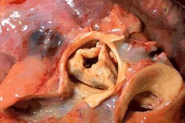
Stenotic aortic valve (macroscopic appearance).
Click to see larger pictureClick to see detailView Full Size Image
Media type:  Photo


  • Agarwal A, Kini AS, Attanti S, et al. Results of repeat balloon valvuloplasty for treatment of aortic stenosis in patients aged 59 to 104 years. Am J Cardiol. Jan 1 2005;95(1):43-7. [Medline].
  • Bauer F, Eltchaninoff H, Tron C, et al. Acute improvement in global and regional left ventricular systolic function after percutaneous heart valve implantation in patients with symptomatic aortic stenosis. Circulation. Sep 14 2004;110(11):1473-6. [Medline].
  • Bonow RO, Carabello B, de Leon AC, et al. Guidelines for the management of patients with valvular heart disease: executive summary. A report of the American College of Cardiology/American Heart Association Task Force on Practice Guidelines. Circulation. Nov 3 1998;98(18):1949-84. [Medline].
  • Braunwald E. Heart disease: a textbook of cardiovascular medicine. In: Braunwald E, ed. Aortic Stenosis. 4th ed. Philadelphia, Pa: WB Saunders; 1992:1035-43.
  • Connolly HM, Oh JK, Orszulak TA, et al. Aortic valve replacement for aortic stenosis with severe left ventricular dysfunction. Prognostic indicators. Circulation. May 20 1997;95(10):2395-400. [Medline].
  • Cribier A, Eltchaninoff H, Bash A, et al. Percutaneous transcatheter implantation of an aortic valve prosthesis for calcific aortic stenosis: first human case description. Circulation. Dec 10 2002;106(24):3006-8. [Medline].
  • Frank S, Johnson A, Ross J Jr. Natural history of valvular aortic stenosis. Br Heart J. Jan 1973;35(1):41-6. [Medline].
  • Hanzel GS, Harrity PJ, Schreiber TL, O'Neill WW. Retrograde percutaneous aortic valve implantation for critical aortic stenosis. Catheter Cardiovasc Interv. Mar 2005;64(3):322-6. [Medline].
  • Hughes BR, Chahoud G, Mehta JL. Aortic stenosis: is it simply a degenerative process or an active atherosclerotic process?. Clin Cardiol. Mar 2005;28(3):111-4. [Medline].
  • Messika-Zeitoun D, Aubry MC, Detaint D, et al. Evaluation and clinical implications of aortic valve calcification measured by electron-beam computed tomography. Circulation. Jul 20 2004;110(3):356-62. [Medline].
  • Novaro GM. Electron beam computed tomography: the latest "stethoscope" for calcific aortic valve disease. Mayo Clin Proc. Oct 2004;79(10):1239-41. [Medline].
  • O'Rourke RA. Aortic valve stenosis: a common clinical entity. Curr Probl Cardiol. Aug 1998;23(8):434-71. [Medline].
  • Otto CM, Burwash IG, Legget ME, et al. Prospective study of asymptomatic valvular aortic stenosis. Clinical, echocardiographic, and exercise predictors of outcome. Circulation. May 6 1997;95(9):2262-70. [Medline].
  • Otto CM. Aortic stenosis and hyperlipidemia: establishing a cause-effect relationship. Am Heart J. May 2004;147(5):761-3. [Medline].
  • Passik CS, Ackermann DM, Pluth JR, Edwards WD. Temporal changes in the causes of aortic stenosis: a surgical pathologic study of 646 cases. Mayo Clin Proc. Feb 1987;62(2):119-23. [Medline].
  • Pellikka PA, Nishimura RA, Bailey KR, Tajik AJ. The natural history of adults with asymptomatic, hemodynamically significant aortic stenosis. J Am Coll Cardiol. Apr 1990;15(5):1012-7. [Medline].
  • Prasad Y, Bhalodkar NC. Aortic sclerosis--a marker of coronary atherosclerosis. Clin Cardiol. Dec 2004;27(12):671-3. [Medline].
  • Rahimtoola SH. The year in valvular heart disease. J Am Coll Cardiol. Jan 4 2005;45(1):111-22. [Medline].
  • Rajamannan NM, Otto CM. Targeted therapy to prevent progression of calcific aortic stenosis. Circulation. Sep 7 2004;110(10):1180-2. [Medline].
  • Rosenhek R, Rader F, Loho N, et al. Statins but not angiotensin-converting enzyme inhibitors delay progression of a