Practice Essentials
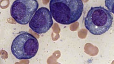
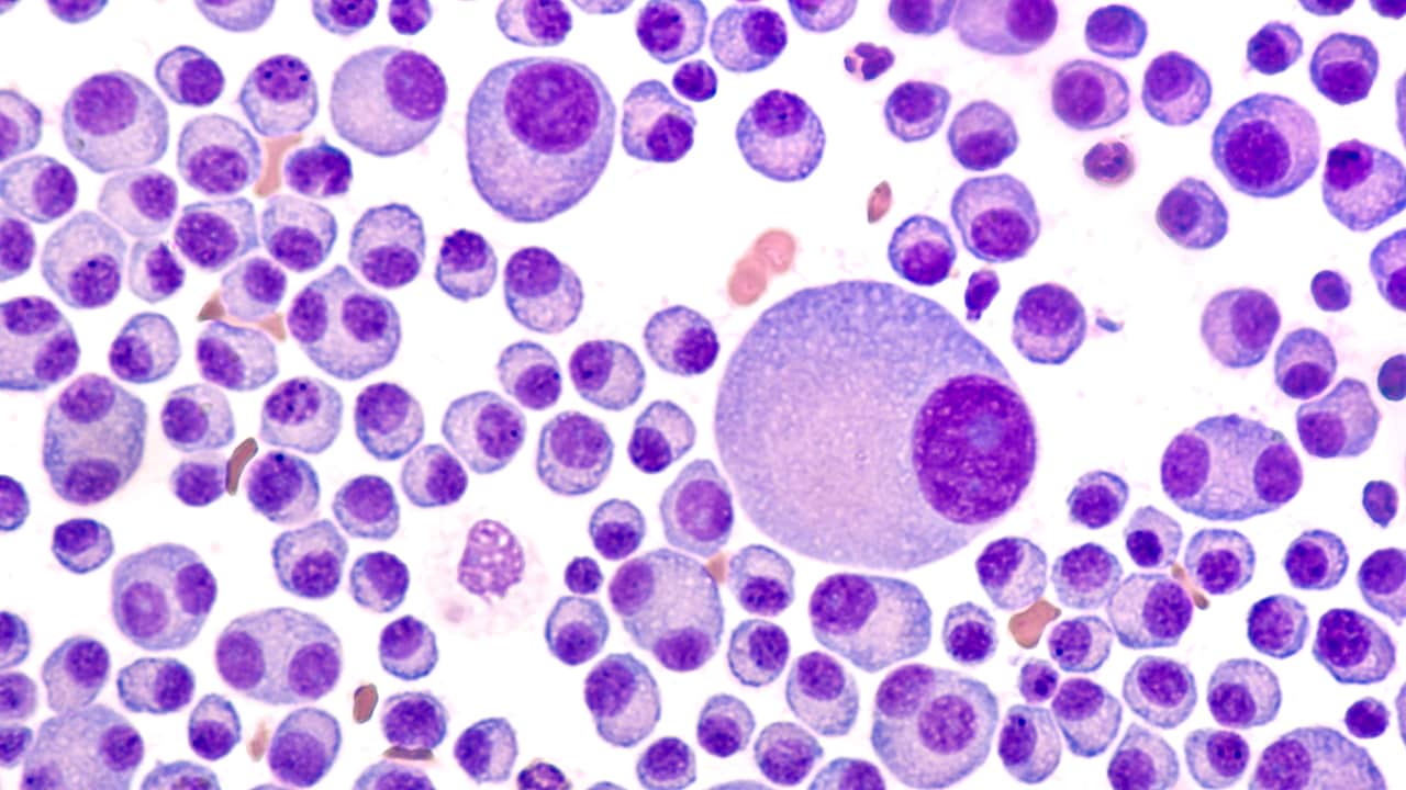
Monoclonal gammopathy of undetermined significance (MGUS) is the most common of a spectrum of diseases called plasma cell dyscrasias. The term MGUS denotes the presence of a monoclonal immunoglobulin (Ig), also called an M-protein, in the serum or urine in persons without evidence of multiple myeloma (MM), Waldenström macroglobulinemia (WM), amyloidosis (AL) or other lymphoproliferative disorders. [1]
Immunoglobulin involvement may be IgM, non-IgM (ie, IgA, IgG, or, rarely, IgD or IgE), or light chain. [2] All pose a risk, albeit varying, of progression to a malignant disorder. Typically, IgG and IgA MGUS progress to MM, IgM MGUS progresses to WM or other lymphoproliferative disorders, and light-chain MGUS is the precursor of light-chain MM. [3]
More recently, MGUS has been differentiated from monoclonal gammopathy of renal significance (MGRS), which includes kidney and sometimes systemic lesions in addition to the hematologic findings of MGUS. [4] Early recognition of MGRS is critical, as suppression of monoclonal immunoglobulin secretion by chemotherapy often improves outcomes. [5, 6] Similarly, MGUS with peripheral neuropathy has been designated as monoclonal gammopathy of neurologic significance; this condition occurs more often in IgM MGUS and typically involves slowly progressive, symmetrical, length-dependent sensory deficits. [7] Recognition that monoclonal gammopathies can involve not only kidneys and nerves but skin and sometimes other organs led to coining of the term monoclonal gammopathies of clinical significance (MGCS). [6, 8]
The cause of MGUS is unknown, though the same theories that apply to the pathogenesis of MM may be valid in MGUS. MM is almost always preceded by MGUS. [9]
Distinguishing between MM and MGUS is critical because patients with MGUS are conservatively treated and do not need chemotherapy. In contrast, MM is a uniformly lethal disease that does not need therapy in its initial stages but progresses to an advanced or aggressive stage that requires therapy. [10]
Pathophysiology
The reason for the monoclonal expansion of a single Ig-secreting plasma cell population in what appears to be a nonmalignant manner is unknown in most cases. Risk factors for MGUS have been identified. Nonmodifiable risk factors include the following [11] :
-
Older age
-
Male sex
-
Black race
-
Family history of MGUS and related diseases
Other risk factors include the following [11] :
-
Specific prior infections (eg, pneumonia, hepatitis, meningitis, HIV [12] )
-
Inflammatory disorders
-
Autoimmune disease
-
Smoking
-
Pesticide exposure (eg, aldrin, dieldrin, permethrin) [13]
Overweight/obesity has been inconsistently linked to the development of MGUS and its transition to multiple myeloma (MM). [11, 14]
Most cases of MGUS involve IgG or IgA monoclonal cell populations. About 15-20% are composed of IgM monoclonal cells. Kyle et al reported that the cells in IgG and IgA MGUS arise from a mature, somatically mutated, postswitch plasma cell. About 50% of cases have evidence of translocation in the Ig heavy-chain region at 14q32. In contrast, IgM MGUS is described as arising from somatically mutated, postgerminal center B lymphocytes that have not undergone isotype class switching and therefore do not have the 14q32 translocation. These translocations are thought to be important in initiating and sustaining clonal proliferation. [15]
Several studies have confirmed that characteristic genetic abnormalities of MM are present in patients with MGUS. [16, 17, 18] Gene-expression profiling has also led to a group with MGUS-like features. This group of patients had a lower complete remission rate, yet also had a lower-risk clinical course and superior survival. [19]
Nagoshi et al identified a possible secondary genetic change involving MGUS; they found transcriptional dysregulation of the deleted in colorectal carcinoma (DCC) gene in 25% of MGUS cases studied, and in 57% of MM. The DCC gene encodes a tumor suppressor that prevents cell growth; allele loss or decreased expression of DCC has been associated with the progression of solid tumors and hematologic malignancies. [20]
The risk of progression to MM or other lymphoproliferative disorder is present at a constant rate throughout the remainder of a patient's life. This observation suggests that the second event responsible for progression is a random event and not cumulative.
In patients with symptomatic MM, the development of a new monoclonal protein following therapy is associated with better outcomes. A study by Mullikin et al in patients with biclonal gammopathy of undetermined significance found that the rate of progression was similar to that seen in MGUS with one monoclonal protein, suggesting that multiple monoclonal proteins do not clinically impact one another. Of the 393 patients in the study, six progressed to smoldering MM, 11 to MM, three to amyloidosis, and two to Waldenström macroglobulinemia. [21]
Epidemiology
MGUS represents two thirds of all plasma cell dyscrasias. The incidence increases with age. In a study of residents of Olmsted County, Minnesota, MGUS was found in 3.2% of persons 50 years of age or older, 5.3% of those 70 years of age or older, and 7.5% of those 85 years of age or older. [22] Subsequently, however, Murray et al used mass spectrometry to retest the baseline samples in 300 of the Olmsted County residents who had a negative work-up for monoclonal proteins but later developed MGUS. This more sensitive assay revealed a prevalence of MGUS of 5.1% among persons 50 years of age and older. [23]
In a study of 154,597 persons in Beijing, China who underwent annual medical checkups, median age at presentation with MGUS was 58 years (range, 25–96). The prevalence of MGUS increased with increasing age: the overall prevalence was 1.11% among participants aged ≥50 years, 2.57% among those aged ≥70 years, and 3.77% in those ≥80 years. [24]
The prevalence of MGUS is higher in HIV-infected patients, although it decreased with the adoption of antiretroviral therapy. In a study of 383 French HIV-infected patients, 359 of whom were on antiretroviral therapy for a median duration of 105 months, there were 12 (3.1%) cases of MGUS, including five IgG kappa cases, 5 IgG lambda cases, one biclonal (2 IgG kappa) case, and one case with three monoclonal immunoglobulins (IgG kappa×2+IgG lambda). In all cases, the monoclonal immunoglobulin levels were low, and the level was below 1 g/L in all cases except two. No factors were found to be predictive of MGUS. [12]
In a prospective cohort study in Vietnam war veterans, the crude prevalence of overall MGUS was 7.1% (34 of 479) in veterans involved in the spraying of Agent Orange, versus 3.1% (15 of 479) in veterans who were not involved in herbicide spray missions. After adjustment for factors including age, race, and body mass index, this translated into a 2.4-fold increased risk for MGUS in exposed veterans (adjusted odds ratio, 2.37; 95% CI, 1.27-4.44; P = 0.007). [25]
In a study of firefighters involved in rescue and/or recovery work at the New York World Trade Center after the September 11, 2001 attack, Landdgren et al reported an age-standardized prevalence rate of MGUS and light-chain MGUS combined of 7.63 per 100 persons (95% CI, 5.45-9.81), which is 1.8-fold higher than rates from a reference population; the rate of light-chain MGUS was more than 3-fold higher than in the same reference population. Of the 781 firefighters studied, 16 had been diagnosed with multiple myeloma (7 of them with light-chain disease), at a median age of 57 years. [26]
Mortality/Morbidity
Patients with MGUS tend to do well when treated conservatively. [27] Regular surveillance is required to assess for progression to either a lymphoproliferative disorder or to MM. [28] This risk has been quantified at 1% per year. [29]
Race-, Sex-, and Age-related Variances
A retrospective study by the US Department of Veterans Affairs revealed that the age-adjusted prevalence ratio of MGUS in black patients was 3.0 compared with white patients. [30] For a discussion of possible genetic factors in the pathogenesis of MGUS, see Landgren et al. [30, 31]
MGUS occurs more commonly in men than in women, and the prognosis for men was worse than that of women in some studies.
Age-related differences in incidence are as follows:
-
The median age of patients with the disease is 70 years; however, most physicians are observing patients younger than this, possibly because of improved screening rather than an increased incidence of the process.
-
The incidence of MGUS is higher in older patients than in younger patients; of patients older than 80 years, the available literature suggests as many as 10-15% may have an M-protein.
Prognosis
The annual risk of progression to multiple myeloma (MM), Waldenström macroglobulinemia (WM), amyloidosis (AL), or other lymphoproliferative disorders is approximately 1%. However, the mode and risk of progression vary between IgM MGUS and those with non-IgM MGUS.
An abnormal serum free light-chain ratio (ratio of kappa to lambda free light chains) and a high serum monoclonal protein (M protein) level (≥1.5 g per deciliter) are risk factors for progression. In IgM MGUS, Kyle et al reported a risk of progression at 20 years of 55% in patients with both risk factors, compared with 41% in those with one risk factor and 19% in patients with neither risk factor. In patients with non-IgM MGUS, the risk of progression at 20 years was 30% in those with both risk factors, 20% in those iwth one risk factor, and 7% in those with neither risk factor. [29]
Rajkumar et al found that the risk of progression in patients with an abnormal serum free light chain (FLC) ratio (kappa-lambda ratio < 0.26 or > 1.65) is independent of the size and type of the serum M protein, and that the relative risk of progression is related to the extent to which the ratio is abnormal. These authors proposed a risk-stratification model for the progression of MGUS, using the combination of the size and type of the M protein and the serum FLC ratio. [32]
Pelzer et al concluded that light-chain MGUS—defined as an abnormal FLC ratio, increase of involved FLC with complete loss of immunoglobulin heavy chain, and absence of a history of lymphoproliferative disease—is a relatively benign condition, and that the monoclonal protein often diisappears over time. In their longitudinal analysis of 75 German patients with light-chain MGUS, after a median observation time of 11.5 years, none of the cases had progressed to light-chain multiple myeloma or other lymphoproliferative disorders. On serial analysis, light-chain MGUS could not be confirmed in 17 of 31 cases (55%), and disappearance of the monoclonal protein was associated with low concentrations of the involved FLC. Although patients with light-chain MGUS had a 1.5-fold increased risk of cancer, overall survival and renal function were not different than in patients with normal FLC. [33]
In a study of 728 Swedish MGUS patients followed for up to 30 years, 84 patients developed a lymphoid disorder, representing a cumulative risk of 15.4%. The 30-year cumulative risk for myeloid malignancies was less than 2%. The 30-year cumulative risk for MM, which occurred in 53 patients, was 10.6%, with an approximately 0.5% annual risk. The following factors were significantly associated with progression [34] :
-
An abnormal FLC ratio of less than 0.26 or more than 1.65
-
An M-protein concentration of 1.5 g/dL or more
-
Reduction of one or two noninvolved immunoglobulin isotype levels
Although autoimmune disease is a well-described risk factor for the development of MGUS, a Swedish population-based study determined that patients with a history of autoimmune disease have a significantly lower risk of progression from MGUS to MM or other lymphoproliferative diseases. The study included 19,303 MGUS patients, 5612 (29.1%) of whom had preceding autoimmune diseases. [35] Similarly, a Mayo Clinic study of 249 young patients with MGUS (age < 40 years), 135 of whom had immune-related conditions, reported a trend toward higher risk of progression in patients without immune-related conditions (hazard ratio 2.36, 95% CI 0.85–6.52, P = 0.088). [36]
Venous thromboembolism
The risk of venous thromboembolism (VTE) is increased in patients with MGUS. [37] In a study by Srkalovic et al, 13% of patients with MGUS developed VTE. [38] Univariate correlates of VTE in patients with MGUS included the following:
-
Family or past history of VTE
-
Immobility
-
Low serum albumin level
-
High leukocyte count
Osteoporosis
Using population-based data from Sweden, Kristinsson et al compared the risks of fractures in 5,326 patients with MGUS diagnosed from 1958-2006 with 20,161 matched controls. [39] Patients with MGUS had an increased risk of any fracture at 5 years (hazard ratio [HR] = 1.74) and 10 (HR = 1.61) years. The risk was significantly higher for axial (skull, vertebral/pelvis, and sternum/costae) compared with distal (arm and leg) fractures (P< 0.001).
A French study found that, in addition to low bone density, patients with MGUS who experienced nontraumatic vertebral fractures were more likely to have a lambda light chain isotype. In this prospective study of 201 patients with incidentally discovered MGUS and no known history of osteoporosis, nontraumatic vertebral fracture was discovered in 8.4% of the patients, with equal distribution between men and women. Patients with lambda light chain had an odds ratio of 4.32 (95% confidence interval 1.80–11.16; P=0.002) for fracture, compared with patients with kappa light chain. [40]
Infection
Kristinsson et al reported that patients with MGUS had a 2-fold increased risk (P < 0.05) of developing any infection. [41] The risk extended to both bacterial and viral infections, and the following specific infections were noted:
-
Pneumonia
-
Osteomyelitis
-
Septicemia
-
Pyelonephritis
-
Cellulitis
-
Endocarditis
-
Meningitis
-
Influenza
-
Herpes zoster
Infection risk was highest in patients with M-protein concentrations over 2.5 g/dL, but was also increased in those with concentrations below 0.5 g/dL. Patients with MGUS who developed infections had no excess risk of progression to related malignancy.