| Patient Education |
|
Click here for patient education.
|
|
AUTHOR AND EDITOR INFORMATION
Section 1 of 11
Author: Sameer Yaseen, MD, Staff Nephrologist, Department of Internal Medicine, Division of Nephrology, Mercy Hospital of Des Moines
Sameer Yaseen is a member of the following medical societies: American College of Physicians, American Heart Association, American Medical Association, American Society of Nephrology, and National Kidney Foundation
Coauthor(s):
Christie Thomas, MD, FACP, FAHA, FASN, Department of Internal Medicine, Division of Nephrology, Professor, University of Iowa Hospitals and Clinics
Editors: Anil Kumar Mandal, MD, Clinical Professor, Department of Internal Medicine, Division of Nephrology, University of Florida School of Medicine; Francisco Talavera, PharmD, PhD, Senior Pharmacy Editor, eMedicine; Eleanor Lederer, MD, Consulting Staff, Louisville VA Hospital; Professor of Medicine, Director of Nephrology Training Program, Kidney Disease Program, University of Louisville School of Medicine; Director, Metabolic Stone Clinic; Rebecca J Schmidt, DO, FACP, FASN, Clinical Associate Professor of Medicine, West Virginia School of Osteopathic Medicine; Professor of Medicine, Section Chief, Department of Medicine, Section of Nephrology, West Virginia University School of Medicine; Vecihi Batuman, MD, Professor of Medicine, Chief, Section of Nephrology, Tulane University School of Medicine; Chief, Renal-Hypertension Section, Department of Medicine, Tulane University Medical Center, New Orleans Veterans Affairs Medical Center
Author and Editor Disclosure
Synonyms and related keywords:
bicarbonate, chloride-resistant alkalosis, chloride-responsive alkalosis
Background
For a general review of acid-base regulation, see Metabolic Acidosis.
Metabolic alkalosis is a primary increase in serum bicarbonate (HCO3-) concentration. This occurs as a consequence of a loss of H+ from the body or a gain in HCO3-. In its pure form, it manifests as alkalemia (pH >7.40). As a compensatory mechanism, metabolic alkalosis leads to alveolar hypoventilation with a rise in arterial carbon dioxide tension (PaCO2), which diminishes the change in pH that would otherwise occur.
Normally, arterial PaCO2 increases by 0.5-0.7 mm Hg for every 1 mEq/L increase in plasma bicarbonate concentration, a compensatory response that is very quick. If the change in PaCO2 is not within this range, then a mixed acid-base disturbance occurs. For example, if the increase in PaCO2 is more than 0.7 times the increase in bicarbonate, then metabolic alkalosis coexists with primary respiratory acidosis. Likewise, if the increase in PaCO2 is less than the expected change, then a primary respiratory alkalosis is also present.
The first clue to metabolic alkalosis is often an elevated bicarbonate concentration that is observed when serum electrolytes are obtained. Remember that an elevated serum bicarbonate concentration may also be observed as a compensatory response to primary respiratory acidosis. However, a bicarbonate concentration greater than 35 mEq/L is almost always caused by metabolic alkalosis.
Calculation of the serum anion gap may also help to differentiate between primary metabolic alkalosis and the metabolic compensation for respiratory acidosis. The anion gap is frequently elevated to a modest degree in metabolic alkalosis because of the increase in the negative charge of albumin and the enhanced production of lactate. However, the only definitive way to diagnose metabolic alkalosis is by performing a simultaneous blood gases analysis, which reveals elevation of both pH and PaCO2 and increased calculated bicarbonate.
Two ways to determine the serum bicarbonate concentration exist. The first method is calculating serum bicarbonate concentration from a blood gas sample using the Henderson-Hasselbalch equation, as follows:
pH = 6.10 + log (HCO3- ÷ .03 X PaCO2)
Alternatively, HCO3- = 24 X PaCO2 ÷ [H+]
Because pH and PaCO2 are directly measured, bicarbonate can be calculated.
The second method is measuring the total carbon dioxide content in serum, which is routinely measured with serum electrolytes obtained from venous blood. In this method, a strong acid is added to serum, which interacts with bicarbonate in the serum sample, forming carbonic acid. Carbonic acid dissociates to carbon dioxide and water; then, carbon dioxide is measured. Note that the carbon dioxide measured includes bicarbonate and dissolved carbon dioxide. The contribution of dissolved carbon dioxide is quite small (0.03 X PaCO2) and is usually ignored, although it accounts for a difference of 1-3 mEq/L between the measured total carbon dioxide content in venous blood and the calculated bicarbonate in arterial blood. Thus, at a PaCO2 of 40, a total carbon dioxide content of 25 means a true bicarbonate concentration of 23.8 (ie, 25 - 0.03 X 40).
Pathophysiology
The organ systems involved are mainly the kidneys and GI tract. The pathogenesis of metabolic alkalosis involves 2 processes, the generation of metabolic alkalosis and the maintenance of metabolic alkalosis, events that usually overlap. The generation of metabolic alkalosis occurs with the loss of acid, the gain of alkali, or the contraction of the extracellular fluid compartment with a consequent change in bicarbonate concentration. The kidneys usually have an enormous capacity to excrete excess bicarbonate generated and to restore normal acid-base balance by the following mechanisms: (1) less reabsorption of bicarbonate because infused sodium bicarbonate (NaHCO3) leads to volume expansion, which reduces reabsorption of sodium ions and bicarbonate in the proximal tubule, and (2) bicarbonate secretion by B-type intercalated cells in the collecting duct that exchange bicarbonate for chloride via the apical chloride/bicarbonate (Cl-/HCO3-) countertransporter. Therefore, to sustain metabolic alkalosis, the kidneys must participate to maintain the alkalosis by overriding these mechanisms. Generation of metabolic alkalosis Metabolic alkalosis may be generated by one of the following mechanisms: Loss of hydrogen ions: Whenever a hydrogen ion is excreted, a bicarbonate ion is gained into the extracellular space. Hydrogen ions may be lost through the kidneys or the GI tract. Vomiting or nasogastric (NG) suction generates metabolic alkalosis by the loss of gastric secretions, which are rich in hydrochloric acid (HCl). Renal losses of hydrogen ions occur whenever the distal delivery of sodium increases in the presence of excess aldosterone, which stimulates the electrogenic epithelial sodium channel (ENaC) in the collecting duct. As this channel reabsorbs sodium ions, the tubular lumen becomes more negative, leading to the secretion of hydrogen ions and potassium ions into the lumen. Shift of hydrogen ions into the intracellular space: This mainly develops with hypokalemia. As the extracellular potassium concentration decreases, potassium ions move out of the cells. To maintain neutrality, hydrogen ions move into the intracellular space. Alkali administration: Administration of sodium bicarbonate in amounts that exceed the capacity of the kidneys to excrete this excess bicarbonate may cause metabolic alkalosis. This capacity is reduced when a reduction in filtered bicarbonate occurs, as observed in renal failure, or when enhanced tubular reabsorption of bicarbonate occurs, as observed in volume depletion (see Maintenance of metabolic alkalosis). Contraction alkalosis: Loss of bicarbonate-poor, chloride-rich extracellular fluid, as observed with thiazide diuretic or loop diuretic therapy or chloride diarrhea, leads to contraction of extracellular fluid volume. Because the original bicarbonate mass is now dissolved in a smaller volume of fluid, an increase in bicarbonate concentration occurs. This increase in bicarbonate causes, at most, a 2- to 4-mEq/L rise in bicarbonate concentration. Maintenance of metabolic alkalosis The following factors are believed to maintain the alkalosis: Decrease in renal perfusion: Decreased perfusion to the kidneys, caused by either volume depletion or a reduction in effective circulating blood volume (eg, edematous states such as heart failure or cirrhosis) stimulates the renin-angiotensin-aldosterone system. This increases renal sodium ion reabsorption throughout the nephron, including the principal cells of the collecting duct. This results in enhanced hydrogen ion secretion via the apical proton pump H+ ATPase in the adjacent A-type intercalated cells. Aldosterone may also independently increase the activity of the apical proton pump. Whenever a hydrogen ion is secreted into the tubular lumen, a bicarbonate ion is gained into the systemic circulation via the basolateral Cl-/HCO3- exchanger. Chloride depletion: Chloride depletion may occur through the GI tract by loss of gastric secretions, which are rich in chloride ions, or through the kidneys with loop diuretics or thiazides. Chloride depletion, even without volume depletion, enhances bicarbonate reabsorption by different mechanisms, as follows:
- Stimulation of the renin-angiotensin-aldosterone system: In the late thick ascending limb and early distal tubule, specialized cells called the macula densa are present. These cells have an Na+/K+/2Cl- cotransporter in the apical membrane, which is mainly regulated by chloride ions. When fewer chloride ions reach this transporter (eg, chloride depletion), the macula densa signals the juxtaglomerular apparatus (ie, specialized cells in the wall of the adjacent afferent arteriole) to secrete renin, which increases aldosterone secretion via angiotensin II.
- Inhibition of bicarbonate secretion by the chloride/bicarbonate exchanger: In alkalemia, the kidneys secrete the excess bicarbonate via the apical chloride/bicarbonate exchanger in the B-type intercalated cells of the collecting duct. In this way, protons are gained to the systemic circulation via the basolateral H+ ATPase. In chloride depletion, fewer chloride ions are available to be exchanged with bicarbonate, and the ability of the kidneys to excrete the excess bicarbonate is impaired.
Hypokalemia: Many of the causes of metabolic alkalosis are also associated with hypokalemia, which maintains metabolic alkalosis by different mechanisms. These include the following:
- Shift of hydrogen ions intracellularly: Intracellular acidosis enhances bicarbonate reabsorption in the collecting duct.
- Stimulation of the apical H+/K+ ATPase in the collecting duct: Increased activity of this ATPase leads to teleologically appropriate potassium ion reabsorption but a corresponding hydrogen ion secretion. This leads to a net gain of bicarbonate, maintaining systemic alkalosis.
- Stimulation of renal ammonia genesis: Ammonium ions (NH4+) are produced in the proximal tubule from the metabolism of glutamine. During this process, alpha-ketoglutarate is produced, the metabolism of which generates bicarbonate that is returned to the systemic circulation.
- Impaired chloride ion reabsorption in the distal nephron: This results in an increase in luminal electronegativity, with subsequent enhancement of hydrogen ion secretion.
- Reduction in glomerular filtration rate (GFR): This has been proven in animal studies. Hypokalemia by unknown mechanisms decreases GFR, which in turn decreases the filtered load of bicarbonate. In the presence of volume depletion, this impairs renal excretion of the excess bicarbonate.
Frequency
United States
Metabolic alkalosis is the most common acid-base disturbance observed in hospitalized patients, accounting for approximately 50% of all acid-base disorders.
Mortality/Morbidity
Severe metabolic alkalosis (ie, blood pH >7.55) is a serious medical problem. Mortality rates have been reported as 45% in patients with an arterial blood pH of 7.55 and 80% when the pH was greater than 7.65.
- Severe alkalosis causes diffuse arteriolar constriction with reduction in tissue perfusion. By decreasing cerebral blood flow, alkalosis may lead to tetany, seizures, and decreased mental status. Metabolic alkalosis also decreases coronary blood flow and predisposes persons to refractory arrhythmias.
-
- Metabolic alkalosis causes hypoventilation, which may cause hypoxemia, especially in patients with poor respiratory reserve, and it may impair weaning from mechanical ventilation.
-
- Alkalosis decreases the serum concentration of ionized calcium by increasing calcium ion binding to albumin. In addition, metabolic alkalosis is almost always associated with hypokalemia, which can cause neuromuscular weakness and arrhythmias, and, by increasing ammonia production, it can precipitate hepatic encephalopathy in susceptible individuals.
History
Symptoms of metabolic alkalosis are not specific. Because hypokalemia is usually present, the patient may experience weakness, myalgia, and polyuria. Hypoventilation develops because of inhibition of the respiratory center in the medulla. Symptoms of hypocalcemia (eg, jitteriness, perioral tingling, muscle spasms) may be present. The clinical history is helpful in establishing the etiology. Important points in the history include the following:
- Vomiting or diarrhea - GI losses of HCl
-
- Age of onset and family history of alkalosis - Familial disorders (eg, Bartter syndrome, which starts during childhood)
-
- Renal failure - Alkali-loading alkalosis only develops when impairment of renal function occurs.
-
- Drug use
-
- Loop or thiazide diuretics
-
- Licorice
-
- Tobacco chewing
-
- Carbenoxolone
-
- Fludrocortisone
-
- Glucocorticoids
-
- Antacids (eg, magnesium hydroxide)
-
- Calcium carbonate
-
Physical
The physical signs of metabolic alkalosis are not specific and depend on the severity of the alkalosis. Because metabolic alkalosis decreases ionized calcium concentration, signs of hypocalcemia (eg, tetany, Chvostek sign, Trousseau sign), change in mental status, or seizures may be present. Physical examination is helpful to establish the cause of metabolic alkalosis. Important aspects of the physical examination include the evaluation of hypertension and volume status assessment. Hypertension accompanies several causes of metabolic alkalosis. See Summary of causes of metabolic alkalosis. Volume status assessment includes evaluation of orthostatic changes in blood pressure and heart rate, mucous membranes, presence or absence of edema, skin turgor, weight change, and urine output. Volume depletion usually accompanies chloride-responsive alkalosis, while volume expansion accompanies chloride-resistant alkalosis.
- Bulimia: Because patients with bulimia frequently self-induce vomiting, they may have erosions of teeth enamel and dental caries because of repeatedly exposing their teeth to gastric acid.
- Cushing syndrome
- Obesity
- Moon face
- Buffalo hump
- Hirsutism
- Violaceous skin striae
- Acne
- Congenital adrenal hyperplasia (CAH): In CAH secondary to 11-hydroxylase deficiency, the infants have hypertension and growth retardation. Male infants have premature sexual development, while female infants develop virilization. In 17-hydroxylase deficiency, males develop sexual ambiguity, while females have sexual infantilism.
Causes
See Summary of causes of metabolic alkalosis for a list of causes and Image 2 for an algorithm to approach metabolic alkalosis. The most common causes of metabolic alkalosis are the use of diuretics and the external loss of gastric secretions. Causes of metabolic alkalosis can be divided into chloride-responsive alkalosis (urine chloride <20 mEq/L), chloride-resistant alkalosis (urine chloride >20 mEq/L), and other causes, including alkali-loading alkalosis.
Chloride-responsive alkalosis (urine chloride <20 mEq/L)Loss of gastric secretions Gastric secretions are rich in HCl. The secretion of HCl by the stomach usually stimulates bicarbonate secretion by the pancreas once HCl reaches the duodenum. Ordinarily, these substances are neutralized, and no net gain or loss of hydrogen ions or bicarbonate occurs. When HCl is lost by vomiting or NG suction, pancreatic secretions are not stimulated and a net gain of bicarbonate into the systemic circulation occurs, generating a metabolic alkalosis. Volume depletion maintains alkalosis. In this case, the hypokalemia is secondary to the alkalosis itself and to renal loss of potassium ions from the stimulation of aldosterone secretion. Ingestion of large doses of nonabsorbable antacids Ingestion of large doses of nonabsorbable antacids (eg, magnesium hydroxide) may generate metabolic alkalosis by a rather complicated mechanism. Upon ingestion of magnesium hydroxide, calcium, or aluminum with base hydroxide or carbonate, the hydroxide anion buffers hydrogen ions in the stomach. The cation binds to bicarbonate secreted by the pancreas, leading to loss of bicarbonate with stools. In this process, both hydrogen ions and bicarbonate are lost, and, usually, no acid-base disturbance occurs. Sometimes, not all the bicarbonate binds to the ingested cation, which means that some bicarbonate is reabsorbed in excess of the lost hydrogen ions. This occurs primarily when antacids are administered with a cation-exchange resin (eg, sodium polystyrene sulfonate [Kayexalate]); the resin binds the cation, leaving bicarbonate unbound. Thiazide or loop diuretics Alkalosis occurs. However, urine chloride may not be less than 20 mEq/L. Thiazides and loop diuretics enhance sodium chloride excretion in the distal convoluted tubule and the thick ascending loop, respectively. These agents cause metabolic alkalosis by chloride depletion and by increased delivery of sodium ions to the collecting duct, which enhances potassium ion and hydrogen ion secretion. Volume depletion also stimulates aldosterone secretion, which enhances sodium ion reabsorption in the collecting duct and increases hydrogen ion and potassium secretion in this segment. Urine chloride is low after discontinuation of diuretic therapy, while it is high during active diuretic use. Miscellaneous causes Villous adenomas (rare cause of diarrhea) usually lead to metabolic acidosis from loss of colonic secretions that are rich in bicarbonate. Occasionally, these tumors cause metabolic alkalosis. The mechanism is not well understood. Some authors opine that the hypokalemia caused by these tumors is the etiology of metabolic alkalosis. Congenital chloridorrhea (see Chloride Diarrhea, Familial. Online Mendelian Inheritance in Man [OMIM]) is a rare form of severe secretory diarrhea that is inherited as an autosomal recessive trait. Mutations in the down-regulated adenoma gene result in defective function of the chloride/bicarbonate exchange in the colon and ileum, leading to increased secretion of chloride and reabsorption of bicarbonate. During respiratory acidosis, the kidneys reabsorb bicarbonate and secrete chloride to compensate for the acidosis. In the posthypercapnic state, urine chloride is high and can lead to chloride depletion. Once the respiratory acidosis is corrected, the kidneys cannot excrete the excess bicarbonate because of the low luminal chloride. Infants with cystic fibrosis (see Cystic Fibrosis [OMIM]) may develop metabolic alkalosis because of loss of chloride in sweat. These infants are also prone to volume depletion.
Chloride-resistant alkalosis (urine chloride >20 mEq/L)Chloride-resistant alkalosis with hypertension An adrenal adenoma (most common), bilateral adrenal hyperplasia, or an adrenal carcinoma may cause primary hyperaldosteronism. Another cause of primary hyperaldosteronism is glucocorticoid-remediable aldosteronism (see Hyperaldosteronism, Familial, Type 1 [OMIM]), an autosomal dominant disorder, in which ectopic production of aldosterone in the zona fasciculata of the adrenal cortex occurs. In this case, aldosterone production is controlled by adrenocorticotropic hormone (ACTH) rather than angiotensin II and potassium, its principal regulators. This type of primary hyperaldosteronism is responsive to glucocorticoid therapy, which inhibits aldosterone secretion by suppressing ACTH. The mineralocorticoid receptor (MR) in the collecting duct usually is responsive to both aldosterone and cortisol. Cortisol has a higher affinity for MR and circulates at a higher concentration than aldosterone. Under physiological conditions, the enzyme 11-beta-hydroxysteroid dehydrogenase type 2 (11B-HSD2) inactivates cortisol to cortisone in the collecting duct, allowing aldosterone free access to its receptor. Deficiency of this enzyme leads to occupation and activation of the MR by cortisol, which, like aldosterone, then stimulates the ENaC. Cortisol behaves as a mineralocorticoid under these circumstances. 11B-HSD2 deficiency may be inherited as an autosomal recessive trait (see Cortisol 11-Beta-Ketoreductase Deficiency [OMIM]), ie, syndrome of apparent mineralocorticoid excess (AME), or the enzyme may be inhibited by the use of licorice, carbenoxolone, or chewing tobacco. The active component in licorice is glycyrrhizic acid. This compound is found in certain candies and some chewing tobaccos. Inhibition or deficiency of 11B-HSD2 causes hypertension with low renin and low aldosterone, hypokalemia, and metabolic alkalosis. Serum cortisol is within the reference range because the negative feedback of cortisol on ACTH is intact. Active use of thiazides or loop diuretics in hypertension is the most common cause of metabolic alkalosis in hypertensive patients. The mechanism of alkalosis is discussed in Thiazide or loop diuretics. The enhanced mineralocorticoid effect in Cushing syndrome is caused by occupation of the MR by the high concentration of cortisol. Hypokalemia and metabolic alkalosis are more common in Cushing syndrome caused by ectopic ACTH production (90%) than in other causes of Cushing syndrome (10%). This difference is related to the higher concentration of plasma cortisol and the defective 11-beta-hydroxysteroid dehydrogenase (11B-HSD) activity found in ectopic ACTH production. Liddle syndrome (see Liddle Syndrome [OMIM]) is a rare autosomal dominant disorder arising from a gain of function mutation in the beta (SCNN1B) or gamma subunit (SCNN1G) of the ENaC in the collecting duct. The channel behaves as if it is permanently open, and unregulated reabsorption of Na+ occurs, leading to volume expansion and hypertension. This unregulated Na+ reabsorption is responsible for secondary renal hydrogen ion and potassium ion losses and persists despite suppression of aldosterone. Significant unilateral or bilateral renal artery stenosis stimulates the renin-angiotensin-aldosterone system, leading to hypertension and hypokalemic metabolic alkalosis. Renin- or deoxycorticosterone-secreting tumors are rare. In renin-secreting tumors, excessive amounts of renin are secreted by tumors in the juxtaglomerular apparatus, stimulating aldosterone secretion. In the latter, deoxycorticosterone (DOC), rather than aldosterone, is secreted by some adrenal tumors and has mineralocorticoid effects. Mutation in mineralocorticoid receptor (see Hypertension, Early-Onset, Autosomal Dominant, with Severe Exacerbation in Pregnancy [OMIM]) is a form of early-onset hypertension with autosomal dominant inheritance that has now been linked to a specific mutation of the MR. This mutation results in constitutive activation of the MR, making the MR responsive to progesterone. Activation of MR leads to unregulated sodium ion reabsorption via the collecting duct sodium ion channel, with accompanying hypokalemia and alkalosis. The disease is characterized by severe exacerbations of hypertension during pregnancy, and spironolactone can exacerbate hypertension. CAH (see Adrenal Hyperplasia, Congenital, Due to 11-Beta-Hydroxylase Deficiency [OMIM] and Adrenal Hyperplasia, Congenital, Due to 17-Alpha-Hydroxylase Deficiency [OMIM]) can be caused by deficiency of either 11-beta-hydroxylase or 17-alpha-hydroxylase. Both enzymes are involved in the synthesis of adrenal steroids. Deficiency of either enzyme leads to increased levels of the mineralocorticoid 11-deoxycortisol, while cortisol and aldosterone production is impaired. 11-Hydroxylase deficiency differs from 17-hydroxylase deficiency by the presence of virilization. Chloride-resistant alkalosis (urine chloride >20 mEq/L) with hypotension or normotension Bartter syndrome (see Hypokalemic Alkalosis with Hypercalciuria [OMIM]) is an inherited autosomal recessive disorder, in which reabsorption of sodium ions and chloride ions in the thick ascending loop of Henle is impaired, leading to their increased delivery to the distal nephron. This condition and the subsequent salt depletion and stimulation of the renin-angiotensin-aldosterone system lead to enhanced secretion of hydrogen and potassium ions. The impaired reabsorption of sodium chloride in the loop of Henle is secondary to loss of function of mutations of 1 of 3 transporters in this site of the nephron: (1) the furosemide-sensitive Na+/K+/2Cl- cotransporter (NKCC2), (2) the basolateral chloride ion channel (CLCNKB), or (3) the inwardly rectifying apical potassium ion channel (ROMK1). Mutations of CLCNKB cause classic Bartter syndrome, while mutations of the other 2 transporters manifest with the antenatal form of Bartter syndrome. Edema and hypertension are absent, and hypercalciuria is common because the impaired reabsorption of sodium chloride inhibits the paracellular reabsorption of calcium. Because loop diuretics inhibit the Na+/K+/2Cl- transporter, the electrolyte abnormalities observed in Bartter syndrome and with loop diuretic use are similar. Gitelman syndrome (see Potassium and Magnesium Depletion [OMIM]) is an inherited autosomal recessive disorder, in which loss of function of the thiazide-sensitive sodium/chloride transporter (NCCT) in the distal convoluted tubule occurs. The subsequent increased distal solute delivery and salt wasting with stimulation of the renin-angiotensin-aldosterone system lead to hypokalemic metabolic alkalosis. Other features of the syndrome are hypocalciuria and hypomagnesemia. The electrolyte abnormalities resemble those caused by thiazide diuretic use. Pure hypokalemia (ie, severe potassium ion depletion) causes mild metabolic alkalosis, but, in combination with hyperaldosteronism, the alkalosis is more severe. Possible mechanisms of alkalosis in hypokalemia are enhanced proximal bicarbonate reabsorption, stimulated renal ammonia genesis, impaired renal chloride reabsorption, reduced GFR (in animals), and intracellular acidosis in the distal nephron with subsequent enhanced hydrogen secretion. Magnesium depletion (ie, hypomagnesemia) may lead to metabolic alkalosis. The mechanism probably is caused by hypokalemia, which is usually caused by or associated with magnesium depletion.
Other causesAlkali-loading alkalosis The kidneys are able to excrete any excess alkali load, whether it is exogenous (eg, infusion of sodium bicarbonate) or endogenous (eg, metabolism of lactate to bicarbonate in lactic acidosis). However, in renal failure or in any condition that maintains the alkalosis, this natural ability of the kidneys to excrete the excess bicarbonate is impaired. Examples include the following: The components of milk alkali syndrome are hypercalcemia, renal insufficiency, and metabolic alkalosis. Before the advent of H2-receptor antagonists, milk alkali syndrome was observed in patients with peptic ulcers who ingested large amounts of milk and antacids. Today, the syndrome is observed mainly in patients who chronically ingest large doses of calcium carbonate, with or without vitamin D. Hypercalcemia that develops in some persons increases renal bicarbonate reabsorption. Renal insufficiency can occur secondary to nephrocalcinosis or hypercalcemia and contributes to maintaining the metabolic alkalosis. Patients with end-stage renal disease (ESRD) are dialyzed with a high concentration of bicarbonate in the dialysate to reverse metabolic acidosis (ie, hemodialysis using high bicarbonate dialysate). Sometimes, this high bicarbonate exceeds is the amount needed to buffer the acidosis. Because the ability of the kidneys to excrete the excess bicarbonate is absent or severely diminished, patients with ESRD maintain the alkalosis temporarily. The degree of alkalosis might be severe if they also have vomiting. Metabolic alkalosis has been reported after regional citrate hemodialysis, which is used instead of heparin in patients who are at increased risk for bleeding. Citrate is infused in the blood inflow line in the hemodialysis circuit, where it prevents clotting by binding calcium. Because the dialyzer does not remove citrate completely, a fraction of the infused citrate might reach the systemic circulation. Citrate in the blood is metabolized to bicarbonate in the liver. The accumulated bicarbonate may lead to metabolic alkalosis. Metabolic alkalosis may be a potential complication of plasmapheresis in patients with renal failure. The source of alkali is the citrate that is used to prevent clotting in the extracorporeal circuit and in the stored blood from which the fresh frozen plasma is prepared. Using heparin as the anticoagulant and using albumin instead of fresh frozen plasma as the replacement solution can prevent the metabolic alkalosis. Recovery from lactic or ketoacidosis in the presence of volume depletion or renal failure typically occurs when exogenous bicarbonate is administered to correct the acidosis. When the patient recovers, the beta-hydroxybutyrate and lactate are metabolized to bicarbonate and the original bicarbonate deficit is recovered. The administered bicarbonate now becomes a surplus. Refeeding with a carbohydrate-rich diet after prolonged fasting results in mild metabolic alkalosis because of enhanced metabolism of ketoacids to bicarbonate. Massive blood transfusion results in mild metabolic alkalosis as the citrate in the transfused blood is converted to bicarbonate. Metabolic alkalosis is more likely to develop in the presence of renal insufficiency. Hypercalcemia Hypercalcemia may cause metabolic alkalosis by volume depletion and enhanced bicarbonate reabsorption in the proximal tubule. However, hypercalcemia from primary hyperparathyroidism is usually associated with a metabolic acidosis. Intravenous penicillin The administration of penicillin, carbenicillin, or other semisynthetic penicillins may cause hypokalemic metabolic alkalosis by distal delivery of nonreabsorbable anions with an absorbable cation such as Na+. Hypoproteinemic alkalosis Metabolic alkalosis has been reported in patients with hypoproteinemia. The mechanism of alkalosis is not clear, but it may be related to loss of negative charges of albumin. A decrease in plasma albumin of 1 g/dL is associated with an increase in plasma bicarbonate of 3.4 mEq/L.
Summary of causes of metabolic alkalosis
- Chloride-responsive alkalosis (urine chloride <20 mEq/L)
- Loss of gastric secretions - Vomiting, NG suction
- Loss of colonic secretions - Congenital chloridorrhea, villous adenoma
- Thiazides and loop diuretics (after discontinuation)
- Posthypercapnia
- Cystic fibrosis
- Chloride-resistant alkalosis (urine chloride >20 mEq/L)
- With hypertension
- Primary hyperaldosteronism - Adrenal adenoma, bilateral adrenal hyperplasia, adrenal carcinoma, glucocorticoid-remediable hyperaldosteronism
- 11B-HSD2 - Genetic, licorice, chewing tobacco, carbenoxolone
- CAH - 11-Hydroxylase or 17-hydroxylase deficiency
- Current use of diuretics in hypertension
- Cushing syndrome
- Exogenous mineralocorticoids or glucocorticoids
- Liddle syndrome
- Renovascular hypertension
- Without hypertension
- Bartter syndrome
- Gitelman syndrome
- Severe potassium depletion
- Current use of thiazides and loop diuretics
- Hypomagnesemia
- Other causes
- Exogenous alkali administration - Sodium bicarbonate therapy in the presence of renal failure, metabolism of lactic acid or ketoacids
- Milk alkali syndrome
- Hypercalcemia
- Intravenous penicillin
- Refeeding alkalosis
- Massive blood transfusion
Respiratory Acidosis
Lab Studies
- Diagnosis is made by obtaining serum electrolytes and an arterial blood gas.
-
- If the etiology of metabolic alkalosis is not clear from the clinical history and physical examination, including drug use and the presence of hypertension, then a urine chloride ion concentration can be obtained. Metabolic alkalosis secondary to volume depletion is usually associated with a low urine chloride ion concentration (<20 mEq/L).
-
- Measurement of urine sodium ion concentration is used in many conditions to determine volume status, especially in patients with oliguria. However, volume depletion in metabolic alkalosis may not lead to low urine sodium. In the first few days of vomiting, the loss of acidic gastric secretions leads to an increase in serum bicarbonate concentration. The kidneys try to excrete the excess bicarbonate as the sodium or potassium salt. Therefore, despite volume depletion, the urine sodium level may be inappropriately high.
-
- Plasma renin activity and aldosterone level
-
- Plasma renin activity and aldosterone level may help to find the etiology of metabolic alkalosis, especially in patients with hypertension, hypokalemic metabolic alkalosis, and renal potassium wasting without diuretic use.
-
- Low renin activity and high plasma aldosterone levels are found in primary hyperaldosteronism, including glucocorticoid-remediable hyperaldosteronism.
-
- Low plasma renin activity and low aldosterone levels are found in Cushing syndrome, exogenous steroid use, CAH, 11B-HSD deficiency, DOC-secreting tumors, and Liddle syndrome.
-
- Both plasma renin activity and aldosterone levels are high in renal artery stenosis, diuretic use, renin-secreting tumors, and in the hypotensive Bartter and Gitelman syndromes.
-
- Primary hyperaldosteronism: Measure aldosterone levels in a 24-hour urine collection after salt loading to diagnose primary hyperaldosteronism.
-
- Cushing syndrome: Evaluate plasma cortisol at midnight during sleep, 24-hour urine free cortisol, or dexamethasone suppression test in Cushing syndrome.
-
- Measuring urine cortisol metabolites in the syndrome of AME: In this syndrome and other causes of 11B-HSD deficiency, the proportion of cortisol to cortisone metabolites is increased (ie, ratio of tetrahydrocortisol and 5-alpha-tetrahydrocortisol to tetrahydrocortisone).
-
- High plasma and urine levels of DOC and 11-deoxycortisol in 11-hydroxylase deficiency: In 17-hydroxylase deficiency, DOC is elevated while 11-deoxycortisol is low. Another important difference between the 2 conditions is the impaired adrenal androgen synthesis in the latter and enhanced synthesis in the former. Therefore, measuring plasma or urine adrenal androgens (eg, dehydroepiandrosterone [DHEA], testosterone) may help to differentiate between the 2 conditions.
-
- Diuretic use: Obtain a urine diuretics screen to exclude surreptitious diuretic use in patients having unexplained hypokalemic metabolic alkalosis.
-
Imaging Studies
- Perform adrenal imaging studies (eg, CT scan, MRI) to find the etiology of primary hyperaldosteronism, Cushing syndrome, and DOC excess.
-
- Renal Doppler ultrasound, captopril renogram, MRI, and renal angiography are helpful in diagnosing renovascular hypertension (ie, significant renal artery stenosis).
-
Other Tests
- Gene analysis is helpful to diagnose inherited causes of hypokalemic alkalosis. Examples are Liddle syndrome, glucocorticoid-remediable hypertension, Bartter syndrome, Gitelman syndrome, syndrome of AME, and CAH.
-
Medical Care
The management of metabolic alkalosis depends primarily on the underlying etiology and on the volume status of the patient. In the case of vomiting, administer antiemetics, if possible. If continuous gastric suction is necessary, gastric acid secretion can be reduced with H2-blockers or more efficiently with proton-pump inhibitors. In patients who are on thiazide or loop diuretics, the dose can be reduced or the drug can be stopped if appropriate. Alternatively, potassium-sparing diuretics or acetazolamide can be added.
- Chloride-responsive alkalosis (general)
-
- If chloride-responsive alkalosis occurs with volume depletion, treat the alkalosis with an intravenous infusion of isotonic sodium chloride solution. Because this type of alkalosis is usually associated with hypokalemia, also use potassium chloride to correct the hypokalemia.
- If chloride-responsive alkalosis occurs in the setting of edematous states (eg, congestive heart failure [CHF]), use potassium chloride to correct the alkalosis instead of sodium chloride to avoid volume overload. If diuresis is needed, a carbonic anhydrase inhibitor (eg, acetazolamide) or a potassium-sparing diuretic (eg, spironolactone, amiloride, triamterene) can be used to correct the alkalosis.
-
- Chloride-resistant metabolic alkalosis (specific) - Depends on underlying cause
-
- Primary hyperaldosteronism: Metabolic alkalosis is corrected with the aldosterone antagonist spironolactone or with other potassium-sparing diuretics (eg, amiloride, triamterene). If the cause of primary hyperaldosteronism is an adrenal adenoma or carcinoma, surgical removal of the tumor should correct the alkalosis. In glucocorticoid-remediable hyperaldosteronism, metabolic alkalosis and hypertension are responsive to dexamethasone.
- Cushing syndrome: Potassium-sparing diuretics should correct the alkalosis until surgical therapy. Definitive therapy includes transsphenoidal microresection of ACTH-producing pituitary adenomas and adrenalectomy for adrenal tumors.
- Syndrome of AME: Metabolic alkalosis may be treated with potassium-sparing diuretics. On the other hand, dexamethasone may be used to suppress cortisol production by inhibiting ACTH. Unlike cortisol and some synthetic glucocorticoids, dexamethasone does not activate the mineralocorticoid receptor.
- Licorice ingestion: Discontinuation of licorice ingestion corrects the alkalosis; however, because full recovery of the enzyme 11B-HSD may occur as long as 2 weeks following long-term licorice use, potassium-sparing diuretics can be used during this interval.
- Bartter syndrome and Gitelman syndrome: Metabolic alkalosis can be corrected partially with potassium supplementation, potassium-sparing diuretics, nonsteroidal anti-inflammatory drugs, or ACE inhibitors.
- Liddle syndrome: Metabolic alkalosis can be treated with amiloride or triamterene but not with spironolactone. Both amiloride and triamterene inhibit the apical sodium ion channel in the collecting duct. Spironolactone, which is an MR antagonist working upstream of the defective sodium ion channel, does not correct the alkalosis or the hypertension.
-
- All metabolic alkalosis (specialized)
-
- Hydrochloric acid: Intravenous HCl is indicated in severe metabolic alkalosis (pH >7.55) or when sodium or potassium chloride cannot be administered because of volume overload or advanced renal failure. HCl may also be indicated if rapid correction of severe metabolic alkalosis is warranted (eg, cardiac arrhythmias, hepatic encephalopathy, digoxin cardiotoxicity). Seek the advice of a nephrologist when severe alkalosis is present and HCl therapy or dialysis is contemplated.
- Dialysis: Both peritoneal dialysis and hemodialysis can be used with certain modifications of the dialysate to correct metabolic alkalosis. The main indication of dialysis in metabolic alkalosis is in patients with advanced renal failure, who usually have volume overload and are resistant to acetazolamide. With hemodialysis, metabolic alkalosis may be corrected by using a low-bicarbonate dialysate (bicarbonate can be as low as 18 mmol/L). Otherwise, acetate-free biofiltration (buffer-free dialysate), in which bicarbonate is not present in the dialysate but is infused separately as needed, may be used. In peritoneal dialysis, dialysis can be performed using isotonic sodium chloride solution as the dialysate.
-
Consultations
Seek the advice of a nephrologist when severe alkalosis is present and HCl therapy or dialysis is contemplated.
Carbonic anhydrase inhibitors and HCl are used to treat metabolic alkalosis.
Drug Category: Carbonic anhydrase inhibitors
Diuretics may be used to treat severe metabolic alkalosis in edematous states (eg, CHF, COPD, right heart failure).
| Drug Name | Acetazolamide (Diamox) |
| Description | Inhibits carbonic anhydrase, the enzyme that catalyzes the hydration of CO2 and dehydration of carbonic acid. Inhibition reduces reabsorption of NaHCO3 in proximal tubule, leading to natriuresis, bicarbonate, diuresis, and a decreased serum bicarbonate. As NaHCO3 delivery to the collecting duct increases, potassium secretion enhances, resulting in hypokalemia. |
| Adult Dose | 250-500 mg PO q6h 5 mg/kg IV qd |
| Pediatric Dose | Not established |
| Contraindications | Documented hypersensitivity; hepatic disease; severe renal disease; adrenocortical insufficiency; severe pulmonary obstruction; volume depletion; severe hypokalemia |
| Interactions | Can decrease lithium levels; alters excretion of certain drugs (eg, amphetamines, quinidine, phenobarbital, salicylates) by causing alkalinization of the urine; increases cyclosporine toxicity; may increase salicylate toxicity |
| Pregnancy | C - Safety for use during pregnancy has not been established.
|
| Precautions | Concomitant administration with aspirin has been reported to cause anorexia, tachypnea, lethargy, coma, and death; caution in COPD or other lung disease associated with CO2 retention (may lead to more CO2 retention and hypoxemia); correct hypokalemia before beginning therapy |
Drug Category: Acids
IV acidic solutions are used to treat severe metabolic alkalosis. Seek the advice of nephrologist in severe alkalosis when HCl therapy or dialysis is contemplated.
| Drug Name | Hydrochloric acid |
| Description | IV HCl may be indicated in severe metabolic alkalosis (pH >7.55) or when NaCl or KCl cannot be administered because of volume overload or advanced renal failure. Also may be indicated if rapid correction of severe metabolic alkalosis is warranted (eg, cardiac arrhythmia, hepatic encephalopathy, digoxin toxicity). Preparations: 0.1-0.2 M, which contain 100 mmol H+/L and 200 mmol H+/L, respectively. |
| Adult Dose | 0.5 X lean body weight (kg) X desired decrement in plasma HCO3- (mEq/L) IV infusion; not to exceed 0.2 mmol/kg/h; 15-20 mEq/h |
| Pediatric Dose | Administer as in adults |
| Contraindications | Administration via a peripheral intravenous line |
| Interactions | Not established |
| Pregnancy | C - Safety for use during pregnancy has not been established.
|
| Precautions | Administer through central line in ICU; monitor ABGs and serum electrolytes |
Drug Category: Potassium-sparing diuretics
May be used to correct potassium deficiency or fluid/electrolyte imbalance.
| Drug Name | Triamterene (Dyrenium) |
| Description | Interferes with potassium/sodium exchange (active transport) in distal tubule, cortical collecting tubule, and collecting duct by inhibiting sodium/potassium ATPase. Decreases calcium excretion and increases magnesium loss. |
| Adult Dose | 100 mg PO bid |
| Pediatric Dose | Not established |
| Contraindications | Documented hypersensitivity; hyperkalemia; diabetes; renal impairment; concurrent use of potassium supplements, amiloride, or spironolactone unless documented evidence of unresponsiveness |
| Interactions | Concurrent use with amiloride, spironolactone, or ACE inhibitors increases risk of hyperkalemia; amantadine plasma levels may increase and urinary excretion may decrease; cimetidine increases bioavailability and decreases clearance; avoid concurrent use with indomethacin because of increased risk of renal failure |
| Pregnancy | B - Usually safe but benefits must outweigh the risks.
|
| Precautions | Caution in severe hepatic encephalopathy, diabetes, renal dysfunction, and history of renal stones; monitor BUN and serum potassium to check kidney function; can cause mild nitrogen retention (reversible upon withdrawal); photosensitization and megaloblastic anemia may occur |
| Drug Name | Spironolactone (Aldactone) |
| Description | Aldosterone antagonist that competitively inhibits binding to aldosterone receptor. Competes for receptor sites in distal renal tubules and increases water excretion while retaining potassium and hydrogen ions needed to restore acid-base balance. |
| Adult Dose | 50-100 mg PO q6h |
| Pediatric Dose | Not established |
| Contraindications | Documented hypersensitivity; anuria; renal failure; hyperkalemia |
| Interactions | May decrease effect of anticoagulants; potassium and potassium-sparing diuretics may increase risk of hyperkalemia |
| Pregnancy | D - Unsafe in pregnancy
|
| Precautions | Caution in renal and hepatic impairment |
| Drug Name | Amiloride (Midamor) |
| Description | A pyrazine-carbonyl-guanidine unrelated chemically to other known antikaliuretic or diuretic agents. Potassium-conserving (antikaliuretic) drug that, compared with thiazide diuretics, possesses weak natriuretic, diuretic, and antihypertensive activity. |
| Adult Dose | 5-20 mg PO qd |
| Pediatric Dose | Not established |
| Contraindications | Documented hypersensitivity; hyperkalemia; potassium supplementation; impaired renal function; concurrent use of potassium-sparing diuretics; diabetic nephropathy |
| Interactions | NSAIDs decrease effects; ACE inhibitors, potassium preparations, and potassium-sparing diuretics increase risk of hyperkalemia; increased toxicity of lithium and amantadine |
| Pregnancy | B - Usually safe but benefits must outweigh the risks.
|
| Precautions | Monitor electrolytes closely with evidence of renal function impairment, ie, BUN >30 mg/100 mL or serum creatinine levels >1.5 mg/100 mL; caution in severe hepatic encephalopathy, diabetes, and potassium retention associated with use of an antikaliuretic agent (accentuated in presence of renal impairment and may result in rapid development of hyperkalemia); monitor BUN and serum potassium; mild hyperkalemia is usually not associated with abnormal findings on ECG; can cause mild nitrogen retention, reversible upon withdrawal; photosensitization |
Drug Category: Angiotensin-converting enzyme inhibitors
Block conversion of angiotensin I to angiotensin II and prevent secretion of aldosterone from the adrenal cortex.
| Drug Name | Captopril (Capoten) |
| Description | Prevents conversion of angiotensin I to angiotensin II, a potent vasoconstrictor, resulting in lower aldosterone secretion. |
| Adult Dose | 6.25-50 mg PO tid |
| Pediatric Dose | Not established |
| Contraindications | Documented hypersensitivity; renal impairment |
| Interactions | NSAIDs may reduce hypotensive effects of captopril; ACE inhibitors may increase digoxin, lithium, and allopurinol levels; rifampin decreases captopril levels; probenecid may increase captopril levels; the hypotensive effects of ACE inhibitors may be enhanced when administered concurrently with diuretics |
| Pregnancy | C - Safety for use during pregnancy has not been established.
|
| Precautions | Caution in renal impairment, valvular stenosis, or severe CHF |
| Drug Name | Enalapril (Vasotec) |
| Description | Competitive inhibitor of ACE. Reduces angiotensin II levels, decreasing aldosterone secretion. |
| Adult Dose | 2.5-20 mg PO qd |
| Pediatric Dose | Not established |
| Contraindications | Documented hypersensitivity |
| Interactions | May increase digoxin, lithium, and allopurinol levels; rifampin decreases levels; probenecid may increase levels; diuretics may enhance hypotensive effects; NSAIDs may reduce hypotensive effects |
| Pregnancy | C - Safety for use during pregnancy has not been established.
|
| Precautions | Caution in renal impairment, valvular stenosis, or severe CHF |
| Drug Name | Lisinopril (Prinivil, Zestril) |
| Description | Prevents conversion of angiotensin I to angiotensin II, a potent vasoconstrictor, resulting in lower aldosterone secretion. |
| Adult Dose | 5-20 mg PO qd |
| Pediatric Dose | Not established |
| Contraindications | Documented hypersensitivity |
| Interactions | May increase digoxin, lithium, and allopurinol levels; probenecid may increase levels; diuretics increase hypotensive effects; NSAIDs may reduce hypotensive effects |
| Pregnancy | C - Safety for use during pregnancy has not been established.
|
| Precautions | Caution in renal impairment, valvular stenosis, or severe CHF |
Drug Category: Potassium supplements
May be used to correct metabolic alkalosis.
| Drug Name | Potassium chloride (K-Dur, Gen-K, Klor-Con) |
| Description | Essential for transmission of nerve impulses, contraction of cardiac muscle, maintenance of intracellular tonicity, skeletal and smooth muscles, and maintenance of normal renal function. |
| Adult Dose | 20-120 mEq PO qd |
| Pediatric Dose | Not established |
| Contraindications | Documented hypersensitivity; hyperkalemia; renal failure; oliguria; azotemia; crush syndrome; anuria; adrenocortical insufficiency; potassium retention |
| Interactions | Concurrent use with ACE inhibitors may result in elevated serum potassium concentrations; potassium-sparing diuretics and potassium-containing salt substitutes can produce severe hyperkalemia; in patients taking digoxin, hypokalemia may result in digoxin toxicity; caution if discontinuing potassium administration in patients maintained on digoxin |
| Pregnancy | A - Safe in pregnancy
|
| Precautions | Do not infuse rapidly; high plasma concentrations may cause death from cardiac depression, arrhythmias, or arrest; plasma levels do not necessarily reflect tissue levels; monitor replacement therapy whenever possible by continuous or serial ECG; when a concentration >40 mEq/L is infused, local pain and phlebitis may follow |
Drug Category: Fluid replacements
Used in chloride-responsive alkalosis with volume depletion.
| Drug Name | Sodium chloride (Adsorbonac, SalineX) |
| Description | Volume expander solution used to correct metabolic imbalances. |
| Adult Dose | Volume status dependent |
| Pediatric Dose | Volume status dependent |
| Contraindications | Poor renal function; inadequate urine output; pulmonary edema (the added fluid promotes more edema); hypernatremia; hypertonic uterus |
| Interactions | May decrease levels of lithium when administered concurrently |
| Pregnancy | A - Safe in pregnancy
|
| Precautions | Interstitial edema; edema in the brain or lungs is potentially fatal; volume overload may occur in poor renal function; caution in CHF, hypertension, liver cirrhosis, and sodium toxicity |
Drug Category: Corticosteroids
Used in glucocorticoid-remediable hyperaldosteronism, metabolic alkalosis, and hypertension.
| Drug Name | Dexamethasone (Decadron, Dexone) |
| Description | Used to suppress cortisol production by inhibiting ACTH. Does not activate mineralocorticoid receptor. |
| Adult Dose | 0.5-4 mg PO bid |
| Pediatric Dose | Not established |
| Contraindications | Documented hypersensitivity; active bacterial or fungal infection |
| Interactions | Effects decrease with coadministration of barbiturates, phenytoin, and rifampin; dexamethasone decreases effect of salicylates and vaccines used for immunization |
| Pregnancy | C - Safety for use during pregnancy has not been established.
|
| Precautions | Increases risk of multiple complications, including severe infections; monitor adrenal insufficiency when tapering drug; abrupt discontinuation of glucocorticoids may cause adrenal crisis; hyperglycemia, edema, osteonecrosis, myopathy, peptic ulcer disease, hypokalemia, osteoporosis, euphoria, psychosis, myasthenia gravis, growth suppression, and infections are possible complications of glucocorticoid use |
Drug Category: Nonsteroidal anti-inflammatory agents
May partially correct metabolic alkalosis in Bartter syndrome and Gitelman syndrome.
| Drug Name | Ibuprofen (Motrin, Advil) |
| Description | Inhibits inflammatory reactions and decreases prostaglandin synthesis. |
| Adult Dose | 400-600 mg PO q6h |
| Pediatric Dose | 5-10 mg/kg PO q6h |
| Contraindications | Documented hypersensitivity; peptic ulcer disease; recent GI bleeding or perforation; renal insufficiency; high risk of bleeding |
| Interactions | Coadministration with aspirin increases risk of inducing serious NSAID-related adverse effects; probenecid may increase concentrations and, possibly, toxicity of NSAIDs; may decrease effect of hydralazine, captopril, and beta-blockers; may decrease diuretic effects of furosemide and thiazides; may increase PT when taking anticoagulants (instruct patients to watch for signs of bleeding); may increase risk of methotrexate toxicity; phenytoin levels may be increased when administered concurrently |
| Pregnancy | B - Usually safe but benefits must outweigh the risks.
|
| Precautions | Pregnancy category D in third trimester; caution in CHF, hypertension, and decreased renal and hepatic function; caution in anticoagulation abnormalities or during anticoagulant therapy |
| Drug Name | Indomethacin (Indocin, Indochron E-R) |
| Description | Rapidly absorbed. Metabolism occurs in liver by demethylation, deacetylation, and glucuronide conjugation. Inhibits prostaglandin synthesis. |
| Adult Dose | 25-50 mg PO q8h |
| Pediatric Dose | 1-3 mg/kg/d PO divided tid |
| Contraindications | Documented hypersensitivity; GI bleeding; renal insufficiency |
| Interactions | Coadministration with aspirin increases risk of inducing serious NSAID-related adverse effects; probenecid may increase concentrations and, possibly, toxicity of NSAIDs; may decrease effect of hydralazine, captopril, and beta-blockers; may decrease diuretic effects of furosemide and thiazides; may increase PT when taking anticoagulants (instruct patients to watch for signs of bleeding); may increase risk of methotrexate toxicity; phenytoin levels may be increased when administered concurrently |
| Pregnancy | B - Usually safe but benefits must outweigh the risks.
|
| Precautions | Pregnancy category D in third trimester; acute renal insufficiency, hyperkalemia, hyponatremia, interstitial nephritis, and renal papillary necrosis may occur; increases risk of acute renal failure in patients with preexisting renal disease or compromised renal perfusion; reversible leukopenia may occur, (discontinue if persistent leukopenia, granulocytopenia, or thrombocytopenia occurs) |
Complications
- Alkalosis may lead to tetany, seizures, and decreased mental status. Metabolic alkalosis also decreases coronary blood flow and predisposes persons to refractory arrhythmias.
-
- Metabolic alkalosis causes hypoventilation, which may cause hypoxemia, especially in patients with poor respiratory reserve, and it may impair weaning from mechanical ventilation.
-
- Metabolic alkalosis is almost always associated with hypokalemia, which can cause neuromuscular weakness and arrhythmias, and, by increasing ammonia production, it can precipitate hepatic encephalopathy in susceptible individuals.
-
Prognosis
- Mortality rates have been reported as 45% in patients with an arterial blood pH of 7.55 and 80% when the pH was greater than 7.65.
-
Medical/Legal Pitfalls
- Severe metabolic alkalosis is a life-threatening condition; recognizing and treating the condition appropriately is important. The diagnosis of metabolic alkalosis is difficult to miss in patients in the ICU because ABGs are performed routinely on most of these patients. In non-ICU patients, metabolic alkalosis is suspected if electrolytes show an elevated carbon dioxide level. An elevated carbon dioxide level may also be secondary to respiratory acidosis. Because treatments for the 2 conditions differ, differentiating between them by reviewing the clinical condition of the patient and performing ABGs if the elevation in carbon dioxide is severe is important. In addition, check serum K+ and ionized Ca2+ because metabolic alkalosis is often associated with hypokalemia and decreased serum ionized Ca2+ levels.
-
| Media file 1:
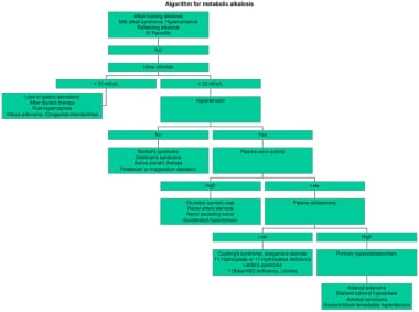
Algorithm for metabolic alkalosis. Same as Image 2 but presented in Adobe Acrobat format for easier printing. |
 | View Full Size Image | |
Media type: Acrobat PDF
|
- Adrogue HJ, Madias NE. Management of life-threatening acid-base disorders. Second of two parts. N Engl J Med. Jan 8 1998;338(2):107-11. [Medline].
- Anderson LE, Henrich WL. Alkalemia-associated morbidity and mortality in medical and surgical patients. Southern Medical Journal. 1987;80:729- 33. [Medline].
- Babior BM. Villous adenoma of the colon. Study of a patient with severe fluid and electrolyte disturbances. American Journal of Medicine. 1966;41:615- 21. [Medline].
- Cruz DN, Perazella MA. Hypertension and hypokalemia: unusual syndromes. Conn Med. Feb 1997;61(2):67-75. [Medline].
- DuBose TD Jr. Metabolic alkalosis. In: Brenner and Rector's The Kidney. 6th ed. Philadelphia: WB Saunders; 2000:971-997.
- Galla JH. Metabolic Alkalosis. Journal of the American Society of Nephrology. 2000;11:369- 75. [Medline].
- Geller DS, Farhi A, Pinkerton N. Activating mineralocorticoid receptor mutation in hypertension exacerbated by pregnancy. Science. Jul 7 2000;289(5476):119-23. [Medline].
- Hixson R, Christmas D. Use of omeprazole in life-threatening metabolic alkalosis. Intensive Care Med. Oct 1999;25(10):1201. [Medline].
- Hodgkin JE, Soeprono FF, Chan DM. Incidence of metabolic alkalemia in hospitalized patients. Critical Care Medicine. 1980;8:725- 8. [Medline].
- Kaplan NM. Hypertension induced by cortisol or deoxycorticosterone. In: Clinical Hypertension. 7th ed. Philadelphia: Williams & Wilkins; 1998:383-392.
- Kaplan NM. Primary aldosteronism. In: Clinical Hypertension. 7th ed. Philadelphia: Williams & Wilkins; 1998:365-79.
- Kelleher SP, Schulman G. Severe metabolic alkalosis complicating regional citrate hemodialysis. Am J Kidney Dis. Mar 1987;9(3):235-6. [Medline].
- Koeppen BM, Stanton BA. Regulation of acid-base balance. In: Vander AJ, ed. Renal Physiology. 2nd ed. New York: McGraw-Hill; 1997:135-55.
- Kunau RT, et al. Acid-base balance. In: MKSAP: Nephrology and Hypertension Book. 1998. 2nd ed. Philadelphia: Lippincott Williams & Wilkins; 252-69.
- Leblanc M, Farah A. Severe metabolic alkalosis corrected by hemodialysis. Clin Nephrol. Jul 1997;48(1):65. [Medline].
- Mauri S, Pedroli G, Rudeberg A. Acute metabolic alkalosis in cystic fibrosis: prospective study and review of the literature. Miner Electrolyte Metab. 1997;23(1):33-7. [Medline].
- Mazur JE, Devlin JW, Peters MJ. Single versus multiple doses of acetazolamide for metabolic alkalosis in critically ill medical patients: a randomized, double-blind trial. Crit Care Med. Jul 1999;27(7):1257-61. [Medline].
- McAuliffe JJ, Lind LJ, Leith DE. Hypoproteinemic alkalosis. Am J Med. Jul 1986;81(1):86-90. [Medline].
- Online Mendelian Inheritance in Man (OMIM). McKusick-Nathans Institute for Genetic Medicine, Johns Hopkins University (Baltimore, MD) and National Center for Biotechnology Information, National Library of Medicine (Bethesda, MD). OMIM. 2000. [Full Text].
- Palmer BF, Alpern RJ. Metabolic alkalosis. J Am Soc Nephrol. Sep 1997;8(9):1462-9. [Medline].
- Rose BD. Acid-base physiology and regulation of acid-base balance. In: Clinical Physiology of Acid-Base and Electrolyte Disorders. 4th ed. New York: McGraw-Hill; 1994:274-339.
- Rose BD. Metabolic alkalosis. In: Clinical Physiology of Acid-Base and Electrolyte Disorders. 4th ed. New York: McGraw-Hill; 1994:515-35.
- Rose BD. Metabolic alkalosis. UpToDate. Available at www.uptodate.com.
- Scheinman SJ, Guay-Woodford LM, Thakker RV. Genetic disorders of renal electrolyte transport. N Engl J Med. Apr 15 1999;340(15):1177-87. [Medline].
- Seldin DW, Rector FC. Symposium on acid-base homeostasis. The generation and maintenance of metabolic alkalosis. Kidney Int. May 1972;1(5):306-21. [Medline].
- Stewart PM. Cortisol as a mineralocorticoid in human disease. J Steroid Biochem Mol Biol. Apr-Jun 1999;69(1-6):403-8. [Medline].
- Stewart PM, Walker BR, Holder G. 11 beta-Hydroxysteroid dehydrogenase activity in Cushing's syndrome: explaining the mineralocorticoid excess state of the ectopic adrenocorticotropin syndrome. J Clin Endocrinol Metab. Dec 1995;80(12):3617-20. [Medline].
- Toto RD, Alpern RJ. Metabolic acid-base disorders. In: Kokko JP, Tannen RL, eds. Fluids and Electrolytes. 3rd ed. Philadelphia: WB Saunders; 1996:201-56.
- Zucchelli P, Santoro A. Correction of acid-base balance by dialysis. Kidney Int Suppl. Jun 1993;41:S179-83. [Medline].
Metabolic Alkalosis excerpt Article Last Updated: May 3, 2007
|