Practice Essentials
Meigs syndrome is defined as the triad of benign ovarian tumor with ascites and pleural effusion that resolves after resection of the tumor. Ovarian fibromas constitute the majority of the benign tumors seen in Meigs syndrome. Meigs syndrome, however, is a diagnosis of exclusion, only after ovarian carcinoma is ruled out. [1]
Signs and symptoms of Meigs syndrome
The chief complaints are vague and generally manifest over time; they include the following:
-
Fatigue
-
Shortness of breath
-
Increased abdominal girth
-
Weight gain/weight loss
-
Nonproductive cough
-
Bloating
-
Amenorrhea in premenopausal women
-
Menstrual irregularity
See Presentation for more detail.
Diagnosis of Meigs syndrome
Laboratory studies
In addition to serum electrolyte levels and a complete blood cell count, the study of interest is the serum cancer antigen 125 (CA-125) test. Tumor marker serum levels of CA-125 can be elevated in Meigs syndrome, but the degree of elevation does not correlate with malignancy.
Imaging studies
Chest radiography confirms pleural effusion. Abdominal and pelvic ultrasonography confirms the ovarian mass and ascites. Computed tomography scanning of the abdomen and pelvis confirms ascites and the presence of an ovarian, uterine, fallopian tube, or broad ligament mass.
See Workup for more detail.
Treatment of Meigs syndrome
Exploratory laparotomy with surgical staging is the treatment of choice. Medical care of patients with Meigs syndrome is intended to provide symptomatic relief of ascites and pleural effusion by means of therapeutic paracentesis and thoracentesis.
See Treatment for more detail.
Background
In 1934, Salmon described the association of pleural effusion with benign pelvic tumors. In 1937, Meigs and Cass described 7 cases of ovarian fibromas associated with ascites and pleural effusion. [2] The syndrome was named as Meigs syndrome by Rhoads and Terrel in 1937. [3] In 1954, Meigs proposed limiting true Meigs syndrome to benign and solid ovarian tumors accompanied by ascites and pleural effusion, with the condition that removal of the tumor cures the patient without recurrence. Histologically, the benign ovarian tumor may be a fibroma, thecoma, cystadenoma, or granulosa cell tumor.
Pseudo-Meigs syndrome consists of pleural effusion (an example of which can be seen in the image below), ascites, and benign tumors of the ovary other than fibromas. These benign tumors include those of the fallopian tube or uterus and mature teratomas, struma ovarii, and ovarian leiomyomas. [4] This terminology sometimes also includes ovarian or metastatic gastrointestinal malignancies.
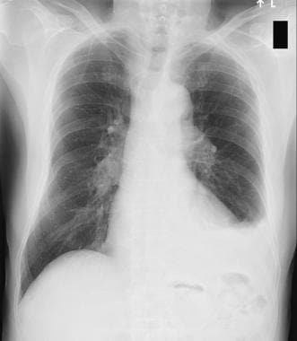 Chest radiograph showing left-sided pleural effusion.
Atypical Meigs syndrome, characterized by a benign pelvic mass with right-sided pleural effusion but without ascites, has been reported at least twice. In a case report of an older woman with atypical Meigs syndrome who presented with right-sided pleural effusion and notable leg edema, heart failure with preserved ejection fraction was initially suspected. [5] As in Meigs syndrome, pleural effusion resolves after removal of the pelvic mass.
Pseudo-pseudo Meigs syndrome includes patients with systemic lupus erythematosus and enlarged ovaries. [6]
Pathophysiology
Ascites is present in 10-15% of cases, and hydrothorax is found in only 1% of cases. [7, 8]
Etiology of ascitic fluid
The pathophysiology of ascites in Meigs syndrome is speculative. Meigs suggested that irritation of the peritoneal surfaces by a hard, solid ovarian tumor could stimulate the production of peritoneal fluid. Samanth and Black studied ovarian tumors accompanied by ascites and found that only tumors larger than 10 cm in diameter with a myxoid component to the stroma are associated with ascites. [9] These authors believe that their observations favor secretion of fluid from the tumor as the source of the ascites.
Other proposed mechanisms are direct pressure on surrounding lymphatics or vessels, hormonal stimulation, and tumor torsion. Development of ascites may be due to release of mediators (eg, activated complements, histamines, fibrin degradation products) from the tumor, leading to increased capillary permeability.
Origin of pleural effusion
The etiology of pleural effusion is unclear. Efskind and Terada et al theorize that ascitic fluid is transferred via transdiaphragmatic lymphatic channels. The size of the pleural effusion is largely independent of the amount of ascites. The pleural fluid may be located on the left side or may be bilateral. [1, 10, 11]
Efskind's study
Efskind injected ink into the lower abdomen of a woman with Meigs syndrome and found that the ink particles accumulated in the lymphatics of the pleural surface within half an hour. Blockage of these lymphatics prevented accumulation of pleural fluid and caused an increase in ascitic fluid.
Terada and colleagues' study
In 1992, Terada and colleagues injected labeled albumin into the peritoneum and found that the maximum concentration was detected in the right pleura within 3 hours.
Nature of the ascitic and pleural fluid
Ascitic fluid and pleural fluid in Meigs syndrome can be either transudative or exudative. [10] Meigs performed electrophoresis on several cases and determined that pleural and ascitic fluids were similar in nature. Tumor size, rather than the specific histologic type, is thought to be the important factor in the formation of ascites and accompanying pleural effusion.
In 2015, the findings of Krenke et al. in their systematic literature review of 541 cases reported with Meig’s syndrome revealed that an exudative origin in pleural effusions was significantly more prevalent than the ones from transudative origin. [12]
Etiology
When an ovarian mass is associated with Meigs syndrome and an elevated CA-125 serum level, a malignant process may be suspected until proven otherwise histologically. A negative cytologic examination result of ascitic effusion, the absence of peritoneal implantation, and benign histology should limit surgical procedures. This decision should be made by an experienced gynecologic surgeon or a gynecologic oncologist.
Note the following:
-
Case reports exist of pseudo-Meigs syndrome associated with malignant struma ovarii and elevated CA-125 levels. [13, 14] The choice of not performing adjuvant therapy is feasible after optimal surgery and adequate staging procedure given to the usually clinical benign course and the low incidence of metastases in malignant struma ovarii. Careful patient counseling is required.
-
Struma ovarii is a rare cause of ascites, hydrothorax, elevated CA-125 levels, and hyperthyroidism. [14] This rare condition should be considered in the differential diagnosis in patients with ascites and pleural effusions but with negative cytologic test results.
-
Liao et al reported a case of right benign struma ovarii in a patient presenting with ascites and CA-125 of 3515 U/Ml, who presented complete remission of symptoms and return to normal CA-125 after surgical resection of the mass. [3]
-
The combination of ascites, pleural effusion, CA-125 level elevation, and no tumor in a patient with systemic lupus erythematosus is either a Tjalma syndrome or due to the migrated Filshie clips a pseudo-Meigs syndrome. [15]
Epidemiology
United States statistics
Ovarian tumors are more prevalent in women in upper socioeconomic groups. Ovarian fibromas represent approximately 2-5% of surgically removed ovarian tumors, and Meigs syndrome occurs in only 1-2% of these cases; thus, it is a rare condition. Ascites is present in 10-15% of women with ovarian fibroma, and hydrothorax is present in 1%, especially those with larger lesions.
International statistics
The international prevalence is unknown.
Age-related demographics
The incidence of ovarian tumor begins to increase in the third decade and increases progressively in postmenopausal women, with an average of about 50 years. [1, 10] Meigs syndrome in prepubertal girls with benign teratomas and cystadenomas has been reported.
Prognosis
Meigs syndrome is a benign disease, if properly treated. No recurrence after sugical removal of the mass has been reported.
Clinicans should be aware of this rare and treatable condition.
Morbidity/mortality
The life expectancy of patients with Meigs syndrome mirrors that of the general population after surgery, and less than 1% of fibromas progress to fibrosarcoma.
Although Meigs syndrome mimics a malignant condition, it is a benign disease and has a very good prognosis if properly managed. Life expectancy after surgical removal of the tumor is the same as the general population. [11]
-
Chest radiograph showing left-sided pleural effusion.

