Continually Updated Clinical Reference
 
 
  All Sources     eMedicine     Medscape     Drug Reference     MEDLINE
 
eMedicine - Gonadotropin-Releasing Hormone Deficiency in Adults : Article by

Quick Find
Authors & Editors
Introduction
Clinical
Differentials
Workup
Treatment
Medication
Follow-up
Miscellaneous
Acknowledgments
Multimedia
References

Related Articles
Follicle-Stimulating Hormone Abnormalities

Infertility

Infertility, Male

Luteinizing Hormone Deficiency

Menopause

Ovarian Failure


Patient Education
Endocrine System Center

Women's Health Center

Pregnancy and Reproduction Center

Hypopituitary Overview

Hypopituitary Causes

Hypopituitary Symptoms

Hypopituitary Treatment

Endocrine System Introduction

Menopause Overview

Amenorrhea Overview

Birth Control Overview

Birth Control FAQs


Author: Vaishali Popat, MD, MPH, Fellow in Endocrinology, National Institutes of Health

Vaishali Popat is a member of the following medical societies: American Association of Clinical Endocrinologists, American Diabetes Association, American Medical Association, and Endocrine Society

Coauthor(s): Karim Anton Calis, PharmD, MPH, FASHP, FCCP, Professor, Medical College of Virginia, Virginia Commonwealth University, Clinical Professor, University of Maryland; Clinical Specialist, Endocrinology and Women's Health, Director, Drug Information Service, Mark O Hatfield Clinical Research Center, National Institutes of Health

Editors: Bruce A Meyer, MD, MBA, Vice President for Medical Affairs, Associate Dean and Director of the Faculty Practice Plan, Professor, Department of Obstetrics and Gynecology, University of Texas Southwestern Medical School; Francisco Talavera, PharmD, PhD, Senior Pharmacy Editor, eMedicine; Antonio V Sison, MD, Program Director, Department of Obstetrics and Gynecology, Robert Wood Johnson University Hospital; Frederick B Gaupp, MD, Consulting Staff, Department of Family Practice, Assumption Community Hospital; Lee P Shulman, MD, Professor of Obstetrics and Gynecology, Feinberg School of Medicine, Northwestern University; Chief, Division of Reproductive Genetics, Department of Obstetrics and Gynecology, Prentice Women's Hospital, Northwestern Memorial Hospital

Author and Editor Disclosure

Synonyms and related keywords: LHRH, LH, gonadotropin-releasing hormone, GnRH, isolated GnRH deficiency with or without associated anosmia, Kallmann syndrome, KS, idiopathic hypogonadotropic hypergonadism, IHH, gonadotropins, fertile eunuch

Background

Gonadotropin-releasing hormone (GnRH) is a neurohormone central to initiation of the reproductive hormone cascade. Pulsatile secretion of GnRH from the hypothalamus is key in establishing normal gonadal function. Failure of this release results in isolated GnRH deficiency that can be distinguished by partial or complete lack of GnRH-induced luteinizing hormone (LH) pulse, normalization with GnRH replacement, and otherwise normal hypothalamic-pituitary neuroanatomy and neurophysiology.

Clinicians and scientists long have been intrigued by the findings of olfactory disturbances and concomitant reproductive dysfunction. Spanish pathologist Maestre de San Juan noted in 1856 the association between the failure of testicular development and the absence of the olfactory bulbs. However, the syndrome is named Kallmann syndrome (KS) after the American geneticist Kallmann. Kallmann, Schoenfeld, and Barrera in 1944 were the first to identify a genetic basis to this disorder. In 1954, de Morsier connected the syndrome of hypogonadism and anosmia with the abnormal development of the anterior portion of the brain. Kallmann syndrome is a rare disorder that occurs in both sexes.

Pathophysiology

GnRH neurons

A fundamental understanding of the anatomy, biochemistry, ontogeny, and physiology of GnRH neurons aids in understanding the pathophysiology, diagnosis, and treatment of KS and idiopathic hypogonadotropic hypogonadism (IHH).

GnRH and GnRH receptors

The decapeptide GnRH is derived from posttranslation processing of a tripartite 92–amino acid (AA) pre-pro-GnRH. The first 23 AA is a signal peptide and the last 56 AA is known as GnRH-associated protein (GAP). GnRH is encoded from a single gene located on the short arm of chromosome 8. Serum levels of GnRH are difficult to obtain due to its short half-life (2-4 min) and complete confinement to the hypophyseal-portal blood supply. Due to the small structure and ease of mutation of GnRH, chemists have created several clinically useful GnRH analogs. GnRH binds with high affinity to cell surface receptors located on the pituitary gonadotrophs. These 7 transmembrane cell surface G protein; coupled receptors activate phospholipase C.

This enzyme leads to the activation of several second messenger molecules, the most important being diacylglycerol (DG) and inositol 1,4,5-trisphosphate (IP3). In turn, DG activates protein kinase C and causes IP3-stimulated release of calcium ions from intracellular pools. The result is the synthesis and release of follicle-stimulating hormone (FSH) and LH from the pituitary gonadotrophs. The released gonadotropins stimulate the gonads to produce steroid hormones and sperm or to release oocytes. Mutated GnRH receptors, as predicted by the biochemistry, could result in clinical manifestation of isolated gonadotropin deficiency. The control of GnRH and its receptor are regulated by many factors; the review of this regulation is beyond the scope of this article.

Ontogeny/function

The migration of GnRH neurons follows a precise path from the nose to the forebrain in humans. The olfactory placode is composed of thickened ectoderm that is lateral to the head of the developing embryo and invaginates to form simple olfactory pits on either side of the nasal septum. The lateral epithelium of the olfactory pits gives rise to olfactory nerves. The medial portion develops into the site of initial GnRH appearance and the terminal nerve. This ganglionated cranial nerve, the exact function of which is unknown, enters the forebrain and serves as a highway for the GnRH neuronal migration.

These migrating neurons do not contain neurosecretory vesicles until they reach the area of the arcuate nucleus in the hypothalamus. For this reason, neurons that do not reach the forebrain are unable to secrete GnRH. The GnRH neurons have been identified in the fetal hypothalamus at 9 weeks' gestation and are connected to the portal system by 16 weeks' gestation. At 10 weeks' gestation, gonadotropins are detectable in the pituitary, and by the 12th week, they are measurable in the bloodstream. Peripheral blood levels peak during the second trimester and decrease by term as the negative feedback mechanism develops.

GnRH is secreted during the neonatal period, resulting in pulsatile LH and FSH secretion, which decreases by age 6 months in boys and by age 1-2 years in girls until puberty. During childhood, GnRH still is secreted in pulsatile fashion but at reduced amplitude. The hypothalamic pulse generator likely is suppressed by a mechanism that inhibits GnRH release but not its synthesis.

This theory has been demonstrated in primates because GnRH messenger RNA (mRNA) and proteins are abundant in the hypothalamus during an equivalent developmental stage. The mechanism that awakens the pubertal onset of GnRH secretion still is unknown. Metabolic enzymes have been implicated, as have norepinephrine, neuropeptide gamma, aspartate, gamma-aminobutyric acid, transforming growth factor-alpha, and leptin. The pubertal period is characterized by an increase in the amplitude of GnRH-induced LH as well as a small increase in the frequency of amplitude increases. Sex steroids are secreted from the gonads in response to this nocturnal increase in gonadotropins. Gonadotropins continue to be secreted in pulsatile fashion during adulthood.

Most studies in males have shown LH pulses to occur every 2 hours, while, in females, GnRH pulse frequency changes throughout the menstrual cycle. In the early follicular phase, GnRH pulse frequency is every 90 minutes and increases to 60 minutes by the late follicular phase. GnRH pulse frequency during the luteal phase varies from 4-8 hours.

Studying GnRH secretion

Studying GnRH physiology in humans has been challenging. GnRH itself is confined almost entirely to the portal blood supply of the pituitary, and direct sampling in humans is not feasible. Samples of GnRH in the periphery are inaccurate because of the rapid 2- to 4-minute half-life. Much of the information known about GnRH has come from animal studies.

Knobil and coworkers in the 1970s demonstrated in rhesus monkeys that pulsatile GnRH is responsible for maintaining gonadotrope function. The researchers were able to differentiate between episodic and continuous stimulation causing maintenance and desensitization, respectively, of the gonadotrope response.

Another animal example in unscrambling the GnRH puzzle is the use of transgenic mice in developing immortalized GnRH cell lines. Interestingly, implantation of these cells into the hypothalamus of GnRH-deficient mice restores their estrus cycle. This has provided an important in vitro tool for studying neuroendocrine function.

Human studies have been limited to frequent sampling studies in healthy and diseased models, the use of pharmacological probes, and genetic studies. LH has long been used as a marker of GnRH pulse activity in humans. Most recently, the glycoprotein free alpha subunit (FAS) has been used as a marker due to its correlation with LH. FAS is useful in tracking GnRH because of its 12- to 15-minute half-life. An estimate of endogenous GnRH can be obtained using GnRH antagonists as probes. Administering a GnRH antagonist induces a GnRH receptor blockade so that the amount of GnRH present is inversely proportional to the amount of LH inhibitor.

Frequency

United States

The incidence in the United States is 1 case per 10,000 men and 1 case per 50,000 women.

International

By examining military records, the incidence of KS has been estimated to be between 1 case per 86,000 in Sardinia and 1 case in 10,000 in France.

Mortality/Morbidity

These patients do not have an increased mortality rate.

Race

Race is not a factor in incidence.

Sex

In the referral population at Massachusetts General Hospital over a 20-year period, the male-to-female ratio was 3.9 to 1.

A spectrum of GnRH deficiency with various secretory patterns ranging from complete lack of LH pulse to diminished amplitude similar to early puberty occurs in both men and women, contributing to the clinical heterogeneity of the disorder. This suggests that multiple genetic determinants may control the expression of GnRH secretion.

Age

The disease comes to attention when the patient fails to begin puberty and develops secondary sexual characteristics.


History

The age of onset, whether congenital or acquired, and the severity, whether complete or partial, determines the phenotypic expression.

  • During the neonatal period, boys present with micropenis. The incomplete descent of the testes and immaturity of the external genitalia are due to the failure of the hypothalamic-pituitary-gonadal axis to activate in the late fetal and neonatal periods. In the embryonic and early fetal periods, fetal testosterone is required for full sexual and external genital development, which is stimulated by maternal human chorionic gonadotropin (hCG) and does not require the stimulation of fetal pituitary gonadotropins. Newborn girls have no obvious abnormalities. Cryptorchidism has been reported in as many as 50% of males with IHH or KS, and microphallus is present in as many as 30% of affected individuals.
  • During childhood, anosmia is the only manifestation in patients with Kallmann syndrome.
  • In most cases, diagnosis is made much later, with absence of pubertal development. Histologically, the ovaries of affected women rarely possess follicles matured past the primordial stages. Hence, most of these women present with primary amenorrhea.
  • Some patients undergo early pubertal development but subsequently develop hypogonadism leading to infertility and sexual dysfunction.

Physical

Most physical findings are related to failure of sexual maturation. These patients have eunuchoidal body habitus, with arm-span greater than height by 5 cm or more. Secondary sexual characteristics are often absent. Women have little or no breast development, and men have little or no beard development. In both genders, pubic hair may be present, as adrenarche may not be affected. Gynecomastia is not a typical feature. GnRH deficiency results in decreased testosterone as well as estrogen production.

Many affected individuals are unaware of their loss of olfaction, especially those with partial defects. The testing with graded dilutions of pure scents is necessary to identify the impaired olfaction. The magnitude of GnRH deficiency appears to correlate to the severity of anosmia. In cases where KS or IHH is suspected but cryptorchidism and microphallus are absent, an MRI may reveal olfactory bulbs, although normal olfactory bulbs have been demonstrated in only 25% of males with KS.

Along with the anosmia, another interesting neurological finding is that of mirror movements related to cerebellar defects. Present in as many as 85% of patients with KS, mirroring is the involuntary movements in a limb that mirror the voluntary movements of the contralateral limb.

Many associated defects have been reported in patients with KS. These can be defined as sporadic and include uterine malformation, congenital heart defects, and dental agenesis. X-linked KS can be associated with another X-linked disorder known as ichthyosis (steroid sulfatase disorder). The finding of renal agenesis/hypoplasia has been noted in some individuals with X-linked KS. Recently, Colquhoun and colleagues (1999) described an Australian family with a high frequency of renal agenesis in the presence or absence of the KAL1 mutation, suggesting an autosomal dominant or X-linked gene, which may independently or codependently contribute to renal agenesis.

Causes

GnRH deficiency is inherited through autosomal dominant, autosomal recessive, and X-linked transmissions. However, more than two thirds of cases are sporadic.

KAL1 gene

The KAL1 gene, described in 1991, is an example of an X-linked gene controlling GnRH secretion. The gene is located on the short arm of the X chromosome at Xp22.3. Deletions produce a syndrome of short stature, mental retardation, ichthyosis, chondroplasia punctata, and Kallmann syndrome. Anosmin, the protein encoded by KAL, is similar in amino acid structure to molecules involved in neural development, such as protease inhibitors, neurophysins, and neural cell adhesion molecules. Anosmin appears to be important to the migration of the GnRH neurons to their resting place in the hypothalamus.

Most of the data on the KAL gene come from studies in chickens. The timing of KAL expression in the chicken has aided in understanding the migration defects of GnRH neurons in human KS. KAL is expressed in 2 distinctly different periods of embryonic development. KAL expression is found in limb buds, facial mesenchyme, and the neurons innervating the extrinsic eye muscles during embryonic days (ED). By ED 5, GnRH neurons migrate along the olfactory nerve and penetrate the olfactory bulb by ED 7-8. KAL expression is increased in the olfactory bulb by ED 7-8. At ED 9-10, KAL expression up-regulates as synapses are formed between the olfactory nerve and the mitral cell layer.

Studies have demonstrated that the migration of the nerves is controlled by the olfactory epithelium. When the olfactory placode is destroyed in the chick, KAL expression continues in the olfactory bulb. This suggests that KAL expression and olfactory nerve innervation are independent of one another. In humans, KAL transcripts are not identified at the time of olfactory nerve migration, again suggesting independence between KAL expression and olfactory nerve migration. In KS, a defect in neuron interaction rather than neuron migration has been suggested. In a study of a 19-week fetus with X-linked KS, the olfactory nerves were shown to have arrested within the meninges while the GnRH neurons were arrested in the forebrain, never reaching the hypothalamus. Both groups of neurons passed through the cribriform plate but arrested prematurely. The KAL gene may play a later role, such as controlling the penetration of GnRH neurons into the olfactory bulb.

Without KAL and without functioning synaptic connections, the olfactory nerve might atrophy and degenerate, causing the GnRH defective migration.

The KAL gene also may play a role in the development of other tissues such as facial mesenchyme, fibrous and perichondral cells, blood vessels, renal glomeruli, and developing limb buds. Again, this has been studied in the chicken. In humans, defective KAL expression in the cerebellum may be linked to nystagmus and ataxia observed in some patients with KS. Further correlation with cleft palate and renal agenesis still needs to be examined.

GPR54

In a large consanguineous Saudi family with 6 individuals with IHH, a homozygous single nucleotide change in exon 3 of GPR54 was found in all 6 affected individuals, resulting in substitution of a serine for the normal leucine in the second intracellular loop of the receptor (L148S) (see Image 1). This change did not occur in the homozygous state in any unaffected family members and was not identified in any controls. This 7-transmembrane domain receptor shares highest homology, about 45%, with the galanin subfamily of receptors. The amino acid sequence is highly conserved across species, with 95% homology between the rat and mouse and 82% between mouse and human (98% in the transmembrane domains) (Seminara, 1998).

A GPR54-deficient mouse model resulted in a phenotype similar to that in humans The mice had normal hypothalamic GnRH content, but developed IHH that was responsive to GnRH therapy. This finding suggests that GnRH neurons are present in the hypothalamus and can synthesize the peptide but that GPR54 is necessary for processing or secretion of GnRH. The ligand for GPR54 is identified as metastatin. Kisspeptin, a 145-amino acid precursor, gives rise to a 54 amino acid product termed metastin after cleavage. Together, this ligand/receptor combination (metastin/GPR54) can advance puberty in rodents and can overcome the amenorrhea of congenital leptin and leptin receptor deficiency, and starvation. Thus, it is becoming clear that this system is a major gatekeeper of the pubertal process (Navarro, 2004).

GnRH receptor

The GnRH receptor is a G protein–coupled receptor, which activates phospholipase C, mobilizing intracellular calcium. Mutations in this receptor have been described in families with hypogonadotropic hypogonadism. One case reports phenotypically normal parents heterozygous for a GnRH receptor mutation who had a son with normal puberty and normal olfaction but with 8-mL testes and an abnormal semen analysis. Their daughter had primary amenorrhea and was infertile. LH probe frequency was normal but with low amplitude pulsation.

Other reports describe GnRH receptor mutations causing hypogonadotropic hypogonadism that presents with complete gonadotropin deficiency. An example is a male patient seeking treatment for delayed puberty presenting with no secondary sexual characteristics, cryptorchid testes, low gonadotropins, and low testosterone. The patient did not respond to GnRH, but treatment with gonadotropins corrected testicular growth and descent.

DAX1 gene

Adrenal hypoplasia congenita arises from X-linked or autosomal recessive syndromes and presents in infancy with primary adrenal insufficiency. Treatable with steroids, it has resulted in affected adults developing hypogonadotropic hypogonadism. A pituitary origin for one group with hypogonadotropic hypogonadism has been suggested by the failed attempts in those patients to stimulate LH and FSH with pulsatile GnRH. A smaller group has had gonadotropin responses to GnRH therapy, characterizing a hypothalamic-versus-pituitary defect.

The DAX1 gene has been identified at Xp21 as the gene responsible for adrenal hypoplasia congenita. As with the KAL gene, growing evidence for DAX mutation suggests a wide phenotypic range. Data has suggested that DAX1 mutations impair gonadotropin production at the levels of both the pituitary and the hypothalamus. Steroidogenic factor 1 (SF-1), a nuclear hormone receptor, plays a regulatory role in adrenal development and development of the hypothalamic-pituitary-gonadal axis.

Specifically, SF-1 regulates the expression of the p450 steroid hydroxylase genes in the gonads and the adrenal cortex, regulates the MIS gene, regulates the alpha subunit of the gonadotropins, and regulates the beta subunit of LH. Another suggested role for DAX1 is as a "brake" for normal male maturation while being necessary for normal adrenal and hypothalamic/pituitary development. DAX1 has been shown to block steroidogenesis in adrenal cells by transcriptional repression. Loss of function of this repressor may lead to a host of adrenal, hypothalamic, and pituitary abnormalities.

Evidence suggests that most familial cases of GnRH deficiency are controlled by autosomal inheritance. In a study of 106 patients with GnRH deficiency at Massachusetts General Hospital, only 21% of familial cases were X-linked. Using isolated congenital anosmia as a marker for KS, X-linked and autosomal recessive transmission was 18% and 32%, respectively.

Autosomal dominance accounted for 50% of cases. When delayed puberty was included in the phenotypic analysis, X-linked cases accounted for 11% of cases while autosomal recessive and autosomal dominant cases were 25% and 64%, respectively.


Follicle-Stimulating Hormone Abnormalities
Infertility
Infertility, Male
Luteinizing Hormone Deficiency
Menopause
Ovarian Failure

Other Problems to be Considered

Differential diagnosis

Differentiating between GnRH deficiency and delayed puberty is difficult. A definitive diagnosis of GnRH deficiency is not made before age 18 years in most cases. Constitutional delay in puberty is more common compared with GnRH deficiency. A variety of clinical clues and follow up over a period of time will clarify the diagnosis.

  • A detailed family history is helpful. GnRH deficiency, anosmia, or presence of associated congenital abnormalities in the family is suggestive of GnRH deficiency.
  • Family history of delayed puberty, early development of breast or testes, and then stalled puberty are suggestive of delayed puberty.
  • However, some families of patients with IHH report a history of delayed puberty within the family. In a series of 106 patients with isolated GnRH deficiency, 12% reported family members with a history of delayed puberty without any physical features of IHH. The finding of delayed puberty and isolated GnRH deficiency within the same family may represent 2 ends of a phenotypic spectrum of GnRH deficiency. The incidence of delayed puberty in the general population is less than 1%.
  • Pubic hair may be present in GnRH deficiency as adrenarche is normal, but in delayed puberty, both, adrenarche and sexual development are attenuated.

Confusion may exist with other CNS clinical syndromes that are associated with hypogonadism. A brief review of the clinical manifestations of these disorders is as follows:

Laurence-Moon-Biedl (LMB) syndrome is characterized by obesity, mental retardation, renal abnormalities, polydactyly, pigmented retinal dystrophy, and spastic paraparesis. The Bardet-Biedl form is the same as LMB, but these individuals do not have the spastic paraparesis.

Prader-Willi syndrome (PWS) includes neonatal hypotonia, obesity, mental retardation, and short stature. The extent of sexual maturation in PWS is variable.

Moebius syndrome is characterized by congenital oculofacial paralysis, eye movement disorders, gait disorders, and mental retardation.

Lowe syndrome is composed of oculocerebral-renal disorders, the inheritance of which is X-linked.

LEOPARD syndrome is an acronym for lentigines, electrocardiographic conduction defects, ocular hypertelorism, pulmonic stenosis, abnormal genitalia, retarded growth, and deafness.

Carpenter syndrome is associated with obesity, acrocephaly, agenesis of the hands and feet, and craniosynostosis. Besides the obvious differences in clinical manifestation, the serum levels of steroids and gonadotropins can exclude these syndromes from KS.

Adult-onset GnRH deficiency: Adult onset IHH recently has been reported in men. Normal puberty is followed by a decline in libido and fertility. Testicular size is nearly normal. The biochemical profile includes a pulsatile LH secretion, low serum testosterone and, in 90% of cases, normal restoration of the reproductive axis with exogenous GnRH. Unlike hypothalamic amenorrhea in women, neither an identifiable factor such as stress exercise or weight loss that would impair GnRH secretion nor any spontaneous waxing or waning has occurred.

Variant forms of GnRH deficiency: The fertile eunuch syndrome was described in 1950 as a clinical entity of eunuchoid body type and secondary sexual characteristics with normal testes and normal spermatogenesis. Fertility can be achieved with testosterone or hCG therapy. Based on the lack of response to Clomid and normal response to GnRH, the abnormality was identified in the hypothalamus. Men with the fertile eunuch syndrome possess the pubertal sleep-augmented GnRH-induced LH pattern. This syndrome represents an incomplete form of GnRH deficiency because the GnRH secretion is sufficient to stimulate enough Leydig cell secretion of testosterone to support spermatogenesis and testicular growth but not enough for full virilization.


Lab Studies

  • Along with the above-described clinical manifestations, most patients have low serum levels of basal gonadotropins, estrogen/testosterone, and poor response to GnRH stimulation. The difficulty arises when trying to differentiate between healthy prepubertal males and those with IHH or KS. Patients with KS can be distinguished from prepubertal males aged 12.5 years or older by determining the LH in pooled serum samples collected every 20 minutes for 6 hours commencing 1 hour after sleep onset.

Imaging Studies

  • MRI appears to be the single best study for the diagnosis of KS and exclusion of other CNS disorders associated with hypogonadotrophic hypogonadism.
  • T1-weighted MRI of the inferior frontal region in the coronal plane appears most helpful in examining the olfactory sulci, bulbs, and rhinencephalon.
  • Dual-energy x-ray absorptiometry: Because of the lack of production of sex steroid, men and women with KS and IHH can experience abnormal bone development. The prudent use of a dual-energy x-ray absorptiometry (DEXA) scan to monitor bone mineral density in these individuals is appropriate.

Other Tests

  • Many affected individuals are unaware of their loss of olfaction, especially those with partial defects. Testing with graded dilutions of pure scents is necessary to identify the impaired olfaction. The magnitude of GnRH deficiency appears to correlate with the severity of anosmia.
  • Along with the anosmia, another interesting neurological finding is that of mirror movements related to the cerebellar defects. Present in as many as 85% of patients with KS, mirroring is the involuntary movements in a limb that mirrors voluntary movements of the contralateral limb.


Medical Care

The choice of therapy depends upon the patient's desire to achieve one or more of the following: secondary sex characteristics, fertility, and bone and muscle mass.

Treatment in males

In deciding when and how to provide androgen replacement in males the patient's age, the adverse effects of therapy, and the patient's desire for fertility should be considered. In the prepubertal teenager who has congenital hypogonadism, androgens stimulate penile growth, body and facial hair growth, bone and muscle development, and voice changes. In addition, androgens stimulate growth hormone production, thus contributing to the adolescent growth spurt.

Because males with androgen deficiency can experience significant social ridicule, starting androgen therapy around the age of 14 or 15 years is prudent.

Oral, injectable, transdermal, and implantable (pellets) androgen formulations currently are available for the treatment of males with KS and IHH. The oral 17 androgens should not be used due to their toxic effects on the liver and adverse effects on lipids.

The injectable long-acting testosterone esters (eg, testosterone enanthate/cypionate) are low-cost, relatively safe, and effective, with a proven 50-year record. The disadvantages include intramuscular injection and a nonphysiologic pattern of testosterone over the dosing interval that in some men can cause wide swings in libido and mood. Research currently is being conducted on longer-acting and sustained-release formulation of testosterone injectables.

Transdermal application avoids first-pass metabolism, provides a noninvasive method of replacement, and results in more physiologic serum concentration of testosterone over a daily dosing period. Transdermal patches (scrotal and nonscrotal) and a gel preparation of testosterone are currently available. The most common adverse effect with these formulations is skin reactions at the application site. The gel preparation may be preferred by some patients because it is not visibly apparent and has fewer dermal reactions. However, the gel formulation may result in cross contamination to those in close contact with the patient.

The usual starting dose of 50-75 mg of testosterone injection is used monthly with the expectation of increasing the dose by 50 mg every 4-6 months until sexual maturation has been reached. The troublesome adverse effects of acne and gynecomastia are monitored closely and the dose adjusted accordingly. Injections of hCG (200-500 IU alternate d) can be used in the prepubertal male. Although doses ultimately should be based on clinical response and testosterone levels, a twice-weekly dosing regimen of 100-1500 IU or 200-500 IU on alternate days is typical. The advantages of hCG are the normalization of testosterone levels and stimulation of testicular growth. The cost and numerous injections have resulted in reserving hCG primarily for men attempting fertility.

In males desiring fertility, a different approach to replacement therapy is employed. Spermatogenesis can be restored with combination of hCG and human menopausal gonadotropin (hMG, FSH, and LH), hCG and FSH alone, or GnRH injection. Occasionally, patients may respond to hCG alone. Testicular volumes greater than 3-4 mL can be used to predict those individuals who respond to hCG. Careful monitoring of testicular size is helpful in gauging the effect of treatment. Those individuals who reach testicular sizes of 12-15 mL usually produce sperm after 12 months of treatment initiation. These hCG-only responsive individuals usually represent the fertile eunuch or late-onset adult IHH patients.

Most patients with IHH and KS require a combination of hCG and FSH to stimulate sperm production. The starting dose for hCG is 1000 IU, and FSH is 75-150 IU on alternate days with dosage adjusted based on trough testosterone level, testicular growth, sperm production, and avoidance of adverse effects. The most common adverse effect is gynecomastia, which occurs in as many as 30% of patients. This is related to increased estrogen production from several factors, such as hCG induction of testicular aromatase and increase in the peripheral aromatization of testosterone. These monitoring periods should occur every 3 months until an adequate level of replacement is documented. Pregnancy has occurred with counts as low as 2.5 X 10⁶, but 20-40 X 106/mL produces higher pregnancy rates. The median time to induction of spermatogenesis is 6-8 months.

The pulsatile administration of GnRH is an effective alternative to gonadotropin administration. The dose of GnRH ranges from 25-600 ng/kg every 2 hours delivered subcutaneously using a programmable portable infusion pump. As with gonadotropins, the dose and pulse are alternated based on testicular size, testosterone levels, spermatogenesis, and adverse effects. Once the testis has reached 8 mL, regular semen analysis can be obtained. Most patients require as long as 2 years of therapy before they reach maximal gonadal size and sperm production. GnRH therapy in prepubertal boys to evoke puberty may represent a more physiologic approach because the pulse of GnRH can be altered to mimic the natural process of puberty. Again, the response time appears to be influenced by the initial testicular size; larger testes at the start of therapy result in less time on gonadotropins or GnRH.

Determination of which therapy to use (ie, gonadotropins or GnRH pulses) is related more to preference than science. Therapies appear to be equally effective. The time to full testicular growth and spermatogenesis may be somewhat shorter when using GnRH, although this appears controversial. Some anecdotal evidence suggests that GnRH therapy has proven successful in individuals refractory to gonadotropin treatments. The disadvantage of GnRH, beyond the need to use a pump, is that it is available only at specialized centers pending approval by the Food and Drug Administration (FDA) for this indication.

Treatment in females

In females, as in males, treatment is dictated by the fertility desires of the patient. For the woman not currently desiring fertility, estrogen replacement is required to prevent osteoporosis.

The principal estrogen produced by the functioning premenopausal ovary is estradiol-17b. Daily serum measurements of estradiol in regularly menstruating women indicate that the mean estradiol levels throughout the menstrual cycle are approximately 104 pg/mL (382 pmol/L) (Mishell, 1971). Oral and parenteral preparations (ie, subcutaneous pellets and implants, transdermal patches, vaginal creams and rings) are available for standard hormone replacement therapy in normal postmenopausal women.

Oral estrogens have the disadvantage of the first hepatic passage. Parenteral administration bypasses the intestine, avoids the first pass effect of liver metabolism, and thus prevents the abnormal E2/E1 ratio observed following oral administration (Stevenson, 1996; Jewelewicz, 1997). Transdermally administered 17-b estradiol has been shown to be an effective regimen for preventing bone loss in normal postmenopausal women (Jewelewicz, 1997; Field, 1993). The goal is to replace sex hormones in young women by trying to mimic the normal ovarian function.

All women with intact uterus should receive a cyclical progestin. The 12-day administration of medroxyprogesterone acetate (10 mg) has been shown to adequately protect the endometrium in continuous hormone replacement therapy (The Writing Group for the Pepi Trial, 1996). Alternatively, oral micronized progesterone can be used.

Optimal hormone therapy depends on whether the patient has primary or secondary amenorrhea. Young women with primary amenorrhea in whom secondary sex characteristics have failed to develop should initially be exposed to very low doses of estrogen in an attempt to mimic the gradual pubertal maturation process. A typical regimen is as follows: 0.3 mg of CEE or 25 mcg estradiol patch unopposed (ie, no progestogen) daily for 6 months with incremental dose increases at 6-month intervals until the required maintenance dose is achieved. Gradual dose escalation often results in optimal breast development and allows time for the young woman to adjust psychologically to her physical maturation. Cyclical progestogen therapy, given 12-14 days per month, should be instituted toward the end of the second year of treatment.

Barrier methods of contraception should also be provided in the rare event that one of these patients spontaneously ovulates, as with women with adult-onset IHH.

The woman requesting fertility, similar to her male counterpart, is faced with a much more complicated process. Reports of women with KS achieving pregnancy are rare, with only about 20 described in the literature. However, one can extrapolate the treatment as one would for other hypogonadotropic hypogonadal women. Intravenous pulsatile GnRH is the logical treatment of choice. Intravenous pulsatile GnRH appears to have advantages over gonadotropins because it can be pulsed to mimic the normal menstrual dynamics. The usual starting dose is 75 ng/kg administered at a pulse. In fact, when applied to other IHH conditions, the pregnancy rate, cancellation of cycles, and multiple births rate are improved when compared to gonadotropin therapy. Recently, gonadotropin therapy, including FSH-only treatment, has proven successful in patients with KS and IHH.


The goals of pharmacotherapy are to reduce morbidity, prevent complications, and correct the hormone deficiency.

Drug Category: Hormones

For replacement therapy in hypogonadism associated with a deficiency or absence of endogenous testosterone.

Drug NameTestosterone (Depo-Testosterone, Andro-LA, Delatest, Androderm, AndroGel)
DescriptionMainstay of treatment that is low-cost, safe, and effective. Dosage adjustments are made by monitoring trough levels prior to next injection. The goal is to maintain trough level in the low-normal range.
Adult Dose100-200 mg IM q2-3wk
AndroGel: Apply 5 g (delivers 5 mg systemically) qd to upper arms, shoulders, or abdomen; may increase dose after 14 d if serum testosterone below normal range to 7.5-10 g (ie, 7.5-10 mg systemically)
Transdermal patch:
Androderm: Apply 5–7.5 mg/d patch, rotate application sites on back, abdomen, upper arms, or thighs

Testoderm TTS: Apply 5 mg/d patch, rotate application sites on arms, back, or upper buttocks
Pediatric Dose50-75 mg IM qmo initial; increase by 50 mg q4-6 mo until sexual maturation occurs
Testosterone gel or patch: Not established
ContraindicationsDocumented hypersensitivity; severe cardiac or renal disease; benign prostatic hypertrophy with obstruction; males with carcinoma of the breast or undiagnosed genital bleeding
InteractionsMay increase effects of anticoagulants
PregnancyX - Contraindicated; benefit does not outweigh risk
PrecautionsAnabolic effects may enhance hypoglycemia; monitor hand and wrist q6mo to determine the rate of bone maturation; monitor for weight gain, gynecomastia, breast tenderness, edema, sleep disturbance, mood swings, libido, and benign prostatic hypertrophy; check hemoglobin and hematocrit in older men for polycythemia

Drug Category: Gonadotropins

Stimulate production of gonadal steroid hormones.

Drug NameHuman chorionic gonadotropin (Corex, Choron)
DescriptionSpermatogenesis may be restored. In children, although doses should be based ultimately on clinical response and testosterone levels, a twice-weekly dosing regimen of 100-1500 U or 200-500 U on alternate d is typical. Advantages of hCG are normalization of testosterone levels and stimulation of testicular growth. Cost and numerous injections have led hCG to be reserved for those males attempting fertility.
Adult Dose1000 U IM, alternate with FSH; adjust dose based on trough testosterone level, testicular growth, sperm production, and adverse effects
Pediatric DosePrepubertal male: 200-500 U IM on alternate d
ContraindicationsDocumented hypersensitivity; prostatic carcinoma; precocious puberty
InteractionsNone reported
PregnancyX - Contraindicated; benefit does not outweigh risk
PrecautionsCaution in asthma, seizure disorders, renal disease, and migraine; most common adverse effect is gynecomastia related to increased estrogen production from several factors, such as hCG induction of testicular aromatase and increase in the peripheral aromatization of testosterone; monitor q3mo until adequate level is documented

Drug NameFollicle-stimulating hormone (Fertinex, Follistim)
DescriptionStimulates gonadal steroid production.
Adult Dose75-150 U IM, alternate with hCG
Pediatric DoseNot established
ContraindicationsDocumented hypersensitivity; tumor of the uterus, ovary, hypothalamus, or breast; abnormal vaginal bleeding; pregnancy
InteractionsNone reported
PregnancyX - Contraindicated; benefit does not outweigh risk
PrecautionsSerious respiratory distress, thromboembolic events, and atelectasis may occur

Drug Category: Gonadotropin-releasing hormones

The determination of which therapy to employ, gonadotropins or GnRH pulses, is related more to preference than to science. Therapies appear to be equally effective. Time to full testicular growth and spermatogenesis may be somewhat shorter when using GnRH, although this appears controversial. Some anecdotal evidence suggests that GnRH therapy has proven successful in individuals refractory to gonadotropin treatments.

Drug NameGonadorelin acetate (GnRH, Factrel, Lutrepulse)
DescriptionStimulates pituitary release of luteinizing hormone. Two years of therapy may be required to reach maximal gonadal size and sperm production. Response time is influenced by initial testicular size; larger initial size yields less time on therapy. Once testis has reached 8 mL regular semen, analysis can be obtained. As with gonadotropins, dose and pulse are alternated based on testicular size, testosterone levels, spermatogenesis, and adverse effects.
Therapy in prepubertal boys may represent a more physiologic approach because the pulse of GnRH may be altered to mimic the natural process of puberty. The disadvantage of treatment, other than the need to use a pump, is that it is available at specialized centers only due to pending approval by FDA for this indication.
In females, this appears to be an effective method of stimulation of the ovary.
Adult DoseMales: 25-600 ng/kg SC q2h using a programmable portable infusion pump
Females: 75 ng/kg SC administered as a pulse using a programmable portable infusion pump
Pediatric DoseNot established
ContraindicationsDocumented hypersensitivity; patients who have ovarian cysts or causes of anovulation other than those of hypothalamic origin; any condition that may be worsened by reproductive hormones, such as a hormonally dependent tumor
InteractionsDecreases effects of oral contraceptives, digoxin, phenothiazines, and dopamine antagonists; increases effects of androgens, glucocorticoids, spironolactone, and levodopa; concurrent use of ovulation stimulators
PregnancyB - Fetal risk not confirmed in studies in humans but has been shown in some studies in animals
PrecautionsCaution if pregnancy is suspected; skin reactions (local and generalized), headache, nausea, and rare anaphylactic reactions may occur; multiple pregnancy is a possibility, minimize by careful attention to the recommended doses and ultrasonographic monitoring of the ovarian response to therapy

Drug Category: Oral contraceptives

These agents may be used as hormone replacement therapy.

Drug NameEthinyl estradiol and norethindrone (Ovcon 35, Nordette)
DescriptionIn young females, low-dose oral contraception generally is an excellent method of hormone replacement. Any low-dose combination pill with 35 mcg of ethinyl estradiol or less and any progestin is appropriate. Also useful because, on occasion, these women may spontaneously ovulate and become pregnant.
Adult Dose21-day pack: 1 tab PO qd for 21 d; start new dosing pack 7 d after last tab is taken
28-day pack: 1 tab PO qd
Pediatric DoseNot established
ContraindicationsDocumented hypersensitivity; thromboembolic disorders; cerebrovascular or coronary artery disease; known or suspected breast cancer; undiagnosed abnormal vaginal bleeding; women smokers >35 y; active liver disease; pregnancy
InteractionsMay reduce hypoprothrombinemic effects of anticoagulants; barbiturates, hydantoins, and rifampin may decrease effects; concomitant use with penicillins or tetracyclines may decrease effects; may increase levels of carbamazepine, tricyclic antidepressants, and corticosteroids
PregnancyX - Contraindicated; benefit does not outweigh risk
PrecautionsUse of any progestin during first 4 mo of pregnancy is not recommended; caution in thromboembolism, stroke, MI, liver tumor, or hypertension; also use caution in cardiac, renal, or hepatic insufficiency; risk of cardiovascular adverse effects increases with cigarette use and in women >35 y

Drug NameConjugated equine estrogen and medroxyprogesterone (Prempro)
DescriptionHormone replacement therapy that induces the synthesis of DNA, RNA, and various proteins in target tissues. Promotes development of secondary sex characteristics. Inhibits secretion of pituitary gonadotropins.
Adult Dose0.625 mg conjugated estrogen PO
2.5-5 mg medroxyprogesterone PO qd
Combination CEE and progestin daily
Pediatric DoseNot established
ContraindicationsDocumented hypersensitivity; known or suspected pregnancy; breast cancer, undiagnosed abnormal genital bleeding; active thrombophlebitis or thromboembolic disorders; history of thrombophlebitis, thrombosis, or thromboembolic disorders associated with previous estrogen use (except when used in treatment of breast or prostatic malignancy); cerebral apoplexy; liver dysfunction
InteractionsMay decrease effects of aminoglutethimide; may reduce hypoprothrombinemic effect of anticoagulants; coadministration of barbiturates, rifampin, and other agents that induce hepatic microsomal enzymes may reduce estrogen levels; pharmacologic and toxicologic effects of corticosteroids may occur as a result of estrogen-induced inactivation of hepatic P-450 enzyme; loss of seizure control has been noted when administered concurrently with hydantoins
PregnancyX - Contraindicated; benefit does not outweigh risk
PrecautionsCertain patients may develop undesirable manifestations of excessive estrogenic stimulation such as abnormal or excessive uterine bleeding or mastodynia; estrogens may cause some degree of fluid retention (exercise caution); prolonged unopposed estrogen therapy may increase risk of endometrial hyperplasia; caution in asthma, depression, renal or cardiac dysfunction, or thromboembolic disorders

Drug NameConjugated estrogens (Premarin)
DescriptionInduces the synthesis of DNA, RNA, and various proteins in target tissues. Promotes development of secondary sex characteristics. Titrate dose depending on the hypoestrogenic symptoms
Adult Dose0.625 mg PO qd, depending on tissue response of patient
Pediatric DoseNot established
ContraindicationsDocumented hypersensitivity; known or suspected pregnancy; breast cancer; undiagnosed abnormal genital bleeding; active thrombophlebitis or thromboembolic disorders; history of thrombophlebitis, thrombosis, or thromboembolic disorders associated with previous estrogen use (except when used in treatment of breast or prostatic malignancy)
InteractionsMay reduce hypoprothrombinemic effect of anticoagulants; coadministration of barbiturates, rifampin, and other agents that induce hepatic microsomal enzymes may reduce estrogen levels; pharmacologic and toxicologic effects of corticosteroids may occur as a result of estrogen-induced inactivation of hepatic P450 enzyme; loss of seizure control has been noted when administered concurrently with hydantoins
PregnancyX - Contraindicated; benefit does not outweigh risk
PrecautionsCertain patients may develop undesirable manifestations of excessive estrogenic stimulation such as abnormal or excessive uterine bleeding or mastodynia; estrogens may cause some degree of fluid retention (exercise caution); prolonged unopposed estrogen therapy may increase risk of endometrial hyperplasia

Drug NameEstradiol (Climara Transdermal, Estraderm Transdermal)
DescriptionRestores estrogen levels to concentrations that induce negative feedback at gonadotrophic regulatory centers. Used for the purpose of hormone replacement and induction of puberty. Acts by regulating transcription of a limited number of genes. Estrogens diffuse through cell membranes, distribute themselves throughout the cell, and bind to and activate the nuclear estrogen receptor, a DNA-binding protein found in estrogen-responsive tissues. The activated estrogen receptor binds to specific DNA sequences or hormone-response elements, which enhances transcription of adjacent genes and, in turn, leads to the observed effects.

Continue treatment until breakthrough menstrual bleeding occurs and then initiate cyclical therapy. This can be achieved with any of a variety of PO contraceptives or the addition of medroxyprogesterone 5 mg to an estradiol regimen during the third wk of every mo with no treatment the last wk. PO contraceptive treatment is easier for patient to follow.
Adult DosePatch: 0.05-0.1 mg applied once/twice weekly (application frequency dependent on brand of patch)
Pediatric DoseNot established
ContraindicationsDocumented hypersensitivity; thrombophlebitis, undiagnosed vaginal bleeding
InteractionsMay reduce hypoprothrombinemic effects of anticoagulants; estrogen levels may be reduced with coadministration of barbiturates, rifampin, and other agents that induce hepatic microsomal enzymes; an increase in corticosteroid levels may occur when administered concurrently with ethinyl estradiol; use of ethinyl estradiol with hydantoins may cause spotting, breakthrough bleeding, and pregnancy; increase in fluid retention caused by estrogen intake may reduce seizure control
PregnancyX - Contraindicated; benefit does not outweigh risk
PrecautionsCaution in hepatic impairment, migraine, seizure disorders, cerebrovascular disorders, breast cancer, or thromboembolic disease

Drug NameProgesterone (Prometrium)
DescriptionCan be administered PO, vaginally, or IM. All routes of administration are equally effective. Begin treatment 2-3 d after ovulation and continue until 10th wk of pregnancy.
Adult DoseVaginal suppository: 25 mg bid
Gel 8%: 1 applicator PV qd
Oral micronized: 100 mg PO tid
17-hydroxyprogesterone caproate: 250 mg IM qwk
Pediatric DoseNot established
ContraindicationsDocumented hypersensitivity; thrombophlebitis, carcinoma of the breast, undiagnosed vaginal bleeding
InteractionsAminoglutethimide may decrease effects
PregnancyX - Contraindicated; benefit does not outweigh risk
PrecautionsFluid retention may occur; caution in patients with history of depression, impaired liver function, diabetes, and epilepsy; monitor for loss of vision, proptosis, diplopia, migraine, signs of embolic disorders

Drug NameMedroxyprogesterone acetate (Provera)
DescriptionIncreases central respiratory drive and stimulates ventilation. May increase upper airway muscular tone.

Progestins stimulate central respiratory drive and may be beneficial in patients with hypoventilation.
Adult Dose60 mg PO divided bid/tid
Pediatric DoseNot recommended
ContraindicationsDocumented hypersensitivity; cerebral apoplexy, undiagnosed vaginal bleeding, thrombophlebitis, and liver dysfunction
InteractionsAminoglutethimide may decrease effects by increasing hepatic metabolism of medroxyprogesterone
PregnancyX - Contraindicated; benefit does not outweigh risk
PrecautionsCaution in asthma, depression, renal or cardiac dysfunction, or thromboembolic disorders


Further Outpatient Care

  • The care of the patient depends on the goals of treatment, such as increase in sperm production or the initiation of ovulation. If fertility is not an issue, monitor the patient for problems associated with hypogonadism. One of the primary health concerns is osteoporosis.

Patient Education


Medical/Legal Pitfalls

  • Difficulty in diagnosis arises when trying to differentiate between healthy prepubertal males and those with IHH or KS.
  • Because males with androgen deficiency can experience a significant social ridicule, starting androgen therapy at the age of 14 or 15 years is prudent.


The authors and editors of eMedicine gratefully acknowledge the contributions of previous authors James N Anasti, MD and Michael Cackovic, MD to the development and writing of this article.


Media file 1:  
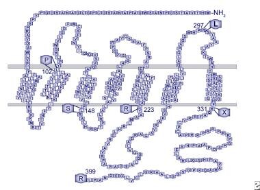
Human GPR54 receptor model. Mutations identified in patients with idiopathic hypogonadotropic hypogonadism are indicated.
Click to see larger pictureClick to see detailView Full Size Image
Media type:  Image

Media file 2:  KiSS-1 protein product model. Amino acids 1-19 are predicted to form a signal peptide. Proteolytic processing is predicted toproduce kisspeptin-54, corresponding to amino acids 68-121. Shown is the C-terminal amidated decapeptide sequence, wherein biologic actively resides.
Click to see larger pictureClick to see detailView Full Size Image
Media type:  Image


  • Adams JM, Taylor AE, Schoenfeld DA. The midcycle gonadotropin surge in normal women occurs in the face of an unchanging gonadotropin-releasing hormone pulse frequency. J Clin Endocrinol Metab. Sep 1994;79(3):858-64. [Medline].
  • Angelopoulos N, Goula A, Tolis G. The role of luteinizing hormone activity in controlled ovarian stimulation. J Endocrinol Invest. Jan 2005;28(1):79-88.
  • Apter D, Cacciatore B, Alfthan H. Serum luteinizing hormone concentrations increase 100-fold in females from 7 years to adulthood, as measured by time-resolved immunofluorometric assay. J Clin Endocrinol Metab. Jan 1989;68(1):53-7. [Medline].
  • Arimura A, Kastin AJ, Gonzalez-Barcena D. Disappearance of LH-releasing hormone in man as determined by radioimmunoassay. Clin Endocrinol (Oxf). Oct 1974;3(4):421-5. [Medline].
  • Backstrom CT, McNeilly AS, Leask RM. Pulsatile secretion of LH, FSH, prolactin, oestradiol and progesterone during the human menstrual cycle. Clin Endocrinol (Oxf). Jul 1 1982;17(1):29-42. [Medline].
  • Baker HW, Santen RJ, Burger HG. Rhythms in the secretion of gonadotropins and gonadal steroids. J Steroid Biochem. May 1975;6(5):793-801. [Medline].
  • Bardin CW, Ross GT, Rifkind AB. Studies of the pituitary-Leydig cell axis in young men with hypogonadotropic hypogonadism and hyposmia: comparison with normal men, prepuberal boys, and hypopituitary patients. J Clin Invest. Nov 1969;48(11):2046-56. [Medline].
  • Barkan AL, Reame NE, Kelch RP. Idiopathic hypogonadotropic hypogonadism in men: dependence of the hormone responses to gonadotropin-releasing hormone (GnRH) on the magnitude of the endogenous GnRH secretory defect. J Clin Endocrinol Metab. Dec 1985;61(6):1118-25. [Medline].
  • Barnhart KM, Mellon PL. The orphan nuclear receptor, steroidogenic factor-1, regulates the glycoprotein hormone alpha-subunit gene in pituitary gonadotropes. Mol Endocrinol. Jul 1994;8(7):878-85. [Medline].
  • Barron JL, Millar RP, Searle D. Metabolic clearance and plasma half-disappearance time of D-TRP6 and exogenous luteinizing hormone-releasing hormone. J Clin Endocrinol Metab. Jun 1982;54(6):1169-73. [Medline].
  • Bartley JA, Miller DK, Hayford JT. Concordance of X-linked glycerol kinase deficiency with X-linked congenital adrenal hypoplasia. Lancet. Oct 2 1982;2(8301):733-6. [Medline].
  • Behre HM, Kliesch S, Leifke E. Long-term effect of testosterone therapy on bone mineral density in hypogonadal men. J Clin Endocrinol Metab. Aug 1997;82(8):2386-90. [Medline].
  • Beighle C, Karp LE, Hanson JW. Small structural changes of chromosome 8. Two cases with evidence for deletion. Hum Genet. Aug 31 1977;38(1):113-21. [Medline].
  • Belchetz PE, Plant TM, Nakai Y. Hypophysial responses to continuous and intermittent delivery of hypopthalamic gonadotropin-releasing hormone. Science. Nov 10 1978;202(4368):631-3. [Medline].
  • Bovet P, Reymond MJ, Rey F. Lack of gonadotropic response to pulsatile gonadotropin-releasing hormone in isolated hypogonadotropic hypogonadism associated to congenital adrenal hypoplasia. J Endocrinol Invest. Mar 1988;11(3):201-4. [Medline].
  • Boyar R, Perlow M, Hellman L. Twenty-four hour pattern of luteinizing hormone secretion in normal men with sleep stage recording. J Clin Endocrinol Metab. Jul 1972;35(1):73-81. [Medline].
  • Boyar RM, Wu RH, Kapen S. Clinical and laboratory heterogeneity in idiopathic hypogonadotropic hypogonadism. J Clin Endocrinol Metab. Dec 1976;43(6):1268-75. [Medline].
  • Boyar RM, Rosenfeld RS, Kapen S. Human puberty. Simultaneous augmented secretion of luteinizing hormone and testosterone during sleep. J Clin Invest. Sep 1974;54(3):609-18. [Medline].
  • Brandenberger AW, Haenggi W, von Fischer B. Kallmann syndrome and associated malformation of the uterus. Fertil Steril. Feb 1994;61(2):395-7. [Medline].
  • Bridges NA, Matthews DR, Hindmarsh PC, et al. Changes in gonadotrophin secretion during childhood and puberty. J Endocrinol. Apr 1994;141(1):169-76. [Medline].
  • Brown DC, Stirling HF, Butler GE. Differentiation of normal male prepuberty and hypogonadotrophic hypogonadism using an ultrasensitive luteinizing hormone assay. Horm Res. 1996;46(2):83-7. [Medline].
  • Calof AL, Chikaraishi DM. Analysis of neurogenesis in a mammalian neuroepithelium: proliferation and differentiation of an olfactory neuron precursor in vitro. Neuron. Jul 1989;3(1):115-27. [Medline].
  • Cariboni A, Pimpinelli F, Colamarino S, et al. The product of X-linked Kallmann''s syndrome gene (KAL1) affects the migratory activity of gonadotropin-releasing hormone (GnRH)-producing neurons. Hum Mol Genet. Nov 15 2004;13(22):2781-91.
  • Castellano JM, Navarro VM, Fernandez-Fernandez R. Changes in hypothalamic KiSS-1 system and restoration of pubertal activation of the reproductive axis by kisspeptin in undernutrition. Endocrinology. Sep 2005;146(9):3917-25.
  • Castillo RH, Matteri RL, Dumesic DA. Luteinizing hormone synthesis in cultured fetal human pituitary cells exposed to gonadotropin-releasing hormone. J Clin Endocrinol Metab. Jul 1992;75(1):318-22. [Medline].
  • Clark SJ, Hauffa BP, Rodens KP. Hormone ontogeny in the ovine fetus: XIX: The effect of a potent luteinizing hormone-releasing factor agonist on gonadotropin and testosterone release in the fetus and neonate. Pediatr Res. Apr 1989;25(4):347-52. [Medline].
  • Clement K, Vaisse C, Lahlou N. A mutation in the human leptin receptor gene causes obesity and pituitary dysfunction. Nature. Mar 26 1998;392(6674):398-401. [Medline].
  • Colquhoun-Kerr JS, Gu WX, Jameson JL. X-linked Kallmann syndrome and renal agenesis occurring together and independently in a large Australian family. Am J Med Genet. Mar 5 1999;83(1):23-7. [Medline].
  • Conn PM, Crowley WF Jr. Gonadotropin-releasing hormone and its analogs. Annu Rev Med. 1994;45:391-405. [Medline].
  • Cortez AB, Galindo A, Arensman FW. Congenital heart disease associated with sporadic Kallmann syndrome. Am J Med Genet. Jun 15 1993;46(5):551-4. [Medline].
  • Crowley WF Jr, Filicori M, Spratt DI. The physiology of gonadotropin-releasing hormone (GnRH) secretion in men and women. Recent Prog Horm Res. 1985;41:473-531. [Medline].
  • Dean JC, Johnston AW, Klopper AI. Isolated hypogonadotrophic hypogonadism: a family with autosomal dominant inheritance. Clin Endocrinol (Oxf). Mar 1990;32(3):341-7. [Medline].
  • Drenth J, Low BW, Richardson JS. The toxin-agglutinin fold. A new group of small protein structures organized around a four-disulfide core. J Biol Chem. Apr 10 1980;255(7):2652-5. [Medline].
  • Duke VM, Winyard PJ, Thorogood P. KAL, a gene mutated in Kallmann''s syndrome, is expressed in the first trimester of human development. Mol Cell Endocrinol. Apr 28 1995;110(1-2):73-9. [Medline].
  • Dungan HM, Clifton DK, Steiner RA. Minireview: kisspeptin neurons as central processors in the regulation of gonadotropin-releasing hormone secretion. Endocrinology. Mar 2006;147(3):1154-8.
  • Edelman GM, Crossin KL. Cell adhesion molecules: implications for a molecular histology. Annu Rev Biochem. 1991;60:155-90. [Medline].
  • Elmquist JK. Anatomic basis of leptin action in the hypothalamus. Front Horm Res. 2000;26:21-41. [Me