Background
Albumin, the body's predominant serum-binding protein, has several important functions.
Albumin comprises 75-80% of normal plasma colloid oncotic pressure and 50% of protein content. When plasma proteins, especially albumin, no longer sustain sufficient colloid osmotic pressure to counterbalance hydrostatic pressure, edema develops. Although primarily in the intravascular space, albumin has a major trafficking function through the interstitium and lymphatics.
Albumin transports various substances, including bilirubin, fatty acids, metals, ions, hormones, and exogenous drugs. One consequence of hypoalbuminemia is that drugs that are usually protein bound are free in the plasma, allowing for higher drug levels, more rapid hepatic metabolism, or both.
Alterations in albumin level affect platelet function.
See the image below.
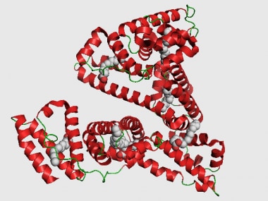 Albumin.
Reference serum values range from 3.5-4.5 g/dL, with a total body content of 300-500 g. Synthesis occurs only in hepatic cells at a rate of approximately 15 g/d in a healthy person, but the rate can vary significantly with various types of physiologic stress. The half-life of albumin is approximately 21 days, with a degradation rate of approximately 4% per day.
Hypoalbuminemia is a common problem among persons with acute and chronic medical conditions. At the time of hospital admission, 20% of patients have hypoalbuminemia. Hypoalbuminemia can be caused by various conditions, including nephrotic syndrome, hepatic cirrhosis, heart failure, and malnutrition; however, most cases of hypoalbuminemia are caused by acute and chronic inflammatory responses.
Serum albumin level is an important prognostic indicator. Among hospitalized patients, lower serum albumin levels correlate with an increased risk of morbidity and mortality.
The presentation, physical examination findings, and laboratory results associated with hypoalbuminemia depending on the underlying disease process.
Pathophysiology
Serum albumin levels are dependent on the rate of synthesis, the amount secreted from the liver cell, the distribution in body fluids, and the level of degradation. Hypoalbuminemia results from a derangement in one or more of these processes.
Synthesis
Albumin synthesis begins in the nucleus, where genes are transcribed into messenger ribonucleic acid (mRNA). The mRNA is secreted into the cytoplasm, where it is bound to ribosomes, forming polysomes that synthesize preproalbumin. Preproalbumin is an albumin molecule with a 24 amino acid extension at the N terminus. The amino acid extension signals insertion of preproalbumin into the membrane of the endoplasmic reticulum. Once inside the lumen of the endoplasmic reticulum, the leading 18 amino acids of this extension are cleaved, leaving proalbumin (albumin with the remaining extension of 6 amino acids). Proalbumin is the principal intracellular form of albumin. Proalbumin is exported to the Golgi apparatus, where the extension of 6 amino acids is removed prior to secretion of albumin by the hepatocyte. Once synthesized, albumin is secreted immediately; it is not stored in the liver.
Distribution
Tracer studies with iodinated albumin show that intravascular albumin is distributed into the extravascular spaces of all tissues, with the majority being distributed in the skin. Approximately 30-40% (210 g) of albumin in the body is found within the vascular compartments of the muscle, skin, liver, gut, and other tissues.
Albumin enters the intravascular space via 2 pathways. First, albumin enters this space by entering the hepatic lymphatic system and moving into the thoracic duct. Second, albumin passes directly from hepatocytes into the sinusoids after traversing the Space of Disse.
After 2 hours, 90% of secreted albumin remains within the intravascular space. The half-life of intravascular albumin is 16 hours. Daily losses of albumin from the intravascular space are approximately 10%. Certain pathological conditions, such as nephrosis, ascites, lymphedema, intestinal lymphangiectasia, and edema, can increase the daily loss of albumin from the plasma.
Albumin distributes into the hepatic interstitial volume, and the concentration of colloids in this small volume is believed to be an osmotic regulator for albumin synthesis. This is the principal regulator of albumin synthesis during normal periods without stress.
Damage
Albumin has four binding sites, one for cobalt (metallic) and the others are for binding to various biologic and foreign molecular species. Albumin can be oxidized in a reversible fashion restored via the glutathione pathway, but in severe end-stage hepatic cirrhosis, it forms a nonreversible oxidized form that has markedly reduced molecular binding capabilities.
Degradation
Degradation of albumin is poorly understood. After secretion into the plasma, the albumin molecule passes into tissue spaces and returns to the plasma via the thoracic duct. Tagged albumin studies suggest that albumin may be degraded within the endothelium of the capillaries, bone marrow, and liver sinuses. Albumin molecules apparently degrade randomly, with no differentiation between old and new molecules.
Etiology
Hypoalbuminemia can result from decreased albumin production, defective synthesis because of hepatocyte damage, deficient intake of amino acids, increased losses of albumin via GI or renal processes, and, most commonly, acute or chronic inflammation. Some of the many causes are discussed below.
Protein malnutrition
Deficient protein intake results in the rapid loss of cellular ribonucleic acid and disaggregation of the endoplasmic reticulum–bound polysomes and, therefore, decreased albumin synthesis. Albumin synthesis can decrease by more than one third during a 24-hour fast. Albumin synthesis may be stimulated by amino acids produced in the urea cycle, such as ornithine.
Defective synthesis
In patients with cirrhosis, synthesis is decreased because of the loss of hepatic cell mass. Also, portal blood flow is often decreased and poorly distributed, leading to maldistribution of nutrients and oxygen. The flow of substrate may affect certain functions of the liver, including protein synthesis, which is decreased in patients with cirrhosis who lack ascites. Albumin synthesis may actually increase in patients with cirrhosis who have ascites, possibly because of a change in hepatic interstitial colloid levels, which may act as an overriding stimulus for albumin production. Although synthesis is increased, the concentration of albumin is decreased because of dilution.
Extravascular protein loss
Nephrotic syndrome
This can produce hypoalbuminemia by massive proteinuria, with 3.5 g or more of protein lost within 24 hours. Albumin is filtered by the glomerulus and catabolized by the renal tubules into amino acids that are recycled. In patients with chronic renal disease, in whom both glomerular and tubular diseases are present, excessive protein filtration may lead to both increased protein loss and increased degradation. Only at higher rates of albuminuria (>100 mg/kg/d) and only when the diet is adequate is albumin synthesis increased.
Protein-losing enteropathy
Under normal conditions, less than 10% of the total albumin is lost through the intestine. This fact has been confirmed by comparing albumin labeled with chromium-51, which helps measure intestinal losses, to albumin labeled with iodine-125, which helps measure overall degradation. In cases of protein-losing enteropathy related to bacterial overgrowth, hypoalbuminemia is exacerbated by peripheral factors that inhibit albumin synthesis by mechanisms similar to those observed with burns, trauma, infection, and carcinoma.
Extensive burns
The skin is the major site for extravascular albumin storage and is the major exchangeable albumin pool needed to maintain plasma levels. Hypoalbuminemia results from direct losses of albumin from tissue damage, from compromised hepatic blood flow due to volume loss, and from inhibitory tissue factors (eg, tumor necrosis factor, interleukin-1, interleukin-6) released at the burn sites.
Lymphatic blockage or mucosal disease
Diseases that result in protein loss from the intestine are divided into 2 main types. The first is lymphatic blockage, which can be caused by constrictive pericarditis, ataxia telangiectasia, and mesenteric blockage due to tumor. The second is mucosal disease with direct loss into the bowel, which is observed with (1) inflammatory bowel disease and sprue and (2) bacterial overgrowth, as in blind loop syndrome after intestinal bypass surgery.
Hemodilution
In the presence of ascites from any cause, the serum albumin level is not a good index of the residual synthetic capacity of the liver unless actual radioisotopic measurements of production are used. With ascites, synthesis may be normal or even increased, but serum levels are low because of the larger volume of distribution. This is true even for ascites due to cirrhosis.
Congestive heart failure
The synthesis of albumin is normal in patients with congestive heart failure. Hypoalbuminemia results from an increased volume of distribution.
Oncotic pressure increase
The serum oncotic pressure partially regulates albumin synthesis. The regulation site may be the oncotic content in the hepatic interstitial volume because albumin synthesis is inversely related to the content of this volume. Conditions that increase other osmotically active substances in the serum tend to decrease the serum albumin concentration by decreasing synthesis. Examples include elevated serum globulin levels in hepatitis and hypergammaglobulinemia.
Acute and chronic inflammation
Albumin levels that are low because of acute inflammation should normalize within weeks of resolution of the inflammation. Persistent hypoalbuminemia beyond this point should prompt an investigation for an ongoing inflammatory process. The cytokines (TNF, IL-6) released as part of the inflammatory response to physiologic stress (infection, surgery, trauma) can decrease serum albumin by the following mechanisms:
-
Increased vascular permeability (allowing albumin to diffuse into the extravascular space)
-
Increased degradation
-
Decreased synthesis (among other mechanisms, by activating TNF-a, which decreases transcription of the albumin gene)
Epidemiology
Frequency
Hypoalbuminemia is more frequent in older patients who are institutionalized, patients who are hospitalized with advanced stages of disease (eg, terminal cancer), and malnourished children.
Race
No race predilection exists.
Sex
No sex predilection exists.
Age
Hypoalbuminemia affects persons of all age groups, depending on the underlying cause.
Prognosis
Low serum albumin levels are an important predictor of morbidity and mortality. A meta-analysis of cohort studies found that, with every 10 g/L decrease in serum albumin, mortality was increased by 137% and morbidity increased by 89%. Patients with serum albumin levels of less than 35 at 3 months following discharge from the hospital have a 2.6 times greater 5-year mortality than those with a serum albumin levels greater than 40.
Hypoalbuminemia has also been studied as an important prognostic factor among subsets of patients, such as patients with severe sepsis, burns, [1] and regional enteritis (Crohn disease) and has recently been associated with an increased risk of reintubation. [2]
Whether or not hypoalbuminemia is merely a marker of severe protein malnutrition, which itself is a cause of increased morbidity and mortality, or an independent risk factor for death, is unclear. However, its association with a poor prognosis remains strong.
Patient Education
Specific dietary recommendations are based on the underlying disease.
-
Albumin.