Overview
Breech presentation is defined as a fetus in a longitudinal lie with the buttocks or feet closest to the cervix. This occurs in 3-4% of all deliveries. The percentage of breech deliveries decreases with advancing gestational age from 22-25% of births prior to 28 weeks' gestation to 7-15% of births at 32 weeks' gestation to 3-4% of births at term. [1]
Predisposing factors for breech presentation include prematurity, uterine malformations or fibroids, polyhydramnios, placenta previa, fetal abnormalities (eg, CNS malformations, neck masses, aneuploidy), and multiple gestations. Fetal abnormalities are observed in 17% of preterm breech deliveries and in 9% of term breech deliveries.
Perinatal mortality is increased 2- to 4-fold with breech presentation, regardless of the mode of delivery. Deaths are most often associated with malformations, prematurity, and intrauterine fetal demise.
Types of breeches
See the list below:
-
Frank breech (50-70%) - Hips flexed, knees extended (pike position)
-
Complete breech (5-10%) - Hips flexed, knees flexed (cannonball position)
-
Footling or incomplete (10-30%) - One or both hips extended, foot presenting
Vaginal Breech Delivery
Historical considerations
Vaginal breech deliveries were previously the norm until 1959 when it was proposed that all breech presentations should be delivered abdominally to reduce perinatal morbidity and mortality. [2]
Vaginal breech delivery
Three types of vaginal breech deliveries are described, as follows:
-
Spontaneous breech delivery: No traction or manipulation of the infant is used. This occurs predominantly in very preterm, often previable, deliveries.
-
Assisted breech delivery: This is the most common type of vaginal breech delivery. The infant is allowed to spontaneously deliver up to the umbilicus, and then maneuvers are initiated to assist in the delivery of the remainder of the body, arms, and head.
-
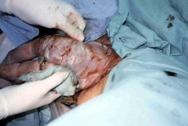
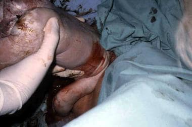
Total breech extraction: The fetal feet are grasped, and the entire fetus is extracted. Total breech extraction should be used only for a noncephalic second twin; it should not be used for a singleton fetus because the cervix may not be adequately dilated to allow passage of the fetal head. Total breech extraction for the singleton breech is associated with a birth injury rate of 25% and a mortality rate of approximately 10%. Total breech extractions are sometimes performed by less experienced accoucheurs when a foot unexpectedly prolapses through the vagina. As long as the fetal heart rate is stable in this situation, it is permissible to manage expectantly to allow the cervix to completely dilate around the breech (see the image below).
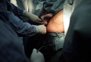 Footling breech presentation. Once the feet have delivered, one may be tempted to pull on the feet. However, a singleton gestation should not be pulled by the feet because this action may precipitate head entrapment in an incompletely dilated cervix or may precipitate nuchal arms. As long as the fetal heart rate is stable and no physical evidence of a prolapsed cord is evident, management may be expectant while awaiting full cervical dilation.

Technique and tips for assisted vaginal breech delivery
The fetal membranes should be left intact as long as possible to act as a dilating wedge and to prevent overt cord prolapse.
Oxytocin induction and augmentation are controversial. In many previous studies, oxytocin was used for induction and augmentation, especially for hypotonic uterine dysfunction. However, others are concerned that nonphysiologic forceful contractions could result in an incompletely dilated cervix and an entrapped head.
An anesthesiologist and a pediatrician should be immediately available for all vaginal breech deliveries. A pediatrician is needed because of the higher prevalence of neonatal depression and the increased risk for unrecognized fetal anomalies. An anesthesiologist may be needed if intrapartum complications develop and the patient requires general anesthesia.
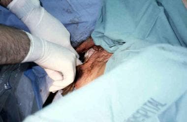
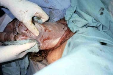
Some clinicians perform an episiotomy when the breech delivery is imminent, even in multiparas, as it may help prevent soft tissue dystocia for the aftercoming head (see the images below).
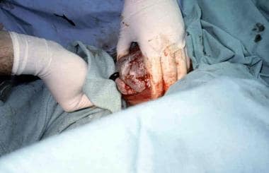
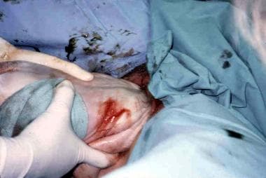
 Assisted vaginal breech delivery. Thick meconium passage is common as the breech is squeezed through the birth canal. This is usually not associated with meconium aspiration because the meconium passes out of the vagina and does not mix with the amniotic fluid.
 Assisted vaginal breech delivery. The Ritgen maneuver is applied to take pressure off the perineum during vaginal delivery. Episiotomies are often performed for assisted vaginal breech deliveries, even in multiparous women, to prevent soft tissue dystocia.
The Pinard maneuver may be needed with a frank breech to facilitate delivery of the legs but only after the fetal umbilicus has been reached. Pressure is exerted in the popliteal space of the knee. Flexion of the knee follows, and the lower leg is swept medially and out of the vagina.
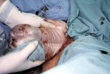
No traction should be exerted on the infant until the fetal umbilicus is past the perineum, after which time maternal expulsive efforts should be used along with gentle downward and outward traction of the infant until the scapula and axilla are visible (see the image below).
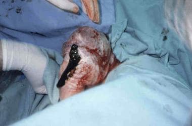 Assisted vaginal breech delivery. No downward or outward traction is applied to the fetus until the umbilicus has been reached.
Use a dry towel to wrap around the hips (not the abdomen) to help with gentle traction of the infant (see the image below).
 Assisted vaginal breech delivery. With a towel wrapped around the fetal hips, gentle downward and outward traction is applied in conjunction with maternal expulsive efforts until the scapula is reached. An assistant should be applying gentle fundal pressure to keep the fetal head flexed.
An assistant should exert transfundal pressure from above to keep the fetal head flexed.
Once the scapula is visible, rotate the infant 90° and gently sweep the anterior arm out of the vagina by pressing on the inner aspect of the arm or elbow (see the image below).
 Assisted vaginal breech delivery. After the scapula is reached, the fetus should be rotated 90° in order to deliver the anterior arm.
 Assisted vaginal breech delivery. The anterior arm is followed to the elbow, and the arm is swept out of the vagina.
Rotate the infant 180° in the reverse direction, and sweep the other arm out of the vagina. Once the arms are delivered, rotate the infant back 90° so that the back is anterior (see the image below).
 Assisted vaginal breech delivery. The fetus is rotated 180°, and the contralateral arm is delivered in a similar manner as the first. The infant is then rotated 90° to the backup position in preparation for delivery of the head.
The fetal head should be maintained in a flexed position during delivery to allow passage of the smallest diameter of the head. The flexed position can be accomplished by using the Mauriceau Smellie Veit maneuver, in which the operator's index and middle fingers lift up on the fetal maxillary prominences, while the assistant applies suprapubic pressure (see the image below).
 Assisted vaginal breech delivery. The fetal head is maintained in a flexed position by using the Mauriceau maneuver, which is performed by placing the index and middle fingers over the maxillary prominence on either side of the nose. The fetal body is supported in a neutral position, with care to not overextend the neck.
Alternatively, Piper forceps can be used to maintain the head in a flexed position (see the image below).
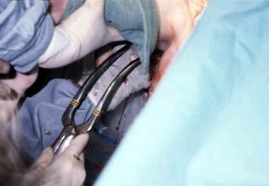 Piper forceps application. Piper forceps are specialized forceps used only for the after-coming head of a breech presentation. They are used to keep the fetal head flexed during extraction of the head. An assistant is needed to hold the infant while the operator gets on one knee to apply the forceps from below.
In many early studies, routine use of Piper forceps was recommended to protect the head and to minimize traction on the fetal neck. Piper forceps are specialized forceps that are placed from below the infant and, unlike conventional forceps, are not tailored to the position of the fetal head (ie, it is a pelvic, not cephalic, application). The forceps are applied while the assistant supports the fetal body in a horizontal plane.
During delivery of the head, avoid extreme elevation of the body, which may result in hyperextension of the cervical spine and potential neurologic injury (see the images below).

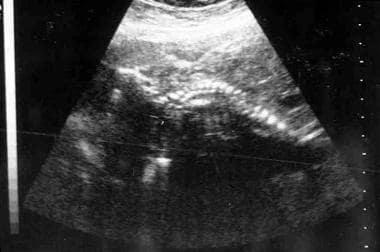 Ultrasound demonstrating a fetus in breech presentation with a hyperextended head (ie, "star gazing").
Risks
Lower Apgar scores, especially at 1 minute, are more common with vaginal breech deliveries. Many advocate obtaining an umbilical cord artery and venous pH for all vaginal breech deliveries to document that neonatal depression is not due to perinatal acidosis.
Fetal head entrapment may result from an incompletely dilated cervix and a head that lacks time to mold to the maternal pelvis. This occurs in 0-8.5% of vaginal breech deliveries. [3] This percentage is higher with preterm fetuses (< 32 wk), when the head is larger than the body. Dührssen incisions (ie, 1-3 cervical incisions made to facilitate delivery of the head) may be necessary to relieve cervical entrapment. However, extension of the incision can occur into the lower segment of the uterus, and the operator must be equipped to deal with this complication. The Zavanelli maneuver has been described, which involves replacement of the fetus into the abdominal cavity followed by cesarean delivery. While success has been reported with this maneuver, fetal injury and even fetal death have occurred.
Nuchal arms, in which one or both arms are wrapped around the back of the neck, are present in 0-5% of vaginal breech deliveries and in 9% of breech extractions. [3] Nuchal arms may result in neonatal trauma (including brachial plexus injuries) in 25% of cases. Risks may be reduced by avoiding rapid extraction of the infant during delivery of the body. To relieve nuchal arms when it is encountered, rotate the infant so that the fetal face turns toward the maternal symphysis pubis (in the direction of the impacted arm); this reduces the tension holding the arm around the back of the fetal head, allowing for delivery of the arm.
Cervical spine injury is predominantly observed when the fetus has a hyperextended head prior to delivery. Ballas and Toaff (1976) reported 20 cases of hyperextended necks, defined as an angle of extension greater than 90° ("star-gazing"), discovered on antepartum radiographs. [4] Of the 11 fetuses delivered vaginally, 8 (73%) sustained complete cervical spinal cord lesions, defined as either transection or nonfunction.
Cord prolapse may occur in 7.4% of all breech labors. This incidence varies with the type of breech: 0-2% with frank breech, 5-10% with complete breech, and 10-25% with footling breech. [3] Cord prolapse occurs twice as often in multiparas (6%) than in primigravidas (3%). Cord prolapse may not always result in severe fetal heart rate decelerations because of the lack of presenting parts to compress the umbilical cord (ie, that which predisposes also protects).
Candidates
Prior to the 2001 recommendations by the American College of Obstetricians and Gynecologists (ACOG), approximately 50% of breech presentations were considered candidates for vaginal delivery. Of these candidates, 60-82% were successfully delivered vaginally.
Candidates can be classified based on gestational age. For pregnancies prior to 26 weeks' gestation, prematurity, not mode of delivery, is the greatest risk factor. Unfortunately, no randomized clinical trials to help guide clinical management have been reported. Vaginal delivery can be considered, but a detailed discussion of the risks from prematurity and the lack of data regarding the ideal mode of delivery should take place with the parent(s). For example, intraventricular hemorrhage, which can occur in an infant of extremely low birth weight, should not be misinterpreted as proof of a traumatic vaginal breech delivery.
For pregnancies between 26 and 32 weeks, retrospective studies suggest an improved outcome with cesarean delivery, although these reports are subject to selection bias. In contrast, between 32 and 36 weeks' gestation, vaginal breech delivery may be considered after a discussion of risks and benefits with the parent(s).
After 37 weeks' gestation, parents should be informed of the results of a recent multicenter randomized clinical trial that demonstrated significantly increased perinatal mortality and short-term neonatal morbidity associated with vaginal breech delivery (see Comparative Studies). For those attempting vaginal delivery, if estimated fetal weight (EFW) is more than 4000 g, some recommend cesarean delivery because of concern for entrapment of the unmolded head in the maternal pelvis, although data to support this practice are limited.
A frank breech presentation is preferred when vaginal delivery is attempted. Complete breeches and footling breeches are still candidates, as long as the presenting part is well applied to the cervix and both obstetrical and anesthesia services are readily available in the event of a cord prolapse.
The fetus should show no neck hyperextension on antepartum ultrasound imaging (see the image below). Flexed or military position is acceptable.
Ultrasound demonstrating a fetus in breech presentation with a hyperextended head (ie, "star gazing").
Regarding prior cesarean delivery, a retrospective study by Ophir et al of 71 women with one prior low transverse cesarean delivery who subsequently delivered a breech fetus found that 24 women had an elective repeat cesarean and 47 women had a trial of labor. [5] In the 47 women with a trial of labor, 37 (78.7%) resulted in a vaginal delivery. Two infants in the trial of labor group had nuchal arms (1 with a transient brachial plexus injury) and 1 woman required a hysterectomy for hemorrhage due to a uterine dehiscence discovered after vaginal delivery. Vaginal breech delivery after one prior cesarean delivery is not contraindicated, though larger studies are needed.
Primigravida versus multiparous
It had been commonly believed that primigravidas with a breech presentation should have a cesarean delivery, although no data (prospective or retrospective) support this view. The only documented risk related to parity is cord prolapse, which is 2-fold higher in parous women than in primigravid women.
Radiographic and CT pelvimetry
Historically, radiograph pelvimetry was believed to be useful to quantitatively assess the inlet and mid pelvis. Recommended pelvimetry criteria included a transverse inlet diameter larger than 11.5 cm, anteroposterior inlet diameter larger than 10.5 cm, transverse midpelvic diameter (between the ischial spines) larger than 10 cm, and anteroposterior midpelvic diameter larger than 11.5 cm. However, radiographic pelvimetry is rarely, if ever, used in the United States.
CT pelvimetry, which is associated with less fetal radiation exposure than conventional radiographic pelvimetry, was more recently advocated by some investigators. It, too, is rarely used today.
Summary
Ultimately, if the obstetrical operator is not experienced or comfortable with vaginal breech deliveries, cesarean delivery may be the best choice. Unfortunately, with the dwindling number of experienced obstetricians who still perform vaginal breech deliveries and who can teach future generations of obstetricians, this technique may soon be lost due to attrition.
Cesarean Delivery
In 1970, approximately 14% of breeches were delivered by cesarean delivery. By 1986, that rate had increased to 86%. In 2003, based on data from the National Center for Health Statistics, the rate of cesarean delivery for all breech presentations was 87.2%. Most of the remaining breeches delivered vaginally were likely second twins, fetal demises, and precipitous deliveries. However, the rise in cesarean deliveries for breeches has not necessarily equated with an improvement in perinatal outcome. Green et al compared the outcome for term breeches prior to 1975 (595 infants, 22% cesarean delivery rate for breeches) with those from 1978-1979 (164 infants, 94% cesarean delivery rate for breeches). [6] Despite the increase in rates of cesarean delivery, the differences in rates of asphyxia, birth injury, and perinatal deaths were not significant.
Maneuvers for cesarean delivery are similar to those for vaginal breech delivery, including the Pinard maneuver, wrapping the hips with a towel for traction, head flexion during traction, rotation and sweeping out of the fetal arms, and the Mauriceau Smellie Veit maneuver.
An entrapped head can still occur during cesarean delivery as the uterus contracts after delivery of the body, even with a lower uterine segment that misleadingly appears adequate prior to uterine incision. Entrapped heads occur more commonly with preterm breeches, especially with a low transverse uterine incision. As a result, some practitioners opt to perform low vertical uterine incisions for preterm breeches prior to 32 weeks' gestation to avoid head entrapment and the kind of difficult delivery that cesarean delivery was meant to avoid. Low vertical incisions usually require extension into the corpus, resulting in cesarean delivery for all future deliveries.
If a low transverse incision is performed, the physician should move quickly once the breech is extracted in order to deliver the head before the uterus begins to contract. If any difficulty is encountered with delivery of the fetal head, the transverse incision can be extended vertically upward (T incision). Alternatively, the transverse incision can be extended laterally and upward, taking great care to avoid trauma to the uterine arteries. A third option is the use of a short-acting uterine relaxant (eg, nitroglycerin) in an attempt to facilitate delivery.
Comparative Studies
Only 3 randomized studies have evaluated the mode of delivery of the term breech. All other studies were nonrandomized or retrospective, which may be subject to selection bias.
In 1980, Collea et al randomized 208 women in labor with term frank breech presentations to either elective cesarean delivery or attempted vaginal delivery after radiographic pelvimetry. [7] Oxytocin was allowed for dysfunctional labor. Of the 60 women with adequate pelves, 49 delivered vaginally. Two neonates had transient brachial plexus injuries. Women randomized to elective cesarean delivery had higher postpartum morbidity rates (49.3% vs 6.7%).
In 1983, Gimovsky et al randomized 105 women in labor with term nonfrank breech presentations to a trial of labor versus elective cesarean delivery. [8] In this group of women, 47 had complete breech presentations, 16 had incomplete breech presentations (hips flexed, 1 knee extended/1 knee flexed), 32 had double-footling presentations, and 10 had single-footling presentations. Oxytocin was allowed for dysfunctional labor. Of the labor group, 44% had successful vaginal delivery. Most cesarean deliveries were performed for inadequate pelvic dimensions on radiographic pelvimetry. The rate of neonatal morbidity did not differ between neonates delivered vaginally and those delivered by cesarean delivery, although a higher maternal morbidity rate was noted in the cesarean delivery group.
In 2000, Hannah and colleagues completed a large, multicenter, randomized clinical trial involving 2088 term singleton fetuses in frank or complete breech presentations at 121 institutions in 26 countries. [9] In this study, popularly known as the Term Breech Trial, subjects were randomized into a planned cesarean delivery group or a planned vaginal birth group. Exclusion criteria were estimated fetal weight (EFW) more than 4000 g, hyperextension of the fetal head, lethal fetal anomaly or anomaly that might result in difficulty with delivery, or contraindication to labor or vaginal delivery (eg, placenta previa).
Subjects randomized to cesarean delivery were scheduled to deliver after 38 weeks' gestation unless conversion to cephalic presentation had occurred. Subjects randomized to vaginal delivery were treated expectantly until labor ensued. Electronic fetal monitoring was either continuous or intermittent. Inductions were allowed for standard obstetrical indications, such as postterm gestations. Augmentation with oxytocin was allowed in the absence of apparent fetopelvic disproportion, and epidural analgesia was permitted.
Adequate labor was defined as a cervical dilation rate of 0.5 cm/h in the active phase of labor and the descent of the breech fetus to the pelvic floor within 2 hours of achieving full dilation. Vaginal delivery was spontaneous or assisted and was attended by an experienced obstetrician. Cesarean deliveries were performed for inadequate progress of labor, nonreassuring fetal heart rate, or conversion to footling breech. Results were analyzed by intent-to-treat (ie, subjects were analyzed by randomization group, not by ultimate mode of delivery).
Of 1041 subjects in the planned cesarean delivery group, 941 (90.4%) had cesarean deliveries. Of 1042 subjects in the planned vaginal delivery group, 591 (56.7%) had vaginal deliveries. Indications for cesarean delivery included: fetopelvic disproportion or failure to progress in labor (226), nonreassuring fetal heart rate tracing (129), footling breech (69), request for cesarean delivery (61), obstetrical or medical indications (45), or cord prolapse (12).
The composite measurement of either perinatal mortality or serious neonatal morbidity by 6 weeks of life was significantly lower in the planned cesarean group than in the planned vaginal group (5% vs 1.6%, P< .0001). Six of 16 neonatal deaths were associated with difficult vaginal deliveries, and 4 deaths were associated with fetal heart rate abnormalities. The reduction in risk in the cesarean group was even greater in participating countries with overall low perinatal mortality rates as reported by the World Health Organization. The difference in perinatal outcome held after controlling for the experience level of the obstetrician. No significant difference was noted in maternal mortality or serious maternal morbidity between the 2 groups within the first 6 weeks of delivery (3.9% vs 3.2%, P = .35).
A separate analysis showed no difference in breastfeeding, sexual relations, or depression at 3 months postpartum, though the reported rate of urinary incontinence was higher in the planned vaginal group (7.3% vs 4.5%).
Based on the multicenter trial, the ACOG published a Committee Opinion in 2001 that stated "planned vaginal delivery of a singleton term breech may no longer be appropriate." This did not apply to those gravidas presenting in advanced labor with a term breech and imminent delivery or to a nonvertex second twin.
A follow-up study by Whyte et al was conducted in 2004 on 923 children who were part of the initial multicenter study. [10] The authors found no differences between the planned cesarean delivery and planned vaginal breech delivery groups with regards to infant death rates or neurodevelopmental delay by age 2 years. Similarly, among 917 participating mothers from the original trial, no substantive differences were apparent in maternal outcome between the 2 groups. [11] No longer-term maternal effects, such as the impact of a uterine scar on future pregnancies, have yet been reported.
A meta-analysis of the 3 above mentioned randomized trials was published in 2015. The findings included a reduction in perinatal/neonatal death, reduced composite short-term outcome of perinatal/neonatal death or serious neonatal morbidity with planned cesarean delivery versus planned vaginal delivery. [12] However, at 2 years of age, there was no significant difference in death or neurodevelopmental delay between the two groups. Maternal outcomes assessed at 2 years after delivery were not significantly different.
With regard to preterm breech deliveries, only one prospective randomized study has been performed, which included only 38 subjects (28-36 wk) with preterm labor and breech presentation. [13] Of these subjects, 20 were randomized to attempted vaginal delivery and 18 were randomized to immediate cesarean delivery. Of the attempted vaginal delivery group, 25% underwent cesarean delivery for nonreassuring fetal heart rate tracings. Five neonatal deaths occurred in the vaginal delivery group, and 1 neonatal death occurred in the cesarean delivery group. Two neonates died from fetal anomalies, 3 from respiratory distress, and 1 from sepsis.
Nonanomalous infants who died were not acidotic at delivery and did not have birth trauma. Differences in Apgar scores were not significant, although the vaginal delivery group had lower scores. The small number of enrolled subjects precluded any definitive conclusions regarding the safety of vaginal breech delivery for a preterm breech.
Retrospective analyses showed a higher mortality rate in vaginal breech neonates weighing 750-1500 g (26-32 wk), but less certain benefit was shown with cesarean delivery if the fetal weight was more than 1500 g (approximately 32 wk). Therefore, this subgroup of very preterm infants (26-32 wk) may benefit from cesarean delivery, although this recommendation is based on potentially biased retrospective data.
A large cohort study was published in 2015 from the Netherlands Perinatal Registry, which included 8356 women with a preterm (26-36 6/7 weeks) breech from 2000 to 2011, over three quarters of whom intended to deliver vaginally. In this overall cohort, there was no significant difference in perinatal mortality between the planned vaginal delivery and planned cesarean delivery groups (adjusted odds ratio 0.97, 95% confidence interval 0.60 – 1.57). However, the subgroup delivering at 28 to 32 weeks had a lower perinatal mortality with planned cesarean section (aOR 0.27, 95% CI 0.10 – 0.77). After adding a composite of perinatal morbidity, planned cesarean delivery was associated with a better outcome than a planned vaginal delivery (aOR 0.77, 95% CI 0.63 – 0.93. [14]
The Maternal-Fetal Medicine Units Network of the US National Institute of Child Health and Human Development considered a multicenter randomized clinical trial of attempted vaginal delivery versus elective cesarean delivery for 24- to 28-week breech fetuses. [15] However, it was not initiated because of anticipated difficulty with recruitment, inadequate numbers to show statistically significant differences, and medicolegal concerns. Therefore, this study is not likely to be performed.
External Cephalic Version
External cephalic version (ECV) is the transabdominal manual rotation of the fetus into a cephalic presentation.
Historical considerations
Initially popular in the 1960s and 1970s, ECV virtually disappeared after reports of fetal deaths following the procedure. Reintroduced to the United States in the 1980s, it became increasingly popular in the 1990s.
Improved outcome may be related to the use of nonstress tests both before and after ECV, improved selection of low-risk fetuses, and Rh immune globulin to prevent isoimmunization.
Procedure
Prepare for the possibility of cesarean delivery. Obtain a type and screen as well as an anesthesia consult. The patient should have nothing by mouth for at least 8 hours prior to the procedure. Recent ultrasonography should have been performed for fetal position, to check growth and amniotic fluid volume, to rule out a placenta previa, and to rule out anomalies associated with breech. Another sonogram should be performed on the day of the procedure to confirm that the fetus is still breech.
A nonstress test (biophysical profile as backup) should be performed prior to ECV to confirm fetal well-being.
Perform ECV in or near a delivery suite in the unlikely event of fetal compromise during or following the procedure, which may require emergent delivery.
ECV can be performed with 1 or 2 operators. Some prefer to have an assistant to help turn the fetus, elevate the breech out of the pelvis, or to monitor the position of the baby with ultrasonography. Others prefer a single operator approach, as there may be better coordination between the forces that are raising the breech and moving the head.
ECV is accomplished by judicious manipulation of the fetal head toward the pelvis while the breech is brought up toward the fundus. Attempt a forward roll first and then a backward roll if the initial attempts are unsuccessful. No consensus has been reached regarding how many ECV attempts are appropriate at one time. Excessive force should not be used at any time, as this may increase the risk of fetal trauma.
Following an ECV attempt, whether successful or not, repeat the nonstress test (biophysical profile if needed) prior to discharge. Also, administer Rh immune globulin to women who are Rh negative. Some physicians traditionally induce labor following successful ECV. However, as virtually all of these recently converted fetuses are unengaged, many practitioners will discharge the patient and wait for spontaneous labor to ensue, thereby avoiding the risk of a failed induction of labor. Additionally, as most ECV’s are attempted prior to 39 weeks, as long as there are no obstetrical or medical indications for induction, discharging the patient to await spontaneous labor would seem most prudent.
In those with an unsuccessful ECV, the practitioner has the option of sending the patient home or proceeding with a cesarean delivery. Expectant management allows for the possibility of spontaneous version. Alternatively, cesarean delivery may be performed at the time of the failed ECV, especially if regional anesthesia is used and the patient is already in the delivery room (see Regional anesthesia). This would minimize the risk of a second regional analgesia.
In those with an unsuccessful ECV, the practitioner may send the patient home, if less than 39 weeks, with plans for either a vaginal breech delivery or scheduled cesarean after 39 weeks. Expectant management allows for the possibility of a spontaneous version. Alternatively, if ECV is attempted after 39 weeks, cesarean delivery may be performed at the time of the failed ECV, especially if regional anesthesia is used and the patient is already in the delivery room (see Regional anesthesia). This would minimize the risk of a second regional analgesia.
Success rate
Success rates vary widely but range from 35% to 86% (average success rate in the 2004 National Vital Statistics was 58%). Improved success rates occur with multiparity, earlier gestational age, frank (versus complete or footling) breech presentation, transverse lie, and in African American patients.
Opinions differ regarding the influence of maternal weight, placental position, and amniotic fluid volume. Some practitioners find that thinner patients, posterior placentas, and adequate fluid volumes facilitate successful ECV. However, both patients and physicians need to be prepared for an unsuccessful ECV; version failure is not necessarily a reflection of the skill of the practitioner.
Zhang et al reviewed 25 studies of ECV in the United States, Europe, Africa, and Israel. [16] The average success rate in the United States was 65%. Of successful ECVs, 2.5% reverted back to breech presentation (other estimates range from 3% to 5%), while 2% of unsuccessful ECVs had spontaneous version to cephalic presentation prior to labor (other estimates range from 12% to 26%). Spontaneous version rates depend on the gestational age when the breech is discovered, with earlier breeches more likely to undergo spontaneous version.
A prospective study conducted in Germany by Zielbauer et al demonstrated an overall success rate of 22.4% for ECV among 353 patients with a singleton fetus in breech presentation. ECV was performed at 38 weeks of gestation. Factors found to increase the likelihood of success were a later week of gestation, abundant amniotic fluid, fundal and anterior placental location, and an oblique lie. [17]
A systematic review in 2015 looked at the effectiveness of ECV with eight randomized trials of ECV at term. Compared to women with no attempt at ECV, ECV reduced non-cephalic presentation at birth by 60% and reduced cesarean sections by 40% in the same group. [18] Although the rate of cesarean section is lower when ECV is performed than if not, the overall rate of cesarean section remains nearly twice as high after successful ECV due to both dystocia and non-reassuring fetal heart rate patterns. [19] Nulliparity was the only factor shown in follow-up to increase the risk of instrumental delivery following successful ECV. [20]
While most studies of ECV have been performed in university hospitals, Cook showed that ECV has also been effective in the private practice setting. [21] Of 65 patients with term breeches, 60 were offered ECV. ECV was successful in 32 (53%) of the 60 patients, with vaginal delivery in 23 (72%) of the 32 patients. Of the remaining breech fetuses believed to be candidates for vaginal delivery, 8 (80%) had successful vaginal delivery. The overall vaginal delivery rate was 48% (31 of 65 patients), with no significant morbidity.
Cost analysis
In 1995, Gifford et al performed a cost analysis of 4 options for breech presentations at term: (1) ECV attempt on all breeches, with attempted vaginal breech delivery for selected persistent breeches; (2) ECV on all breeches, with cesarean delivery for persistent breeches; (3) trial of labor for selected breeches, with scheduled cesarean delivery for all others; and (4) scheduled cesarean delivery for all breeches prior to labor. [22]
ECV attempt on all breeches with attempted vaginal breech delivery on selected persistent breeches was associated with the lowest cesarean delivery rate and was the most cost-effective approach. The second most cost-effective approach was ECV attempt on all breeches, with cesarean delivery for persistent breeches.
Risks
Uncommon risks of ECV include fractured fetal bones, precipitation of labor or premature rupture of membranes, abruptio placentae, fetomaternal hemorrhage (0-5%), and cord entanglement (< 1.5%). A more common risk of ECV is transient slowing of the fetal heart rate (in as many as 40% of cases). This risk is believed to be a vagal response to head compression with ECV. It usually resolves within a few minutes after cessation of the ECV attempt and is not usually associated with adverse sequelae for the fetus.
Trials have not been large enough to determine whether the overall risk of perinatal mortality is increased with ECV. The Cochrane review from 2015 reported perinatal death in 2 of 644 in ECV and 6 of 661 in the group that did not attempt ECV. [18]
Candidates
A 2016 Practice Bulletin by ACOG recommended that all women who are near term with breech presentations should be offered an ECV attempt if there are no contraindications (see Contraindications below). [23] ACOG guidelines issued in 2020 recommend that ECV should be performed starting at 37+0 weeks, in order to reduce the likelihood of reversion and to increase the rate of spontaneous version. [24]
ECV is usually not performed on preterm breeches because they are more likely to undergo spontaneous version to cephalic presentation and are more likely to revert to breech after successful ECV (approximately 50%). Earlier studies of preterm ECV did not show a difference in the rates of breech presentations at term or overall rates of cesarean delivery. Additionally, if complications of ECV were to arise that warranted emergent delivery, it would result in a preterm neonate with its inherent risks. The Early External Cephalic Version (ECV) 2 trial was an international, multicentered, randomized clinical trial that compared ECV performed at 34-35 weeks’ gestation compared with 37 weeks’ gestation or more. [25] Early ECV increased the chance of cephalic presentation at birth; however, no difference in cesarean delivery rates was noted, along with a nonstatistical increase in preterm births.
A systematic review from 2015 looked at 5 studies of ECV completed prior to 37 weeks and concluded that compared with no ECV attempt, ECV commenced before term reduces the non-cephalic presentation at birth, however early ECV may increase the risk of late preterm birth. [26]
Given the increasing awareness of the risks of late preterm birth and early term deliveries, the higher success of earlier ECV should be weighed against the risks of iatrogenic prematurity should a complication arise necessitating delivery.
Contraindications
Absolute contraindications for ECV include multiple gestations with a breech presenting fetus, contraindications to vaginal delivery (eg, herpes simplex virus infection, placenta previa), and nonreassuring fetal heart rate tracing.
Relative contraindications include polyhydramnios or oligohydramnios, fetal growth restriction, uterine malformation, and major fetal anomaly.
Controversial candidates
Women with prior uterine incisions may be candidates for ECV, but data are scant. In 1991, Flamm et al attempted ECV on 56 women with one or more prior low transverse cesarean deliveries. [27] The success rate of ECV was 82%, with successful vaginal births in 65% of patients with successful ECVs. No uterine ruptures occurred during attempted ECV or subsequent labor, and no significant fetal complications occurred.
In 2010 ACOG acknowledged that although there is limited data in both the above study and one more recently, [28] no serious adverse events occurred in these series. A larger prospective cohort study that was published in 2014 reported similar success rates of ECV among women with and without prior cesarean section, although lower vaginal birth rates. There were, however, no cases of uterine rupture or other adverse outcomes. [29]
Another controversial area is performing ECV on a woman in active labor. In 1985, Ferguson and Dyson reported on 15 women in labor with term breeches and intact membranes. [30] Four patients were dilated greater than 5 cm (2 women were dilated 8 cm). Tocolysis was administered, and intrapartum ECV was attempted. ECV was successful in 11 of 15 patients, with successful vaginal births in 10 patients. No adverse effects were noted. Further studies are needed to evaluate the safety and efficacy of intrapartum ECV.
Tocolytics
Data regarding the benefit of intravenous or subcutaneous beta-mimetics in improving ECV rates are conflicting.
In 1996, Marquette et al performed a prospective, randomized, double-blinded study on 283 subjects with breech presentations between 36 and 41 weeks' gestation. [31] Subjects received either intravenous ritodrine or placebo. The success rate of ECV was 52% in the ritodrine group versus 42% in the placebo group (P = .35). When only nulliparous subjects were analyzed, significant differences were observed in the success of ECV (43% vs 25%, P< .03). ECV success rates were significantly higher in parous versus nulliparous subjects (61% vs 34%, P< .0001), with no additional improvement with ritodrine.
A systematic review published in 2015 of six randomized controlled trials of ECV that compared the use of parenteral beta-mimetic tocolysis during ECV concluded that tocolysis was effective in increasing the rate of cephalic presentation in labor and reducing the cesarean delivery rate by almost 25% in both nulliparous and multiparous women. [32] Data on adverse effects and other tocolytics was insufficient. A review published in 2011 on Nifedipine did not show an improvement in ECV success. [33]
Regional anesthesia
Regional analgesia, either epidural or spinal, may be used to facilitate external cephalic version (ECV) success. When analgesia levels similar to that for cesarean delivery are given, it allows relaxation of the anterior abdominal wall, making palpation and manipulation of the fetal head easier. Epidural or spinal analgesia also eliminates maternal pain that may cause bearing down and tensing of the abdominal muscles. If ECV is successful, the epidural can be removed and the patient sent home to await spontaneous labor. If ECV is unsuccessful, a patient can proceed to cesarean delivery under her current anesthesia, if the gestational age is more than 39 weeks.
The main disadvantage is the inherent risk of regional analgesia, which is considered small. Additionally, lack of maternal pain could potentially result in excessive force being applied to the fetus without the knowledge of the operator.
In 1994, Carlan et al retrospectively analyzed 61 women who were at more than 36 weeks' gestation and had ECV with or without epidural. [34] The success rate of ECV was 59% in the epidural group and 24% in the nonepidural group (P< .05). In 7 of 8 women with unsuccessful ECV without epidural, a repeat ECV attempt after epidural was successful. No adverse effects on maternal or perinatal morbidity or mortality occurred.
In 1997, Schorr et al randomized 69 subjects who were at least 37 weeks' gestation to either epidural or control groups prior to attempted ECV. [35] Those in whom ECV failed underwent cesarean delivery. The success rate of ECV was 69% in the epidural group and 32% in the control group (RR, 2.12; 95% CI, 1.24-3.62). The cesarean delivery rate was 79% in the control group and 34% in the epidural group (P = .001). No complications of epidural anesthesia and no adverse fetal effects occurred.
In 1999, Dugoff et al randomized 102 subjects who were at more than 36 weeks' gestation with breech presentations to either spinal anesthesia or a control group. [36] All subjects received 0.25 mg terbutaline subcutaneously. The success rate of ECV was 44% in the spinal group and 42% in the nonspinal group, which was not statistically significant.
In contrast, a 2007 randomized clinical trial of spinal analgesia versus no analgesia in 74 women showed a significant improvement in ECV success (66.7% vs 32.4%, p = .004), with a significantly lower pain score by the patient. [37]
The 2015 systematic review asserted that regional analgesia in combination with a tocolytic was more effective than the tocolytic alone for increasing ECV success; however there was no difference in cephalic presentation in labor. Data from the same review was insufficient to assess regional analgesia without tocolysis [32]
Acoustic stimulation
Johnson and Elliott performed a randomized, blinded trial on 23 subjects to compare acoustic stimulation prior to ECV with a control group when the fetal spine was in the midline (directly back up or back down). [38] Of those who received acoustic stimulation, 12 of 12 fetuses shifted to a spine-lateral position after acoustic stimulation, and 11 (91%) underwent successful ECV. In the control group, 0 of 11 shifts and 1 (9%) successful ECV (P< .0001) occurred. Additional studies are needed.
Amnioinfusion
Although an earlier study reported on the utility of amnioinfusion to successfully turn 6 fetuses who initially failed ECV, [39] a subsequent study was published of 7 women with failed ECV who underwent amniocentesis and amnioinfusion of up to 1 liter of crystalloid. [40] Repeat attempts of ECV were unsuccessful in all 7 cases. Amnioinfusion to facilitate ECV cannot be recommended at this time.
Vaginal delivery rates after successful version
The rate of cesarean delivery ranges from 0-31% after successful external cephalic version (ECV). Controversy has existed on whether there is a higher rate of cesarean delivery for labor dystocia following ECV. In 1994, a retrospective study by Egge et al of 76 successful ECVs matched with cephalic controls by delivery date, parity, and gestational age failed to note any significant difference in the cesarean delivery rate (8% in ECV group, 6% in control group). [41]
However, in 1997, Lau et al compared 154 successful ECVs to 308 spontaneously occurring cephalic controls (matched for age, parity, and type of labor onset) with regard to the cesarean delivery rate. [42] Cesarean delivery rates were higher after ECV (16.9% vs 7.5%, P< .005) because of higher rates of cephalopelvic disproportion and nonreassuring fetal heart rate tracings. This may be related to an increased frequency of compound presentations after ECV. Immediate induction of labor after successful ECV may also contribute to an increase in the cesarean delivery rate due to failed induction in women with unripe cervices and unengaged fetal heads.
Further, in another cohort study from 2015, factors were described which decreased the vaginal delivery rate after successful ECV including labor induction, less than two weeks between ECV and delivery, high body mass index and previous cesarean. [43] The overall caesarean delivery rate in this cohort was 15%.
Conclusions
Vaginal breech delivery requires an experienced obstetrician and careful counseling of the parents. Although studies on the delivery of the preterm breech are limited, the multicenter Term Breech Trial found an increased rate of perinatal mortality and serious immediate perinatal morbidity, though no differences were seen in infant outcome at 2 years of age.
Parents must be informed about potential risks and benefits to the mother and neonate for both vaginal breech delivery and cesarean delivery. Discussion of risks should not be limited only to the current pregnancy. The risks of a cesarean on subsequent pregnancies, including uterine rupture and placental attachment abnormalities (placenta previa, abruption, accreta), as well as maternal and perinatal sequelae from these complications, should be reviewed as well.
It remains concerning that the dearth of experienced physicians to teach younger practitioners will lead to the abandonment of vaginal breeches altogether. For those wishing to learn the art of vaginal breech deliveries, simulation training with pelvic models has been advocated to familiarize trainees with the procedure in a nonthreatening environment. [44] Once comfortable with the appropriate maneuvers, vaginal delivery of the second, noncephalic twin, may be attempted under close supervision by an experienced physician. The cervix will already be fully dilated, and, assuming the second twin is not significantly larger, the successful vaginal delivery rate has been quoted to be as high as 96%.
External cephalic version (ECV) is a safe alternative to vaginal breech delivery or cesarean delivery, reducing the cesarean delivery rate for breech by 50%. ACOG (2016) recommends offering ECV to all women with a breech fetus near term. [23] Adjuncts such as tocolysis, regional anesthesia, and acoustic stimulation when appropriate may improve ECV success rates.
-
Footling breech presentation. Once the feet have delivered, one may be tempted to pull on the feet. However, a singleton gestation should not be pulled by the feet because this action may precipitate head entrapment in an incompletely dilated cervix or may precipitate nuchal arms. As long as the fetal heart rate is stable and no physical evidence of a prolapsed cord is evident, management may be expectant while awaiting full cervical dilation.
-
Assisted vaginal breech delivery. Thick meconium passage is common as the breech is squeezed through the birth canal. This is usually not associated with meconium aspiration because the meconium passes out of the vagina and does not mix with the amniotic fluid.
-
Assisted vaginal breech delivery. The Ritgen maneuver is applied to take pressure off the perineum during vaginal delivery. Episiotomies are often performed for assisted vaginal breech deliveries, even in multiparous women, to prevent soft tissue dystocia.
-
Assisted vaginal breech delivery. No downward or outward traction is applied to the fetus until the umbilicus has been reached.
-
Assisted vaginal breech delivery. With a towel wrapped around the fetal hips, gentle downward and outward traction is applied in conjunction with maternal expulsive efforts until the scapula is reached. An assistant should be applying gentle fundal pressure to keep the fetal head flexed.
-
Assisted vaginal breech delivery. After the scapula is reached, the fetus should be rotated 90° in order to deliver the anterior arm.
-
Assisted vaginal breech delivery. The anterior arm is followed to the elbow, and the arm is swept out of the vagina.
-
Assisted vaginal breech delivery. The fetus is rotated 180°, and the contralateral arm is delivered in a similar manner as the first. The infant is then rotated 90° to the backup position in preparation for delivery of the head.
-
Assisted vaginal breech delivery. The fetal head is maintained in a flexed position by using the Mauriceau maneuver, which is performed by placing the index and middle fingers over the maxillary prominence on either side of the nose. The fetal body is supported in a neutral position, with care to not overextend the neck.
-
Piper forceps application. Piper forceps are specialized forceps used only for the after-coming head of a breech presentation. They are used to keep the fetal head flexed during extraction of the head. An assistant is needed to hold the infant while the operator gets on one knee to apply the forceps from below.
-
Assisted vaginal breech delivery. Low 1-minute Apgar scores are not uncommon after a vaginal breech delivery. A pediatrician should be present for the delivery in the event that neonatal resuscitation is needed.
-
Assisted vaginal breech delivery. The neonate after birth.
-
Ultrasound demonstrating a fetus in breech presentation with a hyperextended head (ie, "star gazing").


