Background
Endoscopic lithotripsy refers to the visualization of a calculus in the urinary tract and the simultaneous application of energy to fragment the stone or stones into either extractable or passable pieces.
Many calculi in the upper urinary tract are treated with extracorporeal shockwave lithotripsy (ESWL). However, for stones that are poor candidates for this modality, endoscopic therapy is indicated. Ureteroscopy is the most common means of visualizing an upper urinary tract calculus. In addition, percutaneous techniques (eg, percutaneous endourology) can also be used.
Depending on stone size and location and associated ureteral obstruction, various treatments can be used. Most ureteral stones are small (< 5 mm) and should pass spontaneously without surgical intervention. Larger stones (< 1.5 cm) that are not associated with complete ureteral or renal obstruction can frequently be treated with ESWL in a noninvasive manner.
Endoscopic treatment is most commonly used to manage obstructive and/or large stones. [1] Most infectious calculi are large and are usually located in the kidney. Thus, these are also commonly treated with endoscopy. In these scenarios, retrograde ureteroscopic lithotripsy or percutaneous nephrostolithotomy is used.
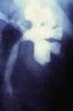 Intravenous pyelography defines a functioning kidney with a large branching intrarenal stone burden.
This article reviews the available endoscopic lithotrites and their clinical applications.
History of the Procedure
Endoscopic lithotrites include ultrasonic, electrohydraulic (EHL), and mechanical devices, as well as various lasers. These instruments are passed through the working channel of the endoscope to fragment stones into extractable pieces. Baskets and graspers are used during lithotripsy to immobilize stones and to remove stone fragments.
Ultrasonic lithotripsy
Ultrasonic lithotripsy was used initially. This modality requires a rigid endoscope and is commonly used via a percutaneous renal approach. It is less useful with ureteroscopy.
Electrohydraulic lithotripsy
EHL probes deliver energy via 2 coaxial electrodes. Ignition creates a small spark of high temperature that vaporizes a small volume of water into a gaseous bubble. The bubble expands circumferentially. Power is proportional to the diameter of the probe. Drawbacks of EHL lithotripsy include its potential for damaging adjacent tissue, producing large fragments, and occasionally failing to fragment the hardest calculi, including calcium oxalate monohydrate.
Mechanical lithotripsy
Pneumatic mechanical devices, such as the Lithoclast, are small endoscopic jackhammers that work best when passed through a straight endoscopic working channel. With reusable stainless steel probes, the Lithoclast can be used through rigid or semirigid endoscopes. The Lithoclast is an efficient and economical means of fragmenting calculi and is particularly useful for managing large and hard stones. It is commonly used for large renal stones (percutaneously) and distal ureteral stones (ureteroscopically).
Laser lithotripsy
Laser lithotripsy was first introduced commercially in the late 1980s with the pulsed-dye laser, which uses 504 nm of light delivered through optical quartz fibers. This was a nonthermal safe laser that produced plasma between the tip of the fiber and the calculus, fragmenting stone with a photo-acoustic effect. The small flexible probes complemented both the semirigid and flexible ureteroscopes and could fragment most urinary calculi, excluding cystine. However, this was not a solid-state laser, and it required frequent maintenance, including changing of the coumarin dye. The energy available at the tip of the fiber is proportional to the fiber diameter. The 200-µm fiber allows the most endoscopic deflection but can deliver only 80 mJ of energy, which is frequently insufficient to fragment calcium oxalate monohydrate calculi.
Advancing laser technology has led to the development of the holmium:YAG (yttrium-aluminum-garnet) laser, which is a thermal laser that uses a 2150-nm wavelength of light. The energy is delivered in a pulsatile fashion through low–water-density quartz fibers. Johnson studied the soft-tissue effects of this laser and found that the thermal effect of this laser within a water-based medium was confined owing to a vaporization bubble formed at the tip of the fiber. [2] In 1995, Matsuoka et al presented the first clinical series of endoscopic lithotripsy with this wavelength and found it to be safe and efficient in treating ureteral stones. [3] As opposed to the coumarin pulsed-dye laser, holmium laser lithotripsy produces smaller fragments that can be, in part, irrigated from the collecting system during treatment.
The energy available at the tip of the holmium laser does not depend on the diameter of the fiber. Techniques used to increase treatment efficiency by varying fiber diameters with complementary endoscopes have been described. These techniques involve larger fibers complemented by increased stiffness, which decrease the flexibility of the endoscope.
For additional information, see Medscape Reference’s Lasers in Urology article.
Indications
Ureteroscopic lithotripsy as a common treatment for distal ureteral stones began in the early 1980s. During the same period, ESWL was introduced as a treatment for uncomplicated, moderately sized renal calculi.
-
While ureteroscopy progressed over the next 10 years, extracorporeal shockwave lithotriptors evolved to second-generation and third-generation devices that required fewer anesthetics during treatment but yielded lower stone-free rates and more related procedures than the first-generation machines.
-
New generators with smaller focal zones had focused shockwaves and required lower overall power.
-
The imaging on the newer extracorporeal lithotriptors allowed easier localization of ureteral stones, and there was great enthusiasm for treating stones throughout the entire upper urinary tract with this modality.
-
In certain cases, ureteral stents were also placed to localize stones in the ureter prior to shockwave lithotripsy and to ensure drainage of an obstructed upper urinary tract.
-
The newest devices did not obtain the success rates of the first-generation Dornier HM3.
In the early 1990s, the American Urological Association (AUA) developed guidelines for treating calculi. The guidelines were based on published clinical experience with ESWL and endoscopic lithotripsy.
-
The first guidelines panel dealt with the treatment of large renal stones (>2.5 cm). In this study, the AUA panel suggested that percutaneous nephrostolithotomy was superior to shockwave lithotripsy for such stones.
-
The second guidelines panel addressed the treatment of ureteral calculi. Treatment was stratified by stone size and location and other considerations. The panel suggested that stones smaller than 5 mm that are unassociated with high-grade upper urinary tract obstruction frequently pass without surgical intervention. The panel also suggested that patients with such stones but without prolonged, symptomatic, or complete upper urinary tract obstruction or associated infection should be monitored clinically.
-
The AUA panel recommended that larger ureteral calculi and those associated with significant obstruction can be treated with either ESWL or ureteroscopic lithotripsy. The most recent clinical series have found shockwave lithotripsy based on the newest extracorporeal lithotriptors to be less invasive and less efficient in treating ureteral stones, with fragment clearance often requiring as many as 4 months of follow-up.
-
Ureteroscopic treatment of renal calculi is gaining popularity because of the recognition of limitations of ESWL. Although ESWL is associated with minimal morbidity, its effectiveness is decreased in the treatment of certain stone compositions (eg, calcium oxalate monohydrate, cysteine), large stones, and stones located in the lower pole.
The 2016 AUA/Endourological Society guidelines provide the following recommendations for surgical treatment [4] :
-
In patients who have mid or distal ureteral stones that require intervention, ureteroscopy should be recommended as first-line therapy
-
For patients who fail or are unlikely to have successful results with shock-wave lithotripsy and/or ureteroscopy, options include percutaneous nephrolithotomy or laparoscopic, open, or robotic-assisted stone removal
-
In symptomatic patients with a total non-lower pole renal stone burden of less than 20 mm, shock-wave lithotripsy or ureteroscopy can be offered
-
In symptomatic patients with a total renal stone burden of more than 20 mm, percutaneous nephrolithotomy is first-line therapy
Flexible ureteroscopy with holmium laser lithotripsy is an attractive alternative to shockwave lithotripsy in the management of renal calculi in anomalous and/or ectopic kidneys (ie, horseshoe kidneys). In addition, ureteroscopy is a primary treatment in select patients with symptomatic stones in pelvic kidneys.
Certain patients or stone characteristics may favor ureteroscopic lithotripsy over ESWL or percutaneous nephrolithotripsy (PCNL). These include the following:
-
Lower-pole stone location
-
Cysteine or calcium oxalate monohydrate stone composition
-
Morbid obesity
-
Uncorrectable bleeding diathesis
-
Stones within a calyceal diverticulum or infundibular stenosis
-
Ectopic kidney
Contraindications
No contraindications to endoscopic lithotripsy exist, with the exception of those associated with endoscopy.
-
Staghorn renal calculus is noted on plain radiograph of the abdomen.
-
Intravenous pyelography defines a functioning kidney with a large branching intrarenal stone burden.
-
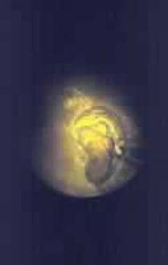
Treatment was based on percutaneous access and rigid endoscopic lithotripsy to clear the central stone burden with the hollow core ultrasonic lithotripter. On this image, a flexible nephroscope and holmium laser were also used to address portions of the stone burden inaccessible to the rigid equipment.
-
This is an endoscopic image of a holmium laser fiber fragmenting a stone burden.
-
Ureteroscopy and laser lithotripsy. Video courtesy of Dennis G Lusaya, MD, and Edgar V Lerma, MD.


