Practice Essentials
Despite the vulnerable position of the testicles, testicular trauma is relatively uncommon. The mobility of the scrotum may be one reason severe injury is rare. Given the importance of preserving fertility, traumatic injuries of the testicle deserve careful attention.
Testicular injuries can be divided into three broad categories based on the mechanism of injury: (1) blunt trauma, (2) penetrating trauma, and (3) degloving trauma. Such injuries are typically seen in males aged 15-40 years.
Blunt trauma refers to injuries sustained from objects applied with any significant force to the scrotum and testicles. Any kind of contact sport, without the use of protective aids, may be associated with genital trauma. Examples include a kick to the groin or a baseball injury. [1] One report even described testicular rupture from a paint ball injury. [2] Also, one study reported an increased incidence of testicular calcifications in extreme mountain bikers over nonbikers, suggesting repeated testicular trauma in these individuals. [3]
Testicular rupture occurs in 50% of cases of direct blunt scrotal trauma. Intense compression of the testis against the inferior pubic ramus or symphysis results in a rupture of the tunica albuginea. A force of approximately 50 kg is necessary to cause testicular rupture. [4]
Penetrating trauma refers to injuries sustained from sharp objects or high-velocity missiles. Examples include gunshot and stab wounds.
Degloving injuries (or avulsion injuries) are less common. With these, scrotal skin is sheared off, for example, when a testicle becomes trapped in heavy machinery. Testicular rupture or fractured testis refers to a rip or tear in the tunica albuginea resulting in extrusion of the testicular contents (see the image below).
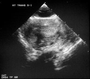 This scrotal sonogram shows a fractured testis with a disrupted tunica albuginea and testicular contents surrounded by tunica vaginalis.
Testicular dislocation is an uncommon and sometimes easily overlooked event that refers to a testis that has been relocated from its orthotopic position to another location secondary to blunt trauma. Indirect inguinal hernias and atrophic testicles may be predisposing factors. Most cases of testicular dislocation are the result of motorcycle crashes, and 25% involve both testicles. [4] This is related to impact with the fuel tank, and the inguinal region is the most frequent site of displacement. [5] Additional dislocation routes include the following:
-
Pubic
-
Preputial
-
Acetabular
-
Canalicular
-
Penile
-
Intra-abdominal
-
Retrovesical
-
Perineal
-
Crural
A thorough history and detailed physical examination are essential for an accurate diagnosis. Ultrasonography is the imaging modality of choice; especially in patients with blunt trauma, scrotal ultrasonography with Doppler flow evaluation can help in determining the nature and extent of injury. [4, 6, 7] The sensitivity and specificity of ultrasonography in this situation has been reported to be 93.5% and 100%, respectively. However, in the setting of a clinically apparent hematocele, some authors question the value of a ultrasonographic examination and feel prompt exploration is more appropriate. [8]
Testicular dislocation is treated with manual closed reduction. Surgical fixation is used if closed reduction is unsuccessful.
Penetrating testicular trauma usually requires scrotal exploration to determine the severity of testicular injury, to assess the structural integrity of the testis, and to control intrascrotal hemorrhage. If the tunica albuginea is violated, early surgical exploration, debridement, and closure of the tunica albuginea are necessary.
Blunt injuries are encountered more often than penetrating injuries and are usually unilateral, whereas penetrating injuries involve both testes in a third of cases. [4] Most cases of blunt trauma to the testicles are minor and usually require only conservative therapy. However, in one study, Buckley and McAninch reported that 46% of patients presenting with blunt scrotal trauma underwent surgical exploration and were found to have rupture of the tunica albuginea. [9] Operative indications for blunt trauma are as follows:
-
Suspicion of rupture
-
Expanding hematomas
-
Dislocation refractory to manual reduction
-
Avulsion
-
Scrotal degloving
However, a study by Chandra et al has suggested that conservative management is an option in blunt trauma patients when ultrasonography demonstrates absence of hematocele, obvious testicular fracture planes, or disruption of the tunica albuginea. [8] In a study of nonoperative management in seven adolescent boys who presented with testicular rupture 1 to 5 days after sustaining blunt scrotal trauma, Cubillos et al reported that none of the patients required orchiectomy or developed atrophy at 6 months of follow-up. [10] Redmond et al reported on 23 patients with significant testicular injury (rupture of tunica albuginea or large hematocele) who were managed conservatively with analgesia, antibiotics and scrotal support, regardless of ultrasound findings. Four patients had evidence of testicular atrophy at their 3-month follow-up appointment. None reported chronic pain or required delayed orchidectomy. Four patients later underwent repair of an asymptomatic post-traumatic hydrocoele. [11] Further investigation is needed before such an approach can be recommended in children or adults.
Genital self-mutilation is another potential source of testicular trauma. These patients are typically psychotic, although nonpsychotic patients practicing autoeroticism, and motivated yet desperate transsexuals, may find themselves requiring an urgent urologic consultation. Most cases of genital self-mutilation involve men castrating themselves. If the patients seek care promptly and the testicles are vital, reimplantation may be considered.
For patient education resources, see Testicular Pain and Injuries.
Relevant Anatomy
To properly evaluate and treat testicular injuries, a thorough knowledge of scrotal and testicular anatomy is required.
The outermost layer of the scrotum is the scrotal skin. The next most superficial layer is the dartos muscle/fascia, which is contiguous with the Scarpa fascia of the abdomen, the Colles fascia of the perineum, and the dartos fascia of the penis. The dartos layer is followed by the external, middle, and internal spermatic fasciae, which are contiguous with the external oblique, internal oblique, and transversalis fasciae, respectively. The middle spermatic fascia forms the cremasteric muscle of the spermatic cord. In most cases, the testicle is tethered to the scrotum inferiorly by the gubernaculum.
The next layer is the tunica vaginalis, which is composed of an outer (parietal) layer and an inner (visceral) layer. The tunica albuginea is a tough, white, fibrous, capsulelike layer surrounding the seminiferous tubules of the testis. The visceral layer of the tunica vaginalis adheres to this layer.
Immediately beneath the tunica albuginea is the final layer, the tunica vascularis, which contains the arterial blood supply to the seminiferous tubules. The tunica albuginea extends inward posteriorly to form the mediastinum testis, the point where vessels and ducts traverse the testicular capsule. The epididymis attaches posterolaterally.
Blood supply to the testes is threefold:
-
The testicular artery is the principal artery, arising from the aorta, just below the renal artery.
-
The cremasteric artery is a branch of the inferior epigastric artery.
-
The deferential artery is a branch of the superior vesical artery.
These 3 vessels collateralize and anastomose in the spermatic cord and near the epididymis.
Pathophysiology
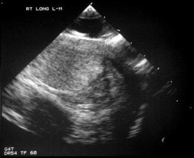
The testis is enveloped by layers of white fibrous connective tissue, the tunica vaginalis and the tunica albuginea. The tunica albuginea is the visceral layer that covers the testis, and the tunica vaginalis is the parietal layer that lines the hydrocele sac. The image below depicts a healthy testis.
The tunica albuginea is the layer that is violated during a testicular rupture. Approximately 50 kg of force is required to rupture the testicle. A tear in the tunica albuginea leads to extrusion of the seminiferous tubules and allows an intratesticular hemorrhage to escape into the tunica vaginalis. This is referred to as a hematocele. Disruption of the tunica vaginalis or extension to the epididymis leads to bleeding into the scrotal wall, resulting in a scrotal hematoma.
Two factors protect the testes from minor external trauma. First, a thin layer of serous fluid (ie, physiologic hydrocele) separates the tunica albuginea from the tunica vaginalis and allows the testis to slide freely within the scrotal sac. Second, the testes are suspended within the scrotum by the spermatic cord, allowing them to move freely within the genital area. In cases of penetrating trauma or severe blunt trauma, these protective features are insufficient to prevent injury to the testis proper.
Etiology
The most common cause of blunt testicular trauma is sports injuries. For example, a study of rugby players in Australia and New South Wales from 1980 to 1993 revealed 14 players with testicular injuries, with the most unfortunate losing both testicles.
However, the risk of sports-related testicular injury in US children is likely less than previously suspected. Wan et al (2003) reviewed the National Pediatric Trauma Registry for all 50 states and referenced commonly played contact sports. Of 5,439 reported sports injuries, none were testicular injuries. [12] The American Academy of Pediatrics Committee on Sports Medicine and Fitness gives an "unqualified yes" to the question of whether or not a boy with only one testicle can play sports. Protective cups may be required in some instances.
The second most common cause of testicular trauma is a kick to the groin. Less common etiologies include motor vehicle accidents, falls, and straddle injuries.
The most common cause of penetrating testicular injuries is a gunshot wound to the genital area. [13, 14] Other causes include stab wounds, self-mutilation, animal bites (usually dog), and emasculation.
Degloving testicular injuries most commonly result from accidents incurred while operating heavy machinery (eg, industrial or farming accidents).
Epidemiology
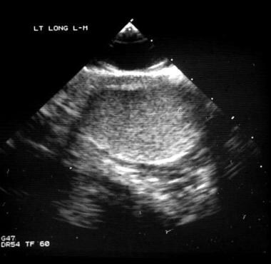
Testicular trauma is relatively uncommon. Blunt trauma accounts for approximately 85% of cases, and penetrating trauma accounts for 15%. As many as 80% of hematoceles (blood in the tunica vaginalis) are associated with testicular rupture. The image below depicts hematoma in testicular fracture.
In a survey of 731 male high school and college athletes, 18% reported having had a testicular injury during sports and 36.4% had observed injuries in other team members. Only 12.9% of respondents reported use of athletic cups. Prevalence rates by sport of reported testicular injuries were as follows [15] :
-
Lacrosse, 48.5%
-
Wrestling, 32.8%
-
Baseball, 21%
-
Football, 17.8%
Testicular dislocation is seen in less than 0.5% of cases of abdominal trauma. One retrospective review of emergency department records found that all instances were missed initially, even with CT scanning demonstrating an empty scrotum and displaced testis. The average delay in diagnosis was 19 days. [16]
In combat operations, wounds to the genitourinary (GU) structures have historically been less common than extremity and penetrating abdominal trauma. However, the use of improvised explosive devices (IEDs) has resulted in a significant increase in GU wounds since 2001. Studies report that 33-36% of GU injuries involved the testes. [17, 18]
Prognosis
Traumatic testicular injuries are relatively uncommon. When present, they are most often caused by blunt trauma. History, physical examination, and scrotal ultrasonography with Doppler studies are important in diagnosing and staging these injuries.
Surgical exploration of all testicular penetrating injuries and many blunt injuries has proven to increase testicular salvage rates and decrease morbidity. Early surgical intervention leads to higher salvage rates, shorter hospitalizations, and a more rapid return to baseline activity. Phonsombat et al (2008) found that testicular salvage rates are significantly higher with gunshot wound injuries than with stab wounds and lacerations, as gunshot wounds less commonly involve the spermatic cord. [19]
Following repair of penetrating testicular trauma caused by conventional bullet wounds, fertility results are approximately 62%. Fertility rates are much lower with high-velocity gunshot wounds.
Complications associated with untreated testicular injuries are significant and include the following:
-
Testicular infarction
-
Testicular torsion
-
Testicular or epididymal abscess
-
Testicular necrosis
-
Testicular atrophy
Complications associated with scrotal exploration and testicular salvage include the following:
-
Bleeding
-
Infection
-
Loss of testis
Nearly all of the aforementioned complications are irreversible. However, Yoshimura et al (2002) reported restoration of spermatogenesis in a patient by orchiopexy 13 years after bilateral traumatic testicular dislocation. Although the patient was azoospermic before surgery and was found to have atrophic testicles rotated 180° intraoperatively, he was able to father a child 10 months later. [20]
Animal-based research has found that grade I unilateral blunt testicular trauma, defined as intratesticular hemorrhage with an intact tunica albuginea, significantly affects germ cell maturation bilaterally and alters the sex hormone profile. Ischemia-reperfusion of the testis, which is possible in a trauma patient, has been shown to cause germ cell–specific apoptosis and subsequent aspermatogenesis. Lysiak et al (2003) suggested that this may be due to a cytokine–stress-related kinase pathway. [21]
Progressive testicular atrophy may occur in spite of a successful repair. Testicular atrophy is most likely the result of the original testicular trauma rather than efforts to salvage the testis. In a follow-up ultrasonographic study of unilateral testicular trauma, Cross and colleagues found that 5 of the 10 patients developed atrophy of the injured testis, defined as a reduction in volume of more than 50% compared with the unaffected side. [22]
Trauma-related torsion was described as early as the 19th century by Mikulicz and Gervais, and recent data suggest that trauma may account for 5%-6% of torsion cases.
-
This scrotal sonogram shows a healthy testis.
-
This scrotal sonogram shows a fractured testis with a disrupted tunica albuginea and testicular contents surrounded by tunica vaginalis.
-
This scrotal sonogram shows intratesticular hematoma in a fractured testis.


