Practice Essentials
Candidiasis is a fungal infection caused by yeasts from the genus Candida. Candida albicans is the predominant cause of the disease.
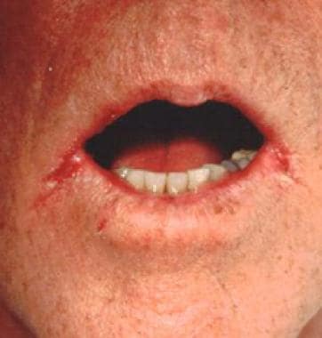 Soreness and cracks at the lateral angles of the mouth (angular cheilitis) are a frequent expression of candidiasis in elderly individuals. Courtesy of Matthew C. Lambiase, DO.
Signs and symptoms
Chronic mucocutaneous candidiasis
Findings reveal disfiguring lesions of the face, scalp, hands, and nails. Chronic mucocutaneous candidiasis occasionally is associated with oral thrush and vitiligo.
Oropharyngeal candidiasis
Individuals with oropharyngeal candidiasis (OPC) usually have a history of HIV infection, wear dentures, have diabetes mellitus, or have been exposed to broad-spectrum antibiotics or inhaled steroids. Although patients frequently are asymptomatic, when symptoms do occur, they can include the following:
-
Sore and painful mouth
-
Burning mouth or tongue
-
Dysphagia
-
Thick, whitish patches on the oral mucosa
Physical examination reveals a diffuse erythema and white patches that appear on the surfaces of the buccal mucosa, throat, tongue, and gums.
The following are the 5 types of OPC:
-
Membranous candidiasis - One of the most common types; characterized by creamy-white, curdlike patches on the mucosal surfaces
-
Chronic atrophic candidiasis (denture stomatitis) - Also thought to be one of the most common forms of the disease; presenting signs and symptoms include chronic erythema and edema of the portion of the palate that comes into contact with dentures
-
Erythematous candidiasis - Associated with an erythematous patch on the hard and soft palates
-
Angular cheilitis - Inflammatory reaction characterized by soreness, erythema, and fissuring at the corners of the mouth
-
Mixed - A combination of any of the above types is possible
Esophageal candidiasis
Patients with esophageal candidiasis may be asymptomatic or may have 1 or more of the following symptoms:
-
Normal oral mucosa (>50% of patients)
-
Dysphagia
-
Odynophagia
-
Retrosternal pain
-
Epigastric pain
-
Nausea and vomiting
Physical examination almost always reveals oral candidiasis.
Nonesophageal gastrointestinal candidiasis
The following symptoms may be present:
-
Epigastric pain
-
Nausea and vomiting
-
Abdominal pain
-
Fever and chills
-
Abdominal mass (in some cases)
Genitourinary tract candidiasis
The types of genitourinary tract candidiasis are as follows:
-
Vulvovaginal candidiasis (VVC) - Erythematous vagina and labia; a thick, curdlike discharge; and a normal cervix upon speculum examination [1]
-
Candida balanitis - Penile pruritus and whitish patches on the penis
-
Candida cystitis - Many patients are asymptomatic, but bladder invasion may result in frequency, urgency, dysuria, hematuria, and suprapubic pain
-
Asymptomatic candiduria - Most catheterized patients with persistent candiduria are asymptomatic
-
Ascending pyelonephritis - Flank pain, abdominal cramps, nausea, vomiting, fever, chills and hematuria
-
Fungal balls - Intermittent urinary tract obstruction with subsequent anuria and ensuing renal insufficiency
See Clinical Presentation for more detail.
Diagnosis
Diagnostic tests for candidiasis include the following:
-
Mucocutaneous candidiasis - For a wet mount, scrapings or smears obtained from skin, nails, or oral or vaginal mucosa are examined under the microscope; a potassium hydroxide smear, Gram stain, or methylene blue is useful for direct demonstration of fungal cells
-
Cutaneous candidiasis - Using a wet mount, scrapings or smears obtained from skin or nails can be examined under the microscope; potassium hydroxide smears are also useful
-
Genitourinary candidiasis - A urinalysis should be performed; evidence of white blood cells (WBCs), red blood cells (RBCs), protein, and yeast cells is common; urine fungal cultures are useful
-
Gastrointestinal candidiasis - Endoscopy with or without biopsy
See Workup for more detail.
Management
Management of candidiasis includes the following:
-
Cutaneous candidiasis - Most localized cutaneous candidiasis infections can be treated with any number of topical antifungal agents (eg, clotrimazole, econazole, ciclopirox, miconazole, ketoconazole, nystatin)
-
Chronic mucocutaneous candidiasis - This condition is generally treated with oral azoles
-
Oropharyngeal candidiasis - This can be treated with either topical antifungal agents or systemic oral azoles
-
Esophageal candidiasis - Treatment requires systemic therapy with oral azoles, such as fluconazole, itraconazole, and voriconazole.
-
VVC - Topical antifungal agents or oral fluconazole can be used [2]
-
Candida cystitis - In noncatheterized patients, Candida cystitis should be treated with fluconazole; in catheterized patients, the Foley catheter should be removed or replaced; if the candiduria persists after the catheter change, then patients can be treated with fluconazole
See Treatment and Medication for more detail.
Background
Candidiasis is caused by infection with species of the genus Candida, predominantly with Candida albicans.Candida species are ubiquitous fungi that represent the most common fungal pathogens that affect humans. The growing problem of mucosal and systemic candidiasis reflects the enormous increase in the number of patients at risk and the increased opportunity that exists for Candida species to invade tissues normally resistant to invasion. Candida species are true opportunistic pathogens that exploit recent technological advances to gain access to the circulation and deep tissues.
The increased prevalence of local and systemic disease caused by Candida species has resulted in numerous new clinical syndromes, the expression of which depends primarily on the immune status of the host. Candida species produce a wide spectrum of diseases, ranging from superficial mucocutaneous disease to invasive illnesses, such as hepatosplenic candidiasis, Candida peritonitis, and systemic candidiasis. The management of serious and life-threatening invasive candidiasis remains severely hampered by delays in diagnosis and the lack of reliable diagnostic methods that prevent the early identification of candidemia and invasive candidiasis.
Advances in medical technology, chemotherapeutics, cancer therapy, and organ transplantation have greatly reduced the morbidity and mortality of life-threatening disease. Patients who are critically ill and in medical and surgical ICUs have been the prime targets for opportunistic nosocomial fungal infections, primarily due to Candida species. Studies suggest that the problem is not under control and, in fact, show it is worsening. On a daily basis, virtually all physicians are confronted with a positive Candida isolate obtained from one or more various anatomic sites. High-risk areas for Candida infection include neonatal, pediatric, and adult ICUs, both medical and surgical. [3] Candida infections can involve any anatomic structure.
Pathophysiology
Candida species are yeastlike fungi that can form true hyphae and pseudohyphae. For the most part, Candida species are confined to human and animal reservoirs; however, they are frequently recovered from the hospital environment, including on foods, countertops, air-conditioning vents, floors, respirators, and medical personnel. They are found as commensals of diseased skin and normal mucosal membranes of the gastrointestinal, genitourinary, and respiratory tracts, since early in life.
Candida species also contain their own set of well-recognized but poorly characterized virulence mechanisms that contribute to their ability to cause infection. [4, 5, 6] The main virulence factors include the following:
-
Surface molecules that permit adherence of the organism to other structures (eg, human cells, extracellular matrix, prosthetic devices)
-
Acid proteases and phospholipases that involve penetration and damage of cell envelopes
-
Ability to convert to a hyphal form (phenotypic switching)
-
Upregulation of alternative carbon utilization pathways (metabolic adaptation)
-
Induction of differential stress resistance responses (superoxidedismutases)
-
Masking of cell wall components for immune evasion
As with most fungal infections, host defects play a significant role in the development of candidal infections. Host defense mechanisms against Candida infection and their associated defects that allow infection are as follows:
-
Intact mucocutaneous barriers - Wounds, intravenous catheters, burns, ulcerations
-
Phagocytic cells -Granulocytopenia
-
Polymorphonuclear leukocytes - Chronic granulomatous disease
-
Monocytic cells -Myeloperoxidase deficiency
-
Complement -Hypocomplementemia
-
Immunoglobulins -Hypogammaglobulinemia
-
Cell-mediated immunity - Chronic mucocutaneous candidiasis, diabetes mellitus, cyclosporin A, corticosteroids, HIV infection
-
Mucocutaneous protective bacterial flora - Broad-spectrum antibiotics
Risk factors associated with mucocutaneous, invasive or systemic candidiasis include the following [7, 5, 8, 9] :
-
Granulocytopenia
-
Bone marrow transplantation
-
Solid organ transplantation (liver, kidney)
-
Parenteral hyperalimentation
-
Hematologic malignancies
-
Foley catheters
-
Solid neoplasms
-
Recent chemotherapy or radiation therapy
-
Corticosteroids, TNF-α inhibitors, and IL-17-targeted biologics
-
Clostridioides difficile infection
-
Broad-spectrum antibiotics
-
Burns
-
Prolonged hospitalization
-
Severe trauma
-
Recent bacterial infection
-
Recent surgery
-
Gastrointestinal tract surgery
-
Central intravascular access devices
-
Premature birth
-
Hemodialysis
-
Acute and chronic renal failure
-
Mechanical ventilation for longer than 3 days
The first step in the development of a candidal infection is colonization of the mucocutaneous surfaces. All of the factors outlined above are associated with increased colonization rates. The routes of candidal invasion include (1) disruption of a colonized surface (skin or mucosa), allowing the organisms access to the bloodstream, and (2) persorption via the gastrointestinal wall, which may occur following massive colonization with large numbers of organisms that pass directly into the bloodstream.
Frequency
United States
Candida species are the most common cause of fungal infection in immunocompromised persons. Oropharyngeal colonization is found in 30-55% of healthy young adults, and Candida species may be detected in 40-65% of normal fecal flora.
Three of every 4 women experience at least 1 bout of vulvovaginal candidiasis (VVC) during their lifetime.
More than 90% of persons infected with HIV who are not receiving highly active antiretroviral therapy (HAART) eventually develop oropharyngeal candidiasis (OPC), and 10% eventually develop at least 1 episode of esophageal candidiasis. [10]
In persons with systemic infections, Candida species have been found as the fourth most commonly isolated pathogens from blood cultures. [11]
Clinical and autopsy studies have confirmed the marked increase in the incidence of disseminated candidiasis, reflecting a parallel increase in the frequency of candidemia. This increase is multifactorial in origin and reflects increased recognition of the fungus, a growing population of patients at risk (eg, patients undergoing complex surgical procedures, patients with indwelling vascular devices), and the improved survival rates among patients with underlying neoplasms or collagen-vascular disease and patients who are immunosuppressed.
International
Similar rates of mucocutaneous and systemic candidiasis/candidemia have been observed worldwide. [12, 13] In fact, throughout the world, Candida species have replaced Cryptococcus species as the most common fungal pathogens affecting immunocompromised hosts. The frequency of non-albicans Candida causing candidemia and invasive candidiasis has grown in recent decades, an observation also found in the United States. [14]
Mortality/Morbidity
Mucocutaneous candidiasis: Most candidal infections are mucocutaneous and, as such, do not cause mortality. However, in patients with advanced immunodeficiency due to HIV infection, these mucosal infections can become refractory to antifungal therapy and may lead to severe oropharyngeal and esophageal candidiasis that initiates a vicious cycle of poor oral intake, malnutrition, wasting, and early death.
Candidemia and disseminated candidiasis: Mortality rates associated with these infections have not improved markedly and remain in the range of 30-40%. Systemic candidiasis causes more case fatalities than any other systemic mycosis. More than a decade ago, investigators reported the enormous economic impact of systemic candidiasis in hospitalized patients. Candidemia is associated with considerable prolongation in hospital stays (70 d vs 40 d in comparable patients without fungemia). Although mucocutaneous fungal infections, such as oral thrush and Candidaesophagitis, are extremely common in patients with AIDS, candidemia and disseminated candidiasis are uncommon.
Sex
Neither sex is predisposed to candidal colonization; however, VVC is the second most common cause of vaginitis in women.
Age
Persons at the extremes of age (neonates and adults >65 y) are most susceptible to candidal colonization. Mucocutaneous candidiasis is also more prevalent in neonates and older adults. Very-low-birth-weight and extremely-low-birth-weight infants are at high risk for blood culture–proven late-onset candidiasis (defined as sepsis that develops after age 72 h). [15]
-
A moist, erosive, pruritic patch of perianal skin and perineum (with satellite pustule formation) is demonstrated in this woman with extensive candidiasis. Courtesy of Matthew C. Lambiase, DO.
-
Discrete superficial pustules developed within hours of birth on the hand of an otherwise healthy newborn. A potassium hydroxide preparation revealed spores and pseudomycelium, and culture demonstrated the presence of Candida albicans. Courtesy of Matthew C. Lambiase, DO.
-
Dry, red, superficially scaly, pruritic macules and patches on the penis represent candidal balanitis. Courtesy of Matthew C. Lambiase, DO.
-
White plaques are present on the buccal mucosa and the undersurface of the tongue and represent thrush. When wiped off, the plaques leave red erosive areas. Courtesy of Matthew C. Lambiase, DO.
-
Erythema, maceration, and satellite pustules in the axilla, accompanied by soreness and pruritus, result in a form of intertrigo. Courtesy of Matthew C. Lambiase, DO.
-
A nailfold with candidal infection becomes erythematous, swollen, and tender with occasional discharge. Courtesy of Matthew C. Lambiase, DO.
-
Soreness and cracks at the lateral angles of the mouth (angular cheilitis) are a frequent expression of candidiasis in elderly individuals. Courtesy of Matthew C. Lambiase, DO.
-
Fine, superficial pustules on an erythematous patchy base are suggestive of candidiasis. Courtesy of Matthew C. Lambiase, DO.
-
Candidal infection should be in the differential diagnosis when one or more nails become discolored, have subungual discoloration, have nailplate separation from the nailbed, and lack evidence of a dermatophyte. Courtesy of Matthew C. Lambiase, DO.
-
Candida dermatitis in the diaper area. Courtesy of Hon Pak, MD.