Background
Burns are wounds sustained through thermal injury. These injuries could be heat, chemical, electrical, radiation.
Approximately 500,000 people seek medical attention for burns every year in the United States, 40,000 of whom require hospitalization. [1] Unlike other types of injury, burn wounds induce metabolic and inflammatory alterations that predispose the patient to various complications. Infection is the most common cause of morbidity and mortality in this population, with almost 61% of deaths being caused by infection. [2]
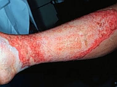 Second-degree burns often are red, wet, and very painful. Their depth, ability to heal, and tendency to result in hypertrophic scar formation vary enormously.
See Thermal Injuries: A Matter of Degree, a Critical Images slideshow, to help identify and treat various degrees and types of burn injuries.
Also, see the 5 Body Modifications and Piercing: Dermatologic Risks and Adverse Reactions slideshow to help recognize various body modifications and the related potential complications.
The skin, one of the largest organs in the body, performs numerous vital functions, including fluid homeostasis, thermoregulation, immunologic functions, neurosensory functions, and metabolic functions (eg, vitamin D synthesis). The skin also provides primary protection against infection by acting as a physical barrier. When this barrier is damaged, pathogens can directly infiltrate the body, resulting in infection. [3]
In addition to the nature and extent of the thermal injury influencing infections, the type and quantity of microorganisms that colonize the burn wound appear to influence the risk of invasive wound infection. The pathogens that infect the wound are primarily gram-positive bacteria such as methicillin-resistant Staphylococcus aureus (MRSA) [4] and gram-negative bacteria such as Acinetobacter baumannii-calcoaceticus complex, Pseudomonas aeruginosa, and Klebsiella species. These latter pathogens are notable for their increasing resistance to a broad array of antimicrobial agents. [5, 6]
Fungal pathogens also can infect burn wounds. These infections more frequently occur after the use of broad-spectrum antibiotics. Among the fungal pathogens, Candida albicans is the most common cause of infection. [7] More recently, a trend toward nosocomially acquired, intrinsically resistant fungal infections (eg, Candida krusei) has been reported. [8]
Factors associated with improved outcome and infection prevention include early burn eschar excision, topical antibiotic therapy, and aggressive infection-control measures. [9]
Pathophysiology
The burn wound typically has 3 characteristic areas of involvement. The first is the zone of coagulation, which is nearest the heat source and includes dead tissue that forms the burn eschar. The second is the zone of stasis, which is adjacent to the area of necrosis; this area is viable but is at a substantial risk for ongoing necrosis and ischemic damage due to perfusion defects. The third is the zone of hyperemia, which includes relatively healthy skin with increased blood flow and vasodilation in response to the injury; the cellular injury in this area is minimal. [3]
Wounds reflect the mechanism of the burn. In thermal burns, the degree of cellular damage varies based on the duration and temperature of exposure. Increasing temperatures alter molecular conformation, destroy cell membranes, denature proteins, and release oxygen-free radicals; all of these phenomena lead to cell death and burn eschar. Types of chemical burns differ and include those due to reducing agents (eg, hydrochloric acid), oxidizing agents (eg, sodium hypochlorite), and corrosive agents (eg, phenol); each causes burn injuries with varying modes of action. [3, 9]
Burns not only alter the innate immune character of the skin but also other arms of the immune system. Overall, T-cell activity is reduced through an increase in the number of T regulatory cells and a decrease in the number of helper cells. The levels of inflammatory cytokines and complement also are decreased. In addition, burns decrease the chemotactic, phagocytic, and bactericidal activity of neutrophils. One of the primary concerns associated with burn injuries is the avascularity of eschar, preventing immune cells and systemically administered antibiotics from being delivered to the infection site. [3]
Immediately following a thermal burn, the surface of the burn wound is free of microorganisms. However, deep cutaneous structures that survive the initial burn injury (eg, sweat glands, hair follicles) often contain staphylococci, which colonize the wound surface during the subsequent 48 hours. Over the next 5-7 days, other microbes, including gram-negative and gram-positive bacteria, colonize the wound. These potential pathogens typically come from the patients’ gastrointestinal tract, upper respiratory tract, or the hospital environment, transferred through contact with healthcare workers. [3]
In animal models, the progression of burn wound infections has been assessed and the following progression observed: burn wound colonization, invasion into subjacent tissue (within 5 days), destruction of granulation tissue, visceral hematogenous lesions, manifestations of septic shock, and death. [10]
Early surgical debridement and skin grafting, use of topical and systemic antimicrobials, and enhanced infection-control practices have led to the replacement of beta-hemolytic streptococci with S aureus and gram-negative bacteria such as P aeruginosa, Klebsiella pneumoniae, and A baumannii as major pathogens in burn wound infections. [11]
In burns older than 7 days, biofilm may be detected in the ulcerated areas of the wounds. As biofilm formation is associated with chronicity of the wound, bacterial colonization is easier to eradicate in the first few days after injury, supporting early excision and closure of the burn wound. [12]
Fungal infections often develop later, after broad-spectrum antibiotics have been administered or after wound care has been delayed. [7, 13] Infections with anaerobes are rare, except after electrical injuries. [14] Infections with viruses such as herpes simplex virus and varicella-zoster virus rarely complicate burn wounds.
Epidemiology
Frequency
United States
According to the National Fire Protection Agency, US fire departments responded to 1.64 million fires during 2006. The total economic impact was estimated at $11.3 billion. [1] The American Burn Association (ABA) National Burn Repository reports that, among burns requiring hospitalizations, the most common place of occurrence was the household (73% of cases). Occupational (8%) and traffic-related injuries (5%) were much less common. One civilian fire death occurs every 2 hours and 41 minutes. The likelihood of a US resident dying of exposure to fire, flames, or smoke is 1 in 1442. [15]
International
At the beginning of the 21st century, the Centre of Fire Statistics estimated that the average number of fires worldwide was 7-8 million, resulting in 70,000-80,000 fire deaths and 500,000-800,000 fire injuries. In Europe, 2-2.5 million fires were reported, resulting in 20,000-25,000 fire deaths and 250,000-500,000 fire injuries. The World Fire Statistics from the Geneva Association reported that, by country, the highest number of fire deaths in 2004 occurred in the United States (4,250), followed by Japan at 2,050 and the United Kingdom at 530. When adjusted for deaths per 100,000 persons between 2002 and 2004, of the 25 countries that reported data, the highest rate was in Hungary (2.1); Japan reported 1.79, the United States reported 1.39, the United Kingdom reported 0.97, Spain reported 0.61, and Singapore reported 0.08.
Mortality/Morbidity
According to the National Burn Repository’s 10-year rolling data collection from January 1, 1996, through June 30, 2006, the mortality rate associated with burns was 5.3% overall, with older age and higher-percentage total body surface area (TBSA) burned correlating with higher mortality rates. The causes of death were reported in 3,463 cases; 27% died of multiple organ failure, 14% died of withheld treatment, 12% died of trauma wounds, 12% died of burn shock, 11% died of pulmonary failure/sepsis, 11% died of cardiovascular failure, 5% died of other causes, and 4% died of burn wound sepsis. Burns covering 1%-10% of the TBSA carried the lowest risk of mortality (0.7%), increasing as the percentage of TBSA burned increased. The mortality rate was 78% in patients with 90% of their TBSA burned. [1]
Among the 19,655 reported complications included in the analysis, pulmonary complications (including pneumonia [3,361], acute respiratory distress syndrome [885], and respiratory failure [1,944]) constituted the greatest percentage of cases (31%). Cellulitis (1,988) and wound infections (1,950) were responsible for 17% of complications. Septicemia (1,672) and other infections (1,250) accounted for 15% of complications. [1]
Socioeconomic Status
The rates of admissions to burn units increase in proportion with diminishing socioeconomic status, with 95% of burn-related deaths occurring in low- and middle-income countries (LMICs). Additionally, there is an association between burn mortality and gross domestic product (GDP). [16] This can be explained as those with the least available resources are exposed to burn hazards such as poor housing quality (eg, a household with no separated kitchen), lack of smoke detectors, cigarette smoking, heavy drinking, solo parenthood, household overcrowding, and low educational level. [17]
Race
According to the American Burn Association, among burn wounds requiring admission, 59% occurred in Whites, 20% in Blacks, 14% in Hispanics, and 7% in other ethnicities. [15]
Sex
Both the National Burn Registry and the American Burn Association report a higher incidence of burn wounds in males than in females, with approximately 69.7% of cases being in males. [1, 15]
Age
Most burns occur in persons aged 5-30 years, with only 8% occurring in persons older than 70 years. Younger individuals are more likely to have scald burns, while older individuals are more likely to be burned by fire. With the same percentage of TBSA burned, older patients have a higher mortality rate. [18]
Prognosis
The overall prognosis depends on numerous factors, including the patient’s age, percentage of TBSA burned, comorbidities, initial management strategies, and the support necessary for long-term rehabilitative care.
Patient Education
For patient education resources, see the First Aid and Injuries Center and the Thermal Heat or Fire) Burns article.
-
Second-degree burns often are red, wet, and very painful. Their depth, ability to heal, and tendency to result in hypertrophic scar formation vary enormously.


