You are in: eMedicine Specialties >
Pulmonology > Obstructive Airways Diseases
Bronchiectasis
Article Last Updated: Jan 31, 2007
AUTHOR AND EDITOR INFORMATION
Section 1 of 11
Author: Ethan E Emmons, MD, Assistant Chief, Department of Medicine, Brooke Army Medical Center
Ethan E Emmons is a member of the following medical societies: American College of Chest Physicians, American Thoracic Society, and Phi Beta Kappa
Editors: Helen M Hollingsworth, MD, Director, Adult Asthma and Allergy Services, Associate Professor, Department of Internal Medicine, Division of Pulmonary and Critical Care, Boston Medical Center; Francisco Talavera, PharmD, PhD, Senior Pharmacy Editor, eMedicine; Daniel R Ouellette, MD, FCCP, Associate Professor of Medicine, Wayne State University School of Medicine; Consulting Staff, Pulmonary Disease and Critical Care Medicine Service, Henry Ford Health System; Timothy D Rice, MD, Associate Professor, Departments of Internal Medicine and Pediatrics and Adolescent Medicine, Saint Louis University School of Medicine; Zab Mosenifar, MD, Director, Division of Pulmonary and Critical Care Medicine, Director, Women's Guild Pulmonary Disease Institute, Executive Vice Chair, Department of Medicine, Cedars Sinai Medical Center; Professor of Medicine, David Geffen School of Medicine at UCLA
Author and Editor Disclosure
Synonyms and related keywords:
bronchial dilatation, dry bronchiectasis, bronchial dilation, dilated bronchus, dilated bronchi, dilatated bronchus, dilatated bronchi, infected bronchi, bronchial infection, chronic obstructive pulmonary lung disease, COPD, bronchial tree infection, cylindrical bronchiectasis, cystic bronchiectasis, varicose bronchiectasis, bronchitis, chronic bronchitis, pulmonary infection, bronchial obstruction, cystic fibrosis, CF, Young syndrome, primary ciliary dyskinesia, allergic bronchopulmonary aspergillosis, bronchopulmonary aspergillosis, alpha-1 antitrypsin deficiency, alpha1-antitrypsin deficiency, rheumatic disease, traction bronchiectasis, congenital bronchiectasis, hemoptysis, bronchial obstruction, chronic aspiration, Pseudomonas aeruginosa, P aeruginosa, non-CF bronchiectasis, atypical mycobacterial infection, Klebsiella, Staphylococcus aureus, Mycobacterium tuberculosis, Mycoplasma pneumoniae, nontuberculous mycobacteria, measles, pertussis,influenza,herpes simplex, respiratory syncytial virus, Mycobacterium avium, S aureus, M tuberculosis, M pneumoniae, M avium
Background
Bronchiectasis is an uncommon disease that results in the abnormal and permanent distortion of one or more of the conducting bronchi or airways, most often secondary to an infectious process. First described by Laennec in 1819, later detailed by Sir William Osler in the late 1800s, and further defined by Reid in the 1950s, bronchiectasis has undergone significant changes in regard to its prevalence, etiology, presentation, and treatment.
Bronchiectasis can be categorized as a chronic obstructive pulmonary lung disease manifested by airways that are inflamed and easily collapsible, resulting in air flow obstruction with shortness of breath, impaired clearance of secretions often with disabling cough, and occasionally hemoptysis.
Bronchiectasis most often presents as (1) a focal process involving a lobe, segment, or subsegment of the lung, or (2) a diffusing process involving both lungs. The former is by far the most common presentation of bronchiectasis, while the latter is most often associated with systemic illnesses and/or sinopulmonary disease and asthma.
Diagnosis is usually based on a compatible clinical history of chronic respiratory symptoms, such as a daily cough and viscid sputum production, as well as radiographically by characteristic findings, such as bronchial wall thickening and luminal dilatation, on CT scan.
Pathophysiology
Bronchiectasis is an abnormal dilation of the proximal and medium-sized bronchi (>2 mm in diameter) caused by destruction of the muscular and elastic components of the bronchial walls. Affected areas may show a variety of changes including transmural inflammation, edema, scarring, ulceration, and other findings. Distal lung parenchyma may also be damaged secondary to persistent microbial infection and frequent postobstructive pneumonia. It can be congenital or acquired but is most often the latter. Congenital bronchiectasis usually affects infants and children and results from developmental arrest of the bronchial tree. The more commonly acquired forms occur in adults and older children and require an infectious insult, impairment of drainage, airway obstruction, and/or a defect in host defense. The tissue is also damaged in part by the host response of neutrophilic proteases, inflammatory cytokines, nitric oxide, and oxygen radicals. This results in damage to the muscular and elastic components of the bronchial wall. Additionally, peribronchial alveolar tissue may be damaged, resulting in diffuse peribronchial fibrosis. The result is abnormal bronchial dilatation with bronchial wall destruction and transmural inflammation. The most important functional finding of altered airway anatomy is severely impaired clearance of secretions from the bronchial tree. Impaired clearance of secretions causes colonization and infection with pathogenic organisms, contributing to the common purulent expectoration observed in patients with bronchiectasis. The result is further bronchial damage and a vicious cycle of bronchial damage, bronchial dilation, impaired clearance of secretions, recurrent infection, and more bronchial damage. 
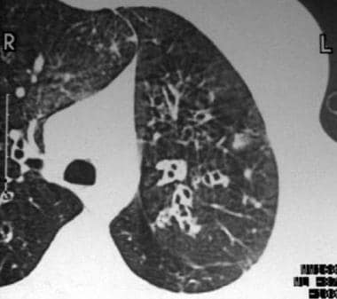
In 1950, Reid characterized bronchiectasis as cylindrical, cystic, or varicose in nature.
- Cylindrical bronchiectasis involves diffuse mucosal edema, with resultant bronchi that are dilated minimally but have straight, regular outlines that end squarely and abruptly (see Image 1).
- Cystic or saccular bronchiectasis has ulceration with bronchial neovascularization and a resultant ballooned appearance that may have air-fluid levels (see Image 2).
- Varicose bronchiectasis has a bulbous appearance with a dilated bronchus and interspersed sites of relative constriction and, potentially, obstructive scarring. The latter may subsequently result in postobstructive pneumonitis and additional parenchymal damage (see Image 3).
Frequency
United States
The prevalence mirrors the socioeconomic conditions of the population under study, being significantly less prevalent in areas where immunizations and antibiotics are readily available. Bronchiectasis is relatively uncommon in the United States, with a prevalence of around 100,000 cases based on data from the 1980s. Bronchiectasis may be underdiagnosed as it is no longer included in survey data and often goes unreported. The exception is bronchiectasis associated with cystic fibrosis (CF); the latter occurs with a prevalence of 1 in 2500 white births. CF is the largest single cause of chronic lung infections and bronchiectasis in industrialized nations. Native Americans in Alaska comprise a subgroup with higher than expected prevalence, with a 4-fold higher incidence of bronchiectasis than the general population. Overall, identifying the true incidence remains a challenge, given the lack of specific symptoms and lack of readily available noninvasive screening tests for population studies.
International
Bronchiectasis is a major cause of morbidity in less-developed countries, especially in countries with limited access to medical care and antibiotic therapy.
Mortality/Morbidity
Mortality is difficult to estimate given the difficulty in identifying prevalence and the lack of definitive studies. One study from Finland identified 842 patients aged 35-74 years with bronchiectasis and followed them for 8-13 years. These patients were also compared with asthma and COPD controls. The mortality was not found to be significantly different among the 3 groups (bronchiectasis, asthma, COPD) with mortality rates of 28%, 20%, and 38% respectively (Keistinen, 1997).
Currently, mortality is more often related to progressive respiratory failure and cor pulmonale than to uncontrolled infection. Life-threatening hemoptysis may also occur but is uncommon.
- Additional complications include chronic bronchial infection, recurrent pneumonia, empyema, pneumothorax, and lung abscess. Amyloidosis and metastatic abscesses occurred in the pre-antibiotic era but are rarely observed today.
Race
No racial predilection exists other than those that may be associated with socioeconomic status.
Sex
Evidence suggests that non-CF related bronchiectasis is more common and more virulent in women. In white women older than 60 years, bronchiectasis is often caused by primary Mycobacterium avium complex.
Age
In the preantibiotic era and in today's less-developed countries, symptoms usually began in the first decade of life. Today, the age of onset, except for those with CF, has moved into adulthood.
Although limited, epidemiologic studies suggest that persons aged 60-80 years have the highest frequency of bronchiectasis. The differences in prevalence between age groups is a direct reflection of the differences in prevalence of the underlying causes of bronchiectasis, lung disease, and/or chronic infections.
History
In clinical practice, the classic manifestations of bronchiectasis are cough and daily mucopurulent sputum production, often lasting months to years. Blood-streaked sputum or hemoptysis may result from airway damage associated with acute infection.
A rare variant known as dry bronchiectasis manifests by episodic hemoptysis with little to no sputum production. Dry bronchiectasis is usually a sequela of tuberculosis and is found in the upper lobes.
Although patients often report repetitive pulmonary infections that require antibiotics over several years, a single episode of a severe infection may result in bronchiectasis, often occurring in childhood. These include tuberculosis, pertussis, or severe bacterial pneumonia. Today, cystic fibrosis is the most common cause of bronchiectasis in children and young adults.
- Less specific symptoms include dyspnea, pleuritic chest pain, wheezing, fever, weakness, and weight loss.
- Patients may relate multiple episodes of bronchitis or pulmonary infections, which are exacerbations of bronchiectasis and often require antibiotics. These acute bacterial infections are often heralded by the onset of increased sputum production over baseline, increased viscidity of sputum, and, occasionally, a foul odor of the sputum. Rarely, low-grade fever may occur.
- Patients may experience an increase in generalized constitutional symptoms, such as fatigue and malaise, as well as increased dyspnea, shortness of breath, wheezing, or pleuritic pain.
- In bronchiectasis, secondary infection, or poorly treated pneumonia, the discrete pathogens are often unknown, but most patients relate a history of childhood infections that may include tuberculosis, pertussis, or Mycoplasma species infection.
- Most individuals have never smoked (55%) or have smoked too little to account for their degree of cough, findings of obstruction on spirometry testing, and daily sputum production.
- Bronchiectasis is a morphologic diagnosis and may exist with relatively few symptoms.
- Chronic productive cough is prominent, occurring in up to 90% of patients. Sputum is produced on a daily basis in 76% of patients.
- Some patients only produce sputum with acute upper respiratory tract infections, but otherwise they have quiescent disease.
- Sputum is typically mucoid and without a rancid odor; however, during infectious exacerbations, sputum becomes purulent and may develop an offensive odor.
- In the past, total daily sputum amount has been used to characterize the severity of bronchiectasis, with less than 10 mL defined as mild bronchiectasis, 10-150 mL defined as moderate bronchiectasis, and greater than 150 mL defined as severe bronchiectasis. Today, bronchiectasis is most often classified by radiographic findings.
- In patients with CF, the volume of sputum produced is generally much greater than that associated with other etiologies of bronchiectasis.
- Hemoptysis occurs in 56-92% of patients with bronchiectasis. Hemoptysis may be massive and life threatening secondary to bronchial artery bleeding.
- Hemoptysis is more commonly observed in dry bronchiectasis, although this presentation of bronchiectasis is rare.
- Hemoptysis is generally mild and manifested by blood flecks in the patient's usual purulent sputum. This is often the factor that leads patients to consult a physician.
- Bleeding usually originates from dilated bronchial arteries, which contain blood at systemic (rather than pulmonary) pressures. Therefore, massive hemoptysis may occur but is rarely a cause of death.
- Dyspnea may occur in as many as 72% of patients but is not a universal finding.
- Dyspnea typically occurs in patients with extensive bronchiectasis observed on chest radiographs.
- Marked dyspnea is more likely to be secondary to a concomitant illness, such as chronic bronchitis or emphysema.
- Wheezing is commonly reported and may be due to airflow obstruction following destruction of the bronchial tree. Like dyspnea, it may also be secondary to concomitant conditions, such as asthma.
- Pleuritic chest pain is an intermittent finding, occurring in 46% of patients in one study. It is most commonly secondary to chronic coughing but also occurs in the setting of acute exacerbation.
- Weight loss often occurs in patients with severe bronchiectasis.
- This is believed to occur secondary to increased caloric requirements associated with the increased work of coughing and clearing secretions.
- Weight loss suggests advanced disease but is not diagnostic of bronchiectasis.
- Fever may occur in the setting of acute infectious exacerbations.
Physical
Findings are nonspecific and may be attributed to other conditions. Most commonly, crackles, rhonchi, wheezing, and inspiratory squeaks may be heard on auscultation. General findings may include digital clubbing, cyanosis, plethora, wasting, and weight loss. Nasal polyps and signs of chronic sinusitis may also be present. In advanced disease, the physical stigmata of cor pulmonale may be observed.
- Crackles and rhonchi are often observed in association with active infections and acute exacerbations.
- Crackles are nonspecific and may occur in as many as 70% of patients.
- Scattered wheezing may be heard in approximately one third of patients. Wheezing may be due to airflow obstruction from secretions, destruction of the bronchial tree leading to airway collapsibility, or concomitant conditions.
- Digital clubbing is an inconsistent finding in approximately 3% of patients. It is more frequent in patients with moderate-to-severe bronchiectasis.
- Cyanosis and plethora are rare findings secondary to polycythemia from chronic hypoxia.
- Wasting and weight loss are suggestive of advanced disease but are not diagnostic of bronchiectasis.
- In severe cases, findings are consistent with cor pulmonale. Right heart failure may be observed, including peripheral edema, hepatomegaly, and hypoxia.
Causes
- Primary infections
- Bronchiectasis may be the sequela of a variety of necrotizing infections that are either poorly treated or not treated at all and are not occurring in the setting of another associated condition. This was particularly common in developed countries prior to the widespread use of antibiotics and today remains an important cause of bronchiectasis in developing countries, where antibiotics are used inconsistently.
- Typical offending organisms include Klebsiella species, Staphylococcus aureus, Mycobacterium tuberculosis, Mycoplasma pneumoniae, nontuberculous mycobacteria, measles, pertussis, influenza, herpes simplex, and certain types of adenovirus.
- Respiratory syncytial virus in childhood may also result in bronchiectasis.
- Mycobacterium avium complex (MAC) deserves special mention for its propensity to occur in the setting of human immunodeficiency virus (HIV) and in the host who is immunocompetent. MAC has been observed, especially in women who are nonsmokers; are older than 60 years; and have a consistent history, positive acid-fast bacilli on sputum smear, and a CT scan with small regular nodules and findings of bronchiectasis.
- Although not a cause of bronchiectasis, patients with non-CF bronchiectasis often develop chronic bronchial infection with Pseudomonas aeruginosa that is related to worsening lung function and increased morbidity and mortality.
- Bronchial obstruction
- Focal postobstructive bronchiectasis may occur in a number of clinical settings (eg, endobronchial tumors, broncholithiasis, bronchial stenosis from infections, encroachment of hilar lymph nodes, foreign body aspiration).
- In adults, foreign body aspiration often takes place in the setting of altered mental status and involves unchewed food. After aspiration, a postobstructive pneumonia may occur, with subsequent development of focal bronchiectasis. Bronchiectasis may also develop in the setting of chronic aspiration.
- Right middle lobe syndrome is a specific type of bronchial obstruction that may result in bronchiectasis. It results from an abnormal angulation of the lobar bronchus at its origin, predisposing it to obstruction, subsequent infection, and development of bronchiectasis.
- Cystic fibrosis
- CF is an autosomal recessive disease affecting approximately 1 in 2,500 whites and 1 in 17,000 blacks in the United States. It is a multisystem disorder that affects the chloride transport system in exocrine tissues, primarily secondary to a defect in the CF transmembrane regulator (CFTR) protein.
- The major pulmonary finding is bronchiectasis, which is an almost universal feature of this disease. It may be the sole feature of CF in adults or those with genetic variations of the disease.
- Bronchiectasis associated with CF is believed to occur secondary to mucous plugging of proximal airways and chronic pulmonary infection, especially with mucoid P aeruginosa.
- Young syndrome
- Young syndrome is clinically similar to CF and may represent a genetic variant of the disease.
- Patients have bronchiectasis, sinusitis, and obstructive azoospermia, but they are not affected with the other findings of CF.
- It is most often observed in middle-aged men.
- The pathogenesis of bronchiectasis is believed to be similar to that of CF.
- Primary ciliary dyskinesia
- Primary ciliary dyskinesia is a group of inherited disorders that may affect 1 in 15,000-30,000 persons. It is manifested by immotile or dyskinetic cilia and/or sperm. This may lead to poor mucociliary clearance, recurrent pulmonary infections, and, ultimately, bronchiectasis.
- A variant of this condition, initially described by Kartagener, encompassed the clinical triad of situs inversus, nasal polyps or sinusitis, and bronchiectasis in the setting of immotile cilia of the respiratory tract.
- Allergic bronchopulmonary aspergillosis
- Allergic bronchopulmonary aspergillosis is a hypersensitivity reaction to inhaled Aspergillus antigen that is characterized by bronchospasm, bronchiectasis, and immunologic evidence of a reaction to Aspergillus species.
- Allergic bronchopulmonary aspergillosis should be suspected in patients with a productive cough who also have a long history of asthma-type symptoms that do not respond to conventional therapy.
- Bronchiectasis is believed to be secondary to airway plugging by viscid secretions containing hyphae of Aspergillus species.
- CT scanning of the chest exhibits central airway bronchiectasis, differentiating this condition from other causes of bronchiectasis.
- Immunodeficiency states
- Immunodeficiency states may occur in the setting of congenital and acquired immunodeficiency. The most common congenital conditions (albeit rare) involve B lymphocyte functions, specifically hypogammaglobulinemia. The latter may involve an immunoglobulin G (IgG) subclass deficiency; X-linked agammaglobulinemia; or selective immunoglobulin A (IgA), immunoglobulin M (IgM), or immunoglobulin E (IgE) deficiency.
- Patients with hypogammaglobulinemia usually present in childhood with repeated sinus or pulmonary infections, although it has been diagnosed in adults who did not have a history of repeated infections. Establishing the diagnosis is important because gammaglobulin replacement may reduce the number of infections and resultant lung injury.
- HIV disease, with resultant acquired immunodeficiency syndrome (AIDS), has been implicated in the development of bronchiectasis and demonstrates the accelerated bronchial damage that may occur from repeated infections in patients who are immunosuppressed. Bronchiectasis in HIV has occurred with and without obvious preceding pulmonary infection and may occur secondary to immunologic dysfunction from the HIV disease itself.
- Congenital anatomic defects include the following:
- Bronchopulmonary sequestration
- Williams-Campbell syndrome (congenital cartilage deficiency)
- Mounier-Kuhn syndrome (tracheobronchomegaly)
- Swyer-James syndrome (unilateral hyperlucent lung)
- Yellow-nail syndrome
- Alpha1-antitrypsin deficiency
- Bronchiectasis has been noted to occur in this rare condition.
- The pathogenesis of bronchiectasis in this setting is unclear and may represent only coincidental findings.
- Autoimmune diseases
- Bronchiectasis has complicated both rheumatoid arthritis and Sjögren syndrome, but the pathogenesis is unclear. It has also been reported in ankylosing spondylitis.
- Rheumatic disease may occur prior to the onset of the rheumatic process.
- Bronchiectasis may also occur as an extraintestinal manifestation of ulcerative colitis, and although not published, it has been observed by this author in Crohn disease.
- Traction bronchiectasis: Traction bronchiectasis is distortion of the airways secondary to mechanical traction on the bronchi from fibrosis of the surrounding lung parenchyma. Although the airways may become dilated in this situation, the other manifestations of bronchiectasis are lacking.
- Toxic gas exposure: Exposure to toxic gas may often cause irreversible damage to the bronchial airways and cystic bronchiectasis. Suspected agents include chlorine gas and ammonia.
- Inflammatory bowel disease has also been associated with increased respiratory infections and bronchiectasis. This is especially true in patients with ulcerative colitis, although bronchiectasis also occasionally occurs in association with Crohn disease.
Alpha1-Antitrypsin Deficiency
Asthma
Bronchitis
Chronic Bronchitis
Chronic Obstructive Pulmonary Disease
Emphysema
Empyema, Pleuropulmonary
Gastroesophageal Reflux Disease
Pneumonia, Aspiration
Pneumonia, Bacterial
Tuberculosis
Other Problems to be Considered
Cystic Fibrosis
Lab Studies
- In a typical patient, bronchiectasis is suspected based on the clinical presentation, especially if purulent sputum is present and other conditions, such as pneumonia and lung abscess, have been ruled out. A sputum analysis may be used to further strengthen clinical suspicion. Radiographic studies, specifically CT scanning, then may be used to confirm the diagnosis. Once the diagnosis is confirmed, additional laboratory testing may be useful to determine the underlying cause. Although many causes are untreatable, identifying treatable conditions is paramount. In a significant percentage of patients, no readily identifiable cause is found. The choice of laboratory tests may vary and should be tailored to the individual patient and clinical situation.
- A sputum analysis may reinforce the diagnosis of bronchiectasis and add significant information regarding potential etiologies.
- Once sputum is allowed to settle, the examination may reveal Dittrich plugs, small white or yellow concretions.
- A Gram stain and culture result may reveal evidence of microorganisms, including mucoid Pseudomonas species and Escherichia coli, which suggest CF but are not diagnostic. Chronic bronchial infection with nonmucoid P aeruginosa is becoming much more common in patients with non-CF bronchiectasis.
- The presence of eosinophils and golden plugs containing hyphae suggests Aspergillus species, although this finding alone is not diagnostic of allergic bronchopulmonary aspergillosis.
- Perform a smear and culture of sputum for mycobacteria and fungi. Atypical mycobacterial infection is a common cause of bronchiectasis in the older population, especially in those with underlying structural lung disease.
- CBC count is often abnormal in patients with bronchiectasis and may be useful.
- Typical findings are nonspecific and include anemia and an elevated white blood cell count with an increased percentage of neutrophils. An increased percentage of eosinophils is one criterion for allergic bronchopulmonary aspergillosis.
- Alternatively, polycythemia secondary to chronic hypoxia may be observed in advanced cases.
- Quantitative immunoglobulin levels, including IgG subclasses, IgM, and IgA, are useful to exclude hypogammaglobulinemia. Note, however, that on rare occasions, bronchiectasis may be seen in patients with antibody production deficiency but normal to low-normal IgG levels. In situations such as these, evaluating antibody response to Haemophilus influenzae and pneumococcal vaccines may be useful.
- Quantitative alpha1-antitrypsin levels are used to rule out deficiency.
- Pilocarpine iontophoresis (sweat test) was the criterion standard test to evaluate for CF. However, genetic analysis has now become much more common and may be performed not only to look for evidence of mutations consistent with CF but also to look for potential variants, such as Young syndrome.
- Aspergillus precipitins and serum total and Aspergillus–specific IgE levels are important in making the diagnosis of allergic bronchopulmonary aspergillosis.
- Rheumatoid factor and/or other autoimmune screening tests may be performed in the appropriate clinical setting.
Imaging Studies
- Imaging studies, especially high-resolution CT (HRCT) scanning, are the cornerstone for the diagnosis of bronchiectasis.
- Additionally, the anatomical distribution of bronchiectasis may be important in helping diagnose any associated condition or cause of bronchiectasis.
- Bronchiectasis as a result of infection generally involves the lower lobes, the right middle lobe, and the lingula.
- Right middle lobe involvement alone suggests right middle lobe syndrome, an anatomic dysfunction, or a neoplastic cause with secondary mechanical obstruction.
- Bronchiectasis caused by CF, M tuberculosis, or chronic fungal infections tends to affect the upper lobes, although this is not universal in the former.
- Allergic bronchopulmonary aspergillosis also affects the upper lobes but usually involves the central bronchi, whereas most other forms of bronchiectasis involve distal bronchial segments.
- Posterior-anterior and lateral chest radiographs should be obtained in all patients.
- Expected general findings include increased pulmonary markings, honeycombing, atelectasis, and pleural changes.
- Specific findings may include linear lucencies and parallel markings radiating from the hila (tram tracking) in cylindrical bronchiectasis, dilated bronchi in varicose bronchiectasis, and clustered cysts in cystic bronchiectasis.
- In occasional patients, the diagnosis of bronchiectasis may be based on chest radiograph findings alone in the context of the appropriate clinical setting.
- CT scanning (see Image 4), particularly HRCT scanning of the chest, has replaced bronchography as the defining modality of bronchiectasis.
- Its reported sensitivity and specificity are 84-97% and 82-99%, respectively, although it may be higher at referral centers.
- Additional advantages of HRCT scanning include noninvasiveness, avoidance of possible allergic reactions to contrast media, and information regarding other pulmonary processes.
- The 3 forms of bronchiectasis in the Reid classification can be visualized by HRCT.
- Cylindrical bronchiectasis has parallel tram track lines, or it may have a signet ring appearance composed of a dilated bronchus cut in a horizontal section with an adjacent pulmonary artery representing the stone.
- Observe the ratio of size of the bronchus lumen versus the size of its adjacent vessel. A luminal airway diameter 1-1.5 times the adjacent vessel is normal. A diameter greater than 1.5 times the adjacent vessel is suggestive of bronchiectasis.
- Varicose bronchiectasis has irregular or beaded bronchi, with alternating areas of dilatation and constriction.
- Cystic bronchiectasis has large cystic spaces and a honeycomb appearance. These cystic changes are in contrast to the blebs of emphysema, which have thinner walls and are not accompanied by proximal airway abnormalities.
Other Tests
- Pulmonary function tests
- Pulmonary function test results may be normal or abnormal and may reflect underlying comorbidities.
- They are useful in obtaining a functional assessment of the patient, as well as allowing for objective determination of the deterioration of a patient's pulmonary function when baseline studies are available.
- The most common abnormality is an obstructive airway defect. This may even be found in patients without a prior smoking history.
- Obstruction is not usually reversible with bronchodilator therapy, although a subgroup of patients may develop hyperreactive airways in conjunction with their bronchiectasis that will respond to bronchodilators.
- Restriction may be observed in patients with severe advanced disease secondary to scarring and atelectasis, but this is not common. Traction bronchiectasis most often occurs in the setting of a restrictive lung defect from underlying fibrosis.
- Electron microscopy: Perform electron microscopic examination of sperm and respiratory epithelium to observe for evidence of primary ciliary structural abnormalities and dyskinesia.
Procedures
- Bronchography, although once common, is now used rarely, having been replaced by HRCT scanning.
- Bronchography is performed by the instillation of contrast material via a catheter or a bronchoscope and plain radiograph imaging. It should only be performed by operators and facilities skilled in its use.
- Currently, it is only of potential value in confirming the location of focal bronchiectasis and in excluding disease elsewhere in the setting of possible surgical resection.
- This procedure carries the risk of acute bronchoconstriction.
- Bronchoscopy is generally not helpful in diagnosing bronchiectasis, but it may be useful in identifying underlying abnormalities, such as tumors, foreign bodies, or other lesions. Bronchoscopy with bronchoalveolar lavage (BAL) may be used to obtain specimens for staining and culture when a primary infectious etiology or a secondary infection is suspected.
Medical Care
The goals of therapy are to improve symptoms, to reduce complications, to control exacerbations, and to reduce morbidity and mortality. Early recognition is essential in bronchiectasis and associated conditions. Additionally, management of underlying conditions, which may include the use of intravenous immunoglobulin or intravenous alpha1-antitrypsin therapy, is essential to the overall treatment.
Antibiotics and chest physiotherapy are the mainstay modalities. Other modalities (beyond those for specific associated conditions) may include bronchodilators, corticosteroid therapy, dietary supplementation, and oxygen or surgical therapies. Admitting patients with severe exacerbations of bronchiectasis to the hospital and treating them with intravenous antibiotics, bronchodilators, aggressive physiotherapy, and supplemental nutrition is not uncommon.
- General therapy
- Patients should stop smoking.
- Patients should avoid second-hand smoke.
- Patients should have adequate nutritional intake with supplementation, if necessary.
- Immunizations for influenza and pneumococcal pneumonia are recommended.
- Immunizations for measles, rubeola, and pertussis should be confirmed.
- Oxygen therapy is reserved for patients who are hypoxemic with severe disease and end-stage complications, such as cor pulmonale.
- Patients with CF should be cared for at specialized CF treatment centers that address all aspects of the disease, including nutritional and psychologic aspects.
- Antibiotics
- Antibiotics have been the mainstay of treatment for more than 40 years.
- Oral, parenteral, and aerosolized antibiotics are used, depending on the clinical situation.
- In acute exacerbation, broad-spectrum antibacterial agents are preferred.
- Acceptable choices for the outpatient who is mild to moderately ill include amoxicillin; tetracycline; trimethoprim-sulfamethoxazole; a newer macrolide, such as azithromycin; a second-generation cephalosporin; or one of the quinolones. In general, the duration is 7-10 days.
- For patients with moderate-to-severe symptoms, parenteral antibiotics, such as an aminoglycoside (gentamicin, tobramycin) and an antipseudomonal synthetic penicillin, a third-generation cephalosporin, or a fluoroquinolone, may be indicated.
- Patients with bronchiectasis from CF are often infected with mucoid Pseudomonas species, and as such, tobramycin is often the drug of choice for acute exacerbation.
- Infection with MAC provides special treatment challenges. For the treatment of MAC in the setting of bronchiectasis, the American Thoracic Society recommends a 3- to 4-drug treatment regimen with clarithromycin, rifampin, ethambutol, and possibly streptomycin that is continued until the patient's culture results are negative for 1 year. The typical duration of therapy may be 18-24 months.
- Additionally, some patients with chronic bronchial infections may need regular antibiotic treatment to control the infectious process. Some clinicians prefer to prescribe antibiotics on a regular basis or for a set number of weeks each month. The oral antibiotics of choice are the same as those mentioned previously. Potential regimens include daily antibiotics for 7-14 days of each month, alternating antibiotics for 7-10 days with antibiotic-free periods of 7-10 days, or a long-term daily dose of antibiotics. For patients with severe CF and bronchiectasis, intermittent courses of intravenous antibiotics are sometimes used.
- Recently, the nebulized route of antibiotic administration has received more attention because it is capable of delivering relatively high concentrations of drugs locally with relatively few systemic adverse effects. Currently, inhaled tobramycin is used widely in the treatment of patients with CF, and recent studies suggest efficacy in patients with non-CF bronchiectasis and chronic infection with P aeruginosa.
- Bronchial hygiene
- With its tenacious sputum and defects in clearance of mucous, good bronchial hygiene is paramount in the treatment of bronchiectasis. Postural drainage with percussion and vibration is used to loosen and mobilize secretions. Other devices available to assist with mucous clearance include flutter devices, intrapulmonic percussive ventilation devices, and incentive spirometry. However, consistent benefits from these techniques are lacking and vary with patient motivation and knowledge. A new device called the Vest system is a pneumatic compression device worn by the patient periodically throughout the day and is relatively technique independent.
- Nebulization with sodium chloride solutions and mucolytics, such as acetylcysteine, are often tried. While some patients seem to benefit from these techniques, a universal benefit does not exist. However, maintaining adequate general hydration, which may improve the viscidity of secretions, is important.
- Aerosolized recombinant DNase, which breaks down by-product DNA from neutrophils, has been shown to benefit patients with CF. However, improvement has not been shown in patients with bronchiectasis from other causes.
- Bronchodilators
- Bronchodilators may help some patients with bronchiectasis, presumably reversing bronchospasm associated with airway hyperreactivity and improving mucociliary clearance.
- Treatment with inhaled bronchodilators may be appropriate, although good, large, randomized clinical trials looking at their use in bronchiectasis have not been performed.
- Anti-inflammatory medication
- The rationale is to modify the inflammatory response caused by the microorganisms associated with bronchiectasis and subsequently reduce the amount of tissue damage. Inhaled corticosteroids, oral corticosteroids, and nonsteroidal anti-inflammatory agents have all been examined. Although evidence tends to support some benefit from the use of these agents, findings are not universally definitive. One recent study did find inhaled corticosteroids beneficial to patients with bronchiectasis when compared with a placebo, particularly those with associated P aeruginosa infections.
- A practical approach is to use tapering oral corticosteroids and antibiotics in the acute exacerbation and to consider inhaled corticosteroids for daily use in patients with significant obstructive physiology on pulmonary function testing and evidence of reversibility suggesting airway hyperreactivity.
Surgical Care
- Surgery is an important adjunct to therapy in some patients with advanced or complicated disease. Surgical resection for bronchiectasis can be performed with acceptable morbidity and mortality at any age. In general, surgery should be reserved for patients who have focal disease that is poorly controlled by antibiotics. The involved bronchiectatic sites should be completely resected for optimal symptom control. Other indications for surgical intervention may include the following:
- Reduction of acute infective episodes
- Reduction of excessive sputum production
- Massive hemoptysis (Alternatively, bronchial artery embolization may be attempted for the control of hemoptysis.)
- Foreign body or tumor removal
- Consideration in the treatment of MAC or Aspergillus species infections
- Complications of surgical intervention include empyema, hemorrhage, prolonged air leak, and persistent atelectasis.
- Patient selection plays an important role in perioperative mortality, which may be as low as 1% in the surgical treatment of segmental or even multisegmental bronchiectasis.
- Single or double lung transplantation has been used as treatment of severe bronchiectasis, predominantly when related to CF. In general, consider patients with CF and bronchiectasis for lung transplantation when forced expiratory volume in 1 second (FEV1) falls below 30% of the predicted value. Female patients and younger patients may need to be considered even sooner.
No specific medical therapy exists for the treatment of bronchiectasis. Therapy is focused on the treatment of infectious exacerbations that the patient commonly experiences, most commonly in the form of an acute bronchitis-type syndrome. Aggressively pursue and treat any associated or known causal condition of the bronchiectasis.
The scope of therapies for these associated medical conditions, such as MAC and CF, is beyond the scope of this article and is found elsewhere in this text.
The remainder of this section focuses on the most widely accepted and commonly used medications in the treatment of acute infectious processes associated with bronchiectasis. These medications include antibiotics, beta-agonists, steroid inhalers, and expectorants. Other more controversial medications have been previously mentioned in this article for completeness but are not discussed here.
Drug Category: Antibiotics
These are the mainstays of treatment of patients with bronchiectasis and infectious exacerbations. The route of antibiotic administration varies with overall clinical condition, with most patients doing well on outpatient regimens. Some patients benefit from a set regimen of antibiotic therapy, such as therapy for 1 week of every month. The choice of antibiotic is provider dependent, but, in general, the antibiotic chosen should have a reasonable spectrum of coverage, including the most common gram-positive and gram-negative organisms.
Treatment of the patient who is more ill or the patient with CF often requires intravenous anti-Pseudomonas species coverage with an aminoglycoside, most often in combination with an antipseudomonal synthetic penicillin or cephalosporin. Recently, aerosolized tobramycin has been found effective in patients with CF.
| Drug Name | Azithromycin (Zithromax) |
| Description | An azalide, a subclass of the macrolide antibiotics. Following oral administration, absorbed rapidly and widely distributed throughout body. Mechanism of action is interference with microbial protein synthesis. Effective against a wide range of organisms, including the most common gram-positive and gram-negative organisms. Has additional coverage of so-called atypical infections, such as Chlamydia, Mycoplasma, and Legionella species. Indicated for treatment of patients with mild-to-moderate infections, including acute bronchitic infections that may be observed with bronchiectasis. |
| Adult Dose | Day 1: 500 mg PO Days 2-5: 250 mg/d PO |
| Pediatric Dose | Day 1: 10 mg/kg PO once; not to exceed 500 mg/d Days 2-5: 5 mg/kg/d PO; not to exceed 250 mg/d |
| Contraindications | Documented hypersensitivity; do not administer with pimozide |
| Interactions | Aluminum- and magnesium-containing antacids reduce peak serum levels; medications not reported to interact with azithromycin but with other macrolides, suggesting careful monitoring, include warfarin, theophylline, digoxin, ergotamine, triazolam, carbamazepine, terfenadine, cyclosporine, and phenytoin; effects are reduced with coadministration of aluminum and/or magnesium antacids; nephrotoxicity and neurotoxicity may occur when coadministered with cyclosporine |
| Pregnancy | B - Usually safe but benefits must outweigh the risks.
|
| Precautions | Site reactions can occur with IV route; bacterial or fungal overgrowth may result with prolonged antibiotic use; may increase hepatic enzymes and cholestatic jaundice; caution in patients with impaired hepatic function, prolonged QT intervals, or pneumonia; caution in patients who are hospitalized, geriatric, or debilitated |
| Drug Name | Trimethoprim and sulfamethoxazole (Septra, Bactrim) |
| Description | Synthetic combination antibiotic. Each tab contains 80 mg of trimethoprim and 400 mg of sulfamethoxazole. Rapidly absorbed after oral administration. Mechanism of action involves blockage of 2 consecutive steps in biosynthesis of nucleic acids and proteins needed by many microorganisms. Coverage for common forms of both gram-positive and gram-negative organisms, including susceptible strains of Streptococcus pneumoniae and H influenzae. Indicated in treatment of acute and chronic bronchitic symptoms in patients with bronchiectasis. |
| Adult Dose | 2 tab PO q12h for 14 d; if used monthly, alternatively may be administered as 10-d course |
| Pediatric Dose | <2 months: Not recommended >2 months: 8 mg/kg TMP and 40 mg/kg SMZ PO per 24 h, administered in 2 divided doses q12h for 10 d |
| Contraindications | Documented hypersensitivity; megaloblastic anemia due to folate deficiency; pregnancy at term; breastfeeding mothers; infants <2 mo |
| Interactions | May have drug-to-drug interactions with thiazide diuretics, warfarin, phenytoin, and methotrexate; may interact with a serum methotrexate assay and may interfere with Jaffe alkaline picrate reaction assay for creatinine |
| Pregnancy | C - Safety for use during pregnancy has not been established.
|
| Precautions | Discontinue at first appearance of skin rash or sign of adverse reaction; obtain CBCs frequently; discontinue therapy if significant hematologic changes occur; goiter, diuresis, and hypoglycemia may occur with sulfonamides; prolonged IV infusions or high doses may cause bone marrow depression (if signs occur, administer 5-15 mg/d leucovorin); caution in folate deficiency (eg, patients with chronic alcoholism, elderly patients, those receiving anticonvulsant therapy, those with malabsorption syndrome); hemolysis may occur in individuals who are G-6-PD deficient; patients with AIDS may not tolerate or respond to TMP-SMZ; caution in renal or hepatic impairment (perform urinalyses and renal function tests during therapy); administer fluids to prevent crystalluria and stone formation |
| Drug Name | Doxycycline (Doryx, Vibra-Tabs, Vibramycin) |
| Description | Broad-spectrum, synthetically derived bacteriostatic antibiotic in the tetracycline class. Almost completely absorbed, concentrates in bile, and is excreted in urine and feces as a biologically active metabolite in high concentrations. Inhibits protein synthesis and, thus, bacterial growth by binding to 30S and possibly 50S ribosomal subunits of susceptible bacteria. May block dissociation of peptidyl tRNA from ribosomes, causing RNA-dependent protein synthesis to arrest. Alternative agent for patients who cannot be given macrolides or penicillins. |
| Adult Dose | 100 mg PO bid for 10 days |
| Pediatric Dose | <8 years: Not recommended >8 years: 2-5 mg/kg/d PO qd or divided bid; not to exceed 200 mg/d |
| Contraindications | Documented hypersensitivity; severe hepatic dysfunction |
| Interactions | Bioavailability decreases with antacids containing aluminum, calcium, magnesium, iron, or bismuth subsalicylate; tetracyclines can increase hypoprothrombinemic effects of anticoagulants; tetracyclines can decrease effects of oral contraceptives, causing breakthrough bleeding and increased risk of pregnancy |
| Pregnancy | D - Unsafe in pregnancy
|
| Precautions | Photosensitivity may occur with prolonged exposure to sunlight or tanning equipment; reduce dose in renal impairment; consider drug serum level determinations in prolonged therapy; tetracycline use during tooth development (last one-half of pregnancy through age 8) can cause permanent discoloration of teeth; Fanconilike syndrome may occur with outdated tetracyclines |
| Drug Name | Gentamicin (Gentacidin, Garamycin) |
| Description | Water-soluble injectable antibiotic of aminoglycoside group. Acts by inhibiting normal protein synthesis; active against variety of pathogenic organisms, including P aeruginosa. When treating Pseudomonas species, often used in combination with an antipseudomonal synthetic penicillin or cephalosporin. In patients with bronchiectasis, gentamicin (or other aminoglycosides) may be indicated in setting of severe respiratory infection or in patients with CF. Dosing regimens are numerous; adjust dose based on CrCl and changes in volume of distribution. May be administered IV/IM. |
| Adult Dose | 3 mg/kg/d IV divided tid in normal renal function; once-a-day dosing also effective; follow each regimen by at least a trough level drawn on the third or fourth dose (0.5 h before dosing); may draw a peak level 0.5 h after 30-min infusion |
| Pediatric Dose | 6-7.5 mg/kg/d IV divided q8h |
| Contraindications | Documented hypersensitivity; non-dialysis-dependent renal insufficiency |
| Interactions | Coadministration with other aminoglycosides, cephalosporins, penicillins, and amphotericin B may increase nephrotoxicity; aminoglycosides enhance effects of neuromuscular blocking agents, thus prolonged respiratory depression may occur; coadministration with loop diuretics may increase auditory toxicity of aminoglycosides; possible irreversible hearing loss of varying degrees may occur (monitor regularly) |
| Pregnancy | C - Safety for use during pregnancy has not been established.
|
| Precautions | Narrow therapeutic index (not intended for long-term therapy); caution in impaired renal function and neuromuscular disorders because may aggravate muscle weakness; serious adverse effects of vestibular and auditory branches of eighth cranial nerve may occur |
| Drug Name | Tobramycin for inhalation (TOBI) |
| Description | Aminoglycoside specifically developed for administration with a nebulizer system. When inhaled, concentrated in airways where antibacterial effect exerted by disrupting protein synthesis. Active against wide range of gram-negative organisms, including P aeruginosa. Indicated for treatment of patients with CF and P aeruginosa infection. |
| Adult Dose | 300-mg dose administered via a nebulizer; recommended treatment regimen is repeated cycles of 28 d of medication q12h, followed by 28 d off |
| Pediatric Dose | <6 years: Not recommended >6 years: Administer as in adults |
| Contraindications | Documented hypersensitivity |
| Interactions | Increases effects of neuromuscular blockers and potentiates effect of extended-spectrum penicillins; concurrent administration with amphotericin B, cephalosporins, and loop diuretics increases risk of nephrotoxicity |
| Pregnancy | D - Unsafe in pregnancy
|
| Precautions | Although not reported to cause same complications as IV aminoglycosides, caution when prescribing to patients with known or suspected renal, auditory, vestibular, or neuromuscular dysfunction |
| Drug Name | Levofloxacin (Levaquin) |
| Description | Fluoroquinolones should be used empirically in patients likely to develop exacerbation due to resistant organisms to other antibiotics. Rapidly becoming a popular choice in pneumonia. This is the L stereoisomer of the D/L parent compound ofloxacin, the D form being inactive. Good monotherapy with extended coverage against Pseudomonas species, as well as excellent activity against pneumococcus. Agent acts by inhibition of DNA gyrase activity. PO form has bioavailability that reportedly is 99%. |
| Adult Dose | 500 mg PO/IV qd |
| Pediatric Dose | <18 years: Not recommended >18 years: Administer as in adults |
| Contraindications | Documented hypersensitivity |
| Interactions | Antacids, iron salts, and zinc salts may reduce serum levels; administer antacids 2-4 h before or after taking fluoroquinolones; cimetidine may interfere with metabolism of fluoroquinolones; levofloxacin reduces therapeutic effects of phenytoin; probenecid may increase levofloxacin serum concentrations |
| Pregnancy | C - Safety for use during pregnancy has not been established.
|
| Precautions | In prolonged therapy, perform periodic evaluations of organ system functions (eg, renal, hepatic, hematopoietic); adjust dose in renal function impairment; superinfections may occur with prolonged or repeated antibiotic therapy |
| Drug Name | Amikacin (Amikin) |
| Description | Irreversibly binds to 30S subunit of bacterial ribosomes; blocks recognition step in protein synthesis; causes growth inhibition. For gram-negative bacterial coverage of infections resistant to gentamicin and tobramycin. Effective against Pseudomonas aeruginosa. Use patient's IBW for dosage calculation. The same principles of drug monitoring for gentamicin apply to amikacin. |
| Adult Dose | 10-15 mg/kg/d IV/IM divided bid/tid; not to exceed 1.5 g/d regardless of higher BW |
| Pediatric Dose | Neonates: Dose variable to postconceptional and postnatal age Children: 15-22.5 mg/kg/d IV/IM divided q8h |
| Contraindications | Documented hypersensitivity |
| Interactions | Coadministration with other aminoglycosides, penicillins, cephalosporins, and amphotericin B increases nephrotoxicity; enhances effects of neuromuscular blocking agents; causes respiratory depression; irreversible hearing loss may occur with coadministration of loop diuretics |
| Pregnancy | D - Unsafe in pregnancy
|
| Precautions | Not intended for long-term therapy; caution in patients with renal failure (not on dialysis), hypocalcemia, myasthenia gravis, and conditions that depress neuromuscular transmission |
Drug Category: Inhaled corticosteroids
Recent studies, although most have been small in numbers, have shown benefit in the use of inhaled steroids. A double-blind, placebo controlled 6-week crossover study with 20 patients using beclomethasone dipropionate (750 mcg bid) showed reduced mean sputum volume and improved FEV1 at 6 weeks. A similar study of 24 patients using fluticasone propionate (500 mcg bid) showed reduced sputum leukocyte density and reduced levels of inflammatory mediators but no change in pulmonary function. A more recent study by Tsang et al showed benefit of inhaled fluticasone in patients with chronic Pseudomonas aeruginosa infection and bronchiectasis (Tsang, 2005). The optimal dosing of inhaled corticosteroid therapy remains to be determined. No significant studies of oral steroid therapy in patients with bronchiectasis have been performed.
| Drug Name | Beclomethasone dipropionate (Beconase AQ Intranasal) |
| Description | Inhibits bronchoconstriction mechanisms, produces direct smooth muscle relaxation, may decrease number and activity of inflammatory cells, in turn decreasing airway hyperresponsiveness. Readily absorbed through nasopharyngeal mucosa and GI tract. Has a weak hypothalamic-pituitary-adrenocortical (HPA) axis inhibitory potency when applied topically. Most reliable during pregnancy because has been in use for many years with no significant problems observed. May decrease number and activity of inflammatory cells, resulting in decreased nasal inflammation. Various dose preparations are available and must be titrated in conjunction with other medications patient is taking; most inhaled PO medications have effect in 24 h. |
| Adult Dose | 504-840 mcg/d (42 mcg per actuation, 12-20 puffs qd) inhaled PO divided tid/qid |
| Pediatric Dose | 336-672 mcg/d (42 mcg per actuation, 8-16 puffs qd) inhaled PO divided tid/qid |
| Contraindications | Documented hypersensitivity, bronchospasm, status asthmaticus, other types of acute episodes of asthma |
| Interactions | Coadministration with ketoconazole may increase plasma levels but do not appear to be clinically significant |
| Pregnancy | C - Safety for use during pregnancy has not been established.
|
| Precautions | Coughing, upper respiratory tract infection, and bronchitis may occur |
| Drug Name | Fluticasone propionate (Flovent) |
| Description | May decrease number and activity of inflammatory cells, in turn decreasing airway hyperresponsiveness. Also has vasoconstrictive activity. Applied as nasal spray. Particularly effective in allergic and vasomotor rhinosinusitis and rhinosinusitis medicamentosa. Used as prophylaxis for nasal polyps. Plasma concentrations very low following intranasal administration in recommended doses. Advise patients to administer spray toward the lateral nasal wall avoiding irritation to septum or having drug run down back of pharynx. Has a weak HPA axis inhibitory potency when applied topically. Studies concerning bioavailability are established; should be considered first line when treating pediatric patients. Not systemically absorbed like other nasal steroids (ie, beclomethasone). Should use nasal steroid spray with fluticasone propionate to help buffer the nose and prevent complications from the spray, such as nasal drying, epistaxis, and, in long-term use, septal perforation. |
| Adult Dose | 110-220 mcg (110 mcg per actuation) inhaled PO bid |
| Pediatric Dose | Administer as in adults |
| Contraindications | Documented hypersensitivity; bronchospasm, status asthmaticus, and other types of acute episodes of asthma |
| Interactions | Coadministration with ketoconazole may increase plasma levels but do not appear to be clinically significant |
| Pregnancy | C - Safety for use during pregnancy has not been established.
|
| Precautions | Coughing, upper respiratory tract infection, and bronchitis may occur |
Drug Category: Inhaled beta-agonists
Although no long-term studies have been performed with inhaled beta-agonists, these medications are routinely used in patients with bronchiectasis for multiple reasons. Bronchiectasis may cause an obstructive defect on pulmonary function testing that may respond to inhaled beta-agonists. Many older patients with bronchiectasis often have a concomitant illness, such as chronic obstructive pulmonary disease, that responds to inhaled beta-agonists. Finally, in the acute infectious bronchitic exacerbation that occurs in patients with bronchiectasis, patients may develop transient obstructive airway physiology that may be improved with an inhaled beta-agonist. Along these same lines, many patients are started on inhaled steroids for long-term airway stabilization, but the efficacy of these medications in bronchiectasis is questionable, and any effect simply may be secondary to the treatment of other concomitant obstructive airway diseases.
| Drug Name | Albuterol sulfate (Proventil, Ventolin, Airet) |
| Description | Relatively selective beta2-adrenergic bronchodilator that, when inhaled, has the effect of causing relaxation of bronchial smooth muscle and inhibiting release of mediators of immediate hypersensitivity from cells, especially mast cells. Administered in metered-dose aerosol unit for oral inhalation; indicated for prevention and relief of bronchospasm from any cause, including those observed in patients with bronchiectasis. |
| Adult Dose | Acute symptoms: 2 inhalations repeated q4-6h |
| Pediatric Dose | <12 years: Not established >12 years: Administer as in adults |
| Contraindications | Documented hypersensitivity |
| Interactions | Do not use other sympathomimetic aerosol bronchodilators concomitantly with albuterol; effects on vascular system may be potentiated by MAOIs or TCAs; beta-receptor blocking agents and albuterol inhibit effect of each other; may lower serum potassium level and be additive to other drugs that also lower serum potassium level |
| Pregnancy | C - Safety for use during pregnancy has not been established.
|
| Precautions | Caution in cardiovascular disorders, including coronary artery disease, hypertension, convulsive disorders, hyperthyroidism, and diabetes mellitus |
Drug Category: Expectorants
One of the hallmarks of bronchiectasis is a chronic, thick, viscid sputum production. In bronchiectasis, it is extremely difficult for the body's natural mucociliary clearance mechanisms to adequately clear the sputum produced. Although definitive evidence is lacking, expectorants are expected to increase respiratory tract fluid secretions and to help loosen phlegm and bronchial secretions. By reducing the viscosity of secretions, this increases the efficacy of mucociliary clearance system. Expectorants are often found in combination with decongestants, which may provide some patients additional relief.
| Drug Name | Guaifenesin (Amibid LA, Guiatuss, Humibid LA) |
| Description | Contains 600 mg of guaifenesin in a sustained-release formulation intended for oral administration. Increases respiratory tract fluid secretions and helps to loosen phlegm and bronchial secretions. Humibid LA and guaifenesin are indicated for patients with bronchiectasis complicated by tenacious mucous and/or mucous plugs. |
| Adult Dose | 600-1200 mg PO q12h; not to exceed 2400 mg/d |
| Pediatric Dose | <2 years: 12 mg/kg/d PO in 6 divided doses 2-6 years: 300 mg PO q12h; not to exceed 600 mg/d 6-12 years: 600 mg PO q12h; not to exceed 1200 mg/d >12 years: Administer as in adults |
| Contraindications | Documented hypersensitivity |
| Interactions | May increase renal clearance of urate and lower serum uric acid levels; may interfere with urine laboratory tests for 5-hydroxyindoleacetic acid and urine testing for catecholamines |
| Pregnancy | C - Safety for use during pregnancy has not been established.
|
| Precautions | When prescribing medication that may suppress cough, important to identify cause of the cough so that suppression does not increase risk of clinical or physiologic complications |
Further Inpatient Care
- Base the need for admission on the severity of exacerbations, the need for intravenous antibiotics, and other comorbid conditions.
Further Outpatient Care
- A pulmonologist or other practitioner skilled in caring for patients with bronchiectasis should be consulted.
- The interval of follow-up care is determined by the patient's clinical condition and associated conditions or causes.
- Patients with CF should optimally be monitored at a center specialized in the care of CF.
In/Out Patient Meds
- Antibiotics and bronchodilators are used.
- Other medications are based on the patient's comorbid conditions.
Complications
- In the pre-antibiotic era, mortality was high and patients most often died within 5 years from the onset of symptoms.
- Today, survival is long, and common complications include recurrent pneumonia requiring hospitalization, empyema, lung abscess, hemoptysis, progressive respiratory failure, and cor pulmonale.
- Progressive respiratory failure and cor pulmonale are the most common causes of pulmonary-related mortality in bronchiectasis. A recent study found age older than 65 years and prior use of long-term oxygen therapy to be risk factors for a poor outcome in patients with bronchiectasis who were admitted to an ICU for respiratory failure.
Prognosis
- Overall, the prognosis is good, but it varies with the underlying or predisposing condition. Bronchiectasis associated with cystic fibrosis may carry a worsened prognosis. A registry study performed in Finland reported no increased mortality in patients with bronchiectasis versus patients with asthma or COPD (Keistinen, 1997).
- In general, patients do well if they are compliant with all treatment regimens and practice routine preventive medicine strategies.
Patient Education
Medical/Legal Pitfalls
- Medicolegal pitfalls center around failure to adequately investigate the possible etiology of a patient's bronchiectasis and, thus, potentially to miss a treatable cause.
- Specifically, allergic bronchopulmonary aspergillosis, atypical mycobacterial infections, immunodeficiency states, and autoimmune diseases are causes of bronchiectasis that may be treated effectively once diagnosed.
- Cystic fibrosis, Young syndrome, primary ciliary dyskinesia, and alpha1-antitrypsin deficiency require aggressive treatment and management once diagnosed and genetic counseling for the patients and their families. Likewise, congenital abnormalities should be identified for the patient and their family as such.
- Foreign body obstruction needs to be excluded as an etiology in all patients.
| Media file 1:
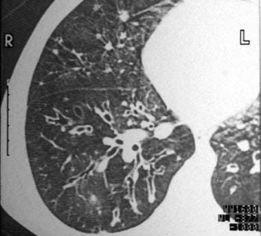
Cylindrical bronchiectasis with signet-ring appearance. Note that the luminal airway diameter is greater than the diameter of the adjacent vessel. |
 | View Full Size Image | |
Media type: CT
|
| Media file 2:
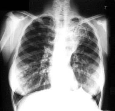
Cystic and cylindrical bronchiectasis of the right lower lobe on a posterior-anterior chest radiograph. |
 | View Full Size Image | |
Media type: X-RAY
|
| Media file 3:
Varicose bronchiectasis with alternating areas of bronchial dilatation and constriction. |
 | View Full Size Image | |
Media type: CT
|
| Media file 4:
This CT scan depicts areas of both cystic bronchiectasis and varicose bronchiectasis. |
 | View Full Size Image | |
Media type: CT
|
- Agasthian T, Deschamps C, Trastek VF, et al. Surgical management of bronchiectasis. Ann Thorac Surg. Oct 1996;62(4):976-8; discussion 979-80. [Medline].
- Balkanli K, Genc O, Dakak M, et al. Surgical management of bronchiectasis: analysis and short-term results in 238 patients. Eur J Cardiothorac Surg. Nov 2003;24(5):699-702. [Medline].
- Barker AF. Bronchiectasis. Semin Thorac Cardiovasc Surg. Apr 1995;7(2):112-8. [Medline].
- Barker AF. Bronchiectasis. N Engl J Med. May 2 2002;346(18):1383-93.
- Davies G, Wilson R. Prophylactic antibiotic treatment of bronchiectasis with azithromycin [Letters to the Editor]. Thorax. 2004;59(6):540-541.
- Drobnic ME, Sune P, Montoro JB, et al. Inhaled tobramycin in non-cystic fibrosis patients with bronchiectasis and chronic bronchial infection with Pseudomonas aeruginosa. Ann Pharmacother. Jan 2005;39(1):39-44. [Medline].
- Dupont M, Gacouin A, Lena H, et al. Survival of patients with bronchiectasis after the first ICU stay for respiratory failure. Chest. May 2004;125(5):1815-20. [Medline].
- Evans DJ, Greenstone M. Long-term antibiotics in the management of non-CF bronchiectasis--do they improve outcome?. Respir Med. Jul 2003;97(7):851-8. [Medline].
- Franco F, Sheikh A, Greenstone M. Short acting beta-2 agonists for bronchiectasis. Cochrane Database Syst Rev. 2003;CD003572.
|