Continually Updated Clinical Reference
 
 
  All Sources     eMedicine     Medscape     Drug Reference     MEDLINE
 
eMedicine - Protein S Deficiency : Article by

Quick Find
Authors & Editors
Introduction
Clinical
Differentials
Workup
Treatment
Medication
Follow-up
Miscellaneous
Multimedia
References

Related Articles
Antiphospholipid Syndrome

Antithrombin Deficiency

Disseminated Intravascular Coagulation

Fetal Growth Restriction

Mesenteric Venous Thrombosis

Metastatic Cancer, Unknown Primary Site

Missed Abortion

Protein C Deficiency


Patient Education
Circulatory Problems Center

Leg Blood Clot Overview

Leg Blood Clot Causes

Leg Blood Clot Symptoms

Leg Blood Clot Treatment


Author: John E Godwin, MD, MS, Professor of Medicine, Chief Division of Hematology/Oncology, Associate Director, Simmons Cooper Cancer Institute, Southern Illinois University School of Medicine

John E Godwin is a member of the following medical societies: American Association for the Advancement of Science, American Heart Association, and American Society of Hematology

Editors: Rodger L Bick, MD, PhD, FACP, Clinical Professor of Medicine, University of Texas Southwestern Medical Center; Director, Dallas and Pacific Thrombosis Hemostasis and Vascular Medicine Clinical Center; Francisco Talavera, PharmD, PhD, Senior Pharmacy Editor, eMedicine; Troy H Guthrie, Jr, MD, Director of Cancer Institute, Baptist Medical Center; Rajalaxmi McKenna, MD, FACP, Consulting Staff, Department of Medicine, Southwest Medical Consultants, SC, Good Samaritan Hospital, Advocate Health Systems; Emmanuel C Besa, MD, Professor, Department of Medicine, Division of Hematologic Malignancies, Kimmel Cancer Center, Thomas Jefferson University

Author and Editor Disclosure

Synonyms and related keywords: purpura fulminans, protein S deficiency, vitamin K–dependent anticoagulant protein, protein S, activated protein C, APC, activated factor V, FVa, activated factor VIII, FVIIIa, thrombosis, venous thromboembolism, VTE, arterial thrombosis

Background

In 1979, researchers in Seattle, Wash, first discovered protein S and arbitrarily named it after the city of its discovery. Protein S is a vitamin K–dependent anticoagulant protein. Its major function is as a cofactor to facilitate the action of activated protein C (APC) on its substrates, activated factor V (FVa) and activated factor VIII (FVIIIa). Protein S deficiencies are associated with thrombosis.

Protein S deficiency may be hereditary or acquired, the latter is usually due to hepatic diseases or a vitamin K deficiency. Protein S deficiency usually manifests clinically as venous thromboembolism (VTE). The association of protein S deficiency with arterial thrombosis appears coincidental or weak at best. Arterial thrombosis is not evident with other hereditary anticoagulant abnormalities (eg, protein C or antithrombin III deficiency, factor V Leiden gene mutation). Protein S deficiency manifests as an autosomal dominant trait; manifestations of thrombosis are observed in both heterozygous and homozygous genetic deficiencies of protein S.

Pathophysiology

To understand how thrombosis occurs in protein S deficiency, its physiological function should be briefly reviewed. Protein S is part of a system of anticoagulant proteins that regulate normal coagulation mechanisms in the body. Under most normal circumstances, the anticoagulant proteins prevail and blood remains in a liquid nonthrombotic state. Whenever procoagulant forces are locally activated to form a physiologic or pathologic clot, protein S participates as part of one mechanism of controlling clot formation.

Protein S functions predominantly as a nonenzymatic cofactor for the action of another anticoagulant protein, activated protein C (APC). This activity occurs via a coordinated system of proteins, termed the protein C system. Image 1 shows a simplified outline of the function of protein S in the protein C system.
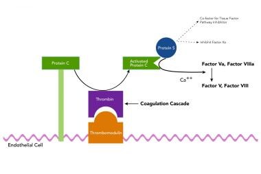
During the process of clotting, multimolecular complexes are formed on membrane surfaces. These membranes are usually negatively charged phospholipids and/or activated platelets. These multimolecular complexes are referred to as the tenase and prothrombinase complexes for their key activities of activation of factor X and prothrombin, respectively. Anchoring these two complexes are the activated form of factor VIII (FVIIa) used for the tenase complex and the activated form of factor V (FVa) for the prothrombinase complex. These two large proteins are homologous in structure and are cofactors, not enzymes, in the clotting process.

In one of many examples of nature's efficiency, the same enzyme that clots blood, thrombin, is converted from clotting to an anticoagulant mechanism on the surface of the endothelium and it then activates protein C to its active enzymatic form, APC. APC requires protein S as a cofactor in its enzymatic action on its 2 substrates, FVa and FVIIIa. Thus, this process is designed to dampen and shut off clotting by switching off the key cofactor proteins FVa and FVIIIa. Protein S and APC are sufficient to inactivate FVa. However, for the inactivation of FVIIIa, APC and protein S require the help of the nonactivated clotting protein, factor V. This is another example of dual use of a protein in this same process.

Factor Va as noted above is cleaved by APC to an inactive form. However, in assisting protein S to inactivate FVIIIa, it is the inactive FV that is cleaved by APC. An important consequence of this dual procoagulant and anticoagulant property of factor V, is that the mutant factor V Leiden, which resists APC cleavage, cannot be switched off but also cannot function here at this step as an anticoagulant protein (see factor V Leiden gene mutation in Race). In addition to its cofactor role in the protein C system, protein S functions independently of protein C to directly inhibit the factor X clotting factor–activating complex and the prothrombin-activating complex.

Furthermore, protein S interacts with the complement system and may play a role in phagocytosis of apoptotic cells. It has been recently observed that protein S binds to phosphatidyl serine on apoptotic cells and stimulates macrophage phagocytosis of early apoptotic cells. The physiological impact of protein S deficiencies on these nonanticoagulant roles of protein S is not yet known.

Protein S is a single-chain glycoprotein, and it is dependent on vitamin K action for posttranslational modification of the protein to a normal functional state. Vitamin K–dependent proteins are synthesized with a unique recognition propeptide piece. The propeptide sequence serves as a recognition site for the vitamin K–dependent gamma-carboxylase enzyme that modifies the nearby glutamic acid residues to gamma-carboxyglutamic acid (Gla) residues. Gla residues are responsible for calcium-dependent binding to membrane surfaces. Structural studies indicate that protein S contains 10-12 Gla residues, a loop region sensitive to thrombin (ie, thrombin-sensitive region [TSR]), 4 epidermal growth factor (EGF)–like modules, and a carboxy-terminal portion that is homologous to a sex hormone-binding globulin (SHBG)–like region.

In blood plasma, protein S exists in both a bound and a free state. A portion of protein S is noncovalently bound with high affinity to the complement regulatory protein C4b-binding protein (C4BP). The C4BP molecule consists of 7 alpha chains that bind to the complement protein, C4b, and one beta chain. The beta chain of the C4bBP molecules contains the binding sites for protein S. The role of protein S in complement regulation by C4BP is not completely understood.

In healthy individuals, approximately 30-40% of total protein S is in the free state. Only free protein S is capable of acting as a cofactor in the protein C system. This distinction between free and total protein S levels is important and gives rise to the current terminology regarding the deficiency states. Type I protein S deficiency is a reduction in the level of free and total protein S. Type III deficiency is a reduction in the level of free protein S only. Type II deficiency is a reduction in the cofactor activity of protein S, with normal antigenic levels.

APC and protein S require negatively charged phospholipids (PL) and Ca2+ for normal anticoagulant activity. Studies of the structure and function relationships of protein S demonstrate that the APC interaction sites are located in the Gla, TSR, and first EGF-like modules of protein S. The binding site for C4BP is located in the SHBG-like region, which is also important for full anticoagulant activity.

Researchers have identified 2 genes for human protein S and both are linked closely on chromosome 3p11.1-3q11.2. One gene is the active gene, PROS-b (ie, PROS1), and the other, PROS-a, is an evolutionarily duplicated nonfunctional gene, which is classified as a pseudogene because it contains multiple coding errors (eg, frameshifts, stop codons). The expressed (alpha) PROS1 gene is more than 80 kb long and contains 15 exons and 14 introns. The protein S pseudogene (beta) has 97% homology to the PROS-a gene.

Molecular studies into the genetic causes of protein S deficiency are complicated by the presence of the pseudogene, PROS-b, and phenotypic variation. Deletions of large portions of the PROS-a gene are associated with protein S deficiency and thrombophilia. Researchers located the first such deletion in the central portion of the PROS- a gene. The second deletion described (5.3 kb) was a deletion of coding exon XIII, which resulted in a truncated protein product.

Family members with either deletion exhibit protein S deficiency and thrombophilia; however, subsequent studies indicate that the most common genetic defects in the protein S gene are point mutations rather than gene deletions. Phenotypic variation has been observed in protein S deficiency. The coexistence of type I deficiency and type III deficiency in families with the same protein S mutation has been shown at least in one family to be due to an age-related increase in total protein S level. In this family, the apparent type III variant with only low free S blood levels, was explained by the age increase in total protein S. Younger persons in the family when tested for blood levels still had low total and free protein S.

Frequency

United States

Congenital protein S deficiency is an autosomal dominant disease, and the heterozygous state occurs in approximately 2% of unselected patients with VTE.

Protein S deficiency is rare in the healthy population without abnormalities. Frequency is approximately 1 out of 700 based on extrapolations from a study of over 9000 blood donors who were tested for protein C deficiency. When looking at a selected group of patients with recurrent thrombosis or family history of thrombosis, the frequency of protein S deficiency increases to 3-6%.

Very rarely, protein S deficiency occurs as a homozygous state, and these individuals have a characteristic thrombotic disorder, purpura fulminans. Purpura fulminans is characterized by small vessel thrombosis with cutaneous and subcutaneous necrosis, and it appears early in life, usually during the neonatal period or within the first year of life.

International

Data for European studies indicated the same frequencies for protein S deficiency as in the United States. Recent studies have indicated that the prevalence of protein S deficiency is particularly high in the Japanese population. In several reported series of patients with VTE in the United States, protein S deficiency was seen in 1-7% of patients. The deficiency is rare in population surveys of Caucasians, at approximately 0.03%. However, Japanese patients with VTE have reported a frequency of approximately 12.7% protein S deficiency and similarly elevated population frequencies of approximately 0.63%.

Mortality/Morbidity

  • VTE develops in 60-80% of patients who are heterozygous for protein S deficiency. The remaining patients are asymptomatic, and some heterozygous individuals never develop VTE. Though it is controversial, no clear association exists between protein S deficiency and arterial thrombosis. Many case reports and small case series describe protein S deficiency as one factor found in patients with arterial thrombosis most commonly stroke. However, prospective and cohort studies have not shown convincing increased risk for arterial thrombosis.
  • Protein S deficiency is also associated with fetal loss in women, in the absence of VTE. Some authors suggest that as many as 40% of women with obstetric complications other than VTE may carry some form of thrombophilia. Protein S deficiency is one of these factors along with several other more common genetic thrombophilic states.
  • Mortality is from pulmonary embolism. In several studies, the 3-month mortality rate of pulmonary embolism ranged from 10.0-17.5%. In a study of Medicare recipients with pulmonary embolism, men had a 13.7% mortality rate compared to 12.8% in women; the mortality rate was 16.1% in blacks, compared to 12.9% in whites.

Race

Race-related variations exist in thrombophilic disorders, as one may expect from genetic-based population traits. In general, a significant difference exists in the frequency of thrombophilic disorders in whites compared with thrombophilic disorders in Japanese (Asian) and black African persons. Current research indicates that protein S deficiency is 5-10 times higher in Japanese populations compared with Caucasians. Protein C deficiency is estimated to be 3 times higher in Japanese populations.

The factor V Leiden mutation is common in white populations and is now known to be the result of a founder effect estimated to be 30,000 years old. This mutation is rare and almost never found in Japanese or Asian populations. In general, black Africans and African Americans with VTE have a lower detection of any of the currently recognized thrombophilic disorders, especially factor V Leiden.

Sex

No difference exists in the male-to-female rate of occurrence.

Age

Protein S deficiency is a hereditary disorder, but the age of onset of thrombosis varies by heterozygous versus homozygous state. Most VTE events in heterozygous protein S deficiency occur in persons younger than 40-45 years. The rare homozygous patients have neonatal purpura fulminans, as described above; onset occurs in early infancy. As noted above in the discussion on genetics, age does affect total protein S antigen levels, but not free protein S levels. Older patients deficient in protein S have low free S levels, even if their total protein S level rises into the normal range.


History

  • Symptoms related to protein S deficiency are those associated with deep venous thrombosis (DVT), thrombophlebitis, or pulmonary embolus. As noted in Deterrence/Prevention, some women may have fetal loss as their only manifestation of a thrombophilic disorder (eg, protein S deficiency).
  • Venous thrombosis of the lower limbs: Lower limb swelling and discomfort are the usual symptoms. Occasionally, redness or discoloration also is present, with or without associated cellulitis.
  • A family history of thrombosis is an important finding, which suggests inherited thrombophilia.
  • Thrombosis at an early age (eg, usually <55 y) or recurrent thrombosis is also frequently indicative of an inherited thrombophilic state (eg, protein S deficiency).

Physical

Direct the examination to identify signs of venous thrombosis or pulmonary embolism. The results of the physical examination are nonspecific and often misleadingly indicate the diagnosis of DVT.

  • Deep vein thrombosis
    • The most common manifestation is venous thrombosis of the lower extremities, and this accounts for approximately 90% of all events.
    • Superficial veins that are obviously thrombosed usually appear distended, firm, and noncompressible (cords), with or without associated redness or pain.
    • Superficial thrombophlebitis can be observed in some cases, with or without DVT.
    • Suspect DVT if identifying signs of venous obstruction and local inflammation are present on examination.
    • The classic presentation of DVT is a triad of calf pain, edema, and pain on dorsiflexion of the foot (ie, Homans sign). However, less than a third of DVT cases exhibit these 3 findings. Physicians observe unilateral leg or calf swelling with mild or moderate pain more often, which suggests DVT. Rarely, calf discomfort without swelling is the only sign of DVT.
    • Differential diagnoses for DVT include muscle strains and tears, passive swelling of a paralyzed or immobilized limb, Baker cyst, cellulitis, knee trauma or derangement, lymphatic obstruction, and postphlebitic syndrome.
  • In postphlebitic syndrome, chronic swelling and pain are present in the limb, and the occurrence of a new venous thrombosis is often impossible to assess without Doppler or venography.
  • Pulmonary embolism
    • Some patients may have associated or isolated pulmonary embolism and may experience dyspnea, chest pain, syncope, or cardiac palpitations. Dyspnea is the most frequent symptom of pulmonary embolism, and tachypnea is the most frequent sign.
    • Some patients with massive pulmonary embolism can present with syncope or cyanosis.
    • Classic pleuritic chest pain, cough, or hemoptysis suggests an embolism with pleural involvement.
    • Acute right-sided heart failure occurs rarely and is associated with massive embolus.
    • Findings of right ventricular dysfunction include bulging neck veins, a left parasternal lift, and an accentuated pulmonic component of the second heart sound. These findings in the chest are also not specific for pulmonary embolism.
  • Unusual sites of thrombosis (eg, mesenteric vein, cerebral sinuses) are rare (<5%) but, when observed, characteristically suggest one of the inherited thrombophilias (eg, protein S deficiency).

Causes

  • The causes of protein S deficiency are due to genetic defects (hereditary) as described above in detail in the Pathophysiology section.
  • Acquired protein S deficiency
    • Rarely, an acquired disorder causes protein S deficiency. Acquired deficiencies of protein S occur with liver disease, vitamin K deficiency, or as a result of antagonism with oral warfarin anticoagulants.
    • Protein S levels decrease in pregnancy and can fall into the abnormal-low laboratory range. These low levels of protein S in pregnancy do not cause thrombosis by themselves.
    • Another seldom recognized cause for acquired protein S deficiency is sickle cell anemia; however, this condition alone does not produce a thrombophilic state.
    • Age affects total protein S but not free protein S levels. Generally, the total protein S level increases in persons older than 50 years. This rise is in association with total increases in the complement binding protein, C4BP. Free protein S levels do not increase with age. These factors may explain the observation that families with the same recognized genetic defect in protein S can have both type I and type III deficiencies. Type I deficiency is a reduction in both total and free protein S. Type III deficiency is isolated reduction in free protein S. When families with the same genetic type I defect are surveyed, older individuals even with deficiency in protein S have an increase in total S and now appear to have type III deficiency.


Antiphospholipid Syndrome
Antithrombin Deficiency
Disseminated Intravascular Coagulation
Fetal Growth Restriction
Mesenteric Venous Thrombosis
Metastatic Cancer, Unknown Primary Site
Missed Abortion
Protein C Deficiency


Lab Studies

  • Protein S deficiency is diagnosed using laboratory tests for the protein S antigen and by using other tests for functional protein S activity (based on clotting assays).
    • Protein S antigen: Laboratories can test protein S antigen as total antigen (ie, protein S bound to C4BP plus free protein) or free protein S antigen. The free form of protein S has functional activity, and researchers have developed assays specifically for the free protein S antigen. Both free and total protein S are measured by ELISA methods in the laboratory.
    • Functional protein S: Assays for functional protein S are indirect and are based on prolongation of blood clotting by the generation of APC and its function in the assay. These functional tests are difficult to perform. In addition, the tests introduce several other factors that can alter the interpretation of test results. Most importantly, a falsely low protein S functional assay value can be observed in patients with factor V Leiden genetic defect, which is another common cause of hereditary thrombophilia that interferes with protein C function. Some new commercial methods for determining protein S deficiency can measure activity in factor V Leiden patients accurately after dilution of test plasma.1
  • Several clinical conditions affect the blood levels of protein S—both antigenic and functional assays. As one would expect, vitamin K deficiency, liver disease, or antagonism with warfarin reduces protein S levels. In the setting of acute thrombosis, protein S levels fall, sometimes into the deficient range. Pregnancy also results in lower blood levels of protein S, especially as measured by functional assays. As noted previously in the section on genetics of protein S, total protein S levels actually rise with age. Free protein S levels are not affected by age.
  • Based on the measurement of free and total protein S antigen and functional protein S activity, scientists classify protein S deficiency into 3 phenotypes using the classification proposed at the 1991 meeting of the Scientific Subcommittee of the International Society on Thrombosis and Haemostasis in Munich, Germany:
    • Type I deficiency is characterized by a decrease in the total protein S antigen and free protein S antigen together (quantitative deficiency).
    • Type II deficiency is characterized by normal total and free antigen levels but reduced protein S activity (functional deficiency).
    • Type III deficiency is characterized by low free protein S levels, whereas the total plasma concentration of protein S is normal.
  • Although reports document a few type II deficiencies, they are rare. The most common types are I and III. The distinction between type I and type III has no clinical implications. In both type I and type III deficiencies, free protein S levels are reduced.
  • Physicians should request free protein S antigen testing for any patient suspected of having deficiencies of protein S because this test detects most cases (ie, type I or III), and the use of a total protein S assay is not routinely needed. Consider use of the functional assay for protein S deficiency if the other test results are normal and a reliable assay can be performed after excluding other interfering defects.


Medical Care

Management of protein S deficiency takes place in the event of acute VTE or in patients with asymptomatic carrier states without a thrombotic event. Following an acute thrombosis, administer heparin therapy and then transition to warfarin oral anticoagulation.

  • Heparin therapy
    • Physicians may administer the initial heparin treatment as intravenous unfractionated heparin or as subcutaneous low molecular weight heparin (LMWH).
    • Heparin should be administered for a minimum of 5 days.
    • Manage heparin with standard protocols. See Deep Venous Thrombosis or Pulmonary Embolism for additional details.
  • Warfarin
    • Warfarin administration can start on day 1 or 2 of heparin therapy. After 2 consecutive therapeutic international normalized ratio (INR) clotting tests and a minimum of 5 days of heparin therapy, the patient can continue on warfarin alone.
    • In most patients, specialists recommend 6-9 months of initial treatment with warfarin.
    • The question of whether to continue lifelong warfarin in patients with identified protein S deficiency after their first thrombotic event is controversial. If the first thrombotic event was life threatening or occurred in multiple or unusual sites (eg, cerebral veins, mesenteric veins), most experts recommend lifelong therapy initially. If precipitated by a strong event (eg, trauma, surgery) and the thrombosis did not meet the criteria of life threatening or multiple or unusual sites, some experts argue that these patients may have a lower risk of recurrence and deserve a trial without warfarin after 9 months.
  • In patients who are asymptomatic carriers of protein S deficiency, the goal of therapy is prevention of the first thrombosis. In such patients, avoid drugs that predispose to thrombosis, including oral contraceptives. In these patients, if surgery or orthopedic injury occurs, prophylaxis with heparin is mandatory.
  • In pregnancy, experts recommend prophylaxis with heparin; however, the timing is controversial. Most experts would treat from the second trimester through 4-6 weeks postpartum.
  • Patient bleeding risks must be assessed on an individual basis for any of these prophylactic recommendations, and no single prescription fits all cases.

Diet

Dietary issues relate to patients with protein S deficiency who are on oral anticoagulation with warfarin. Avoid diets rich in vitamin K foods.

Activity

Restrictions apply to activity shortly after acute venous thrombosis (ie, DVT, pulmonary embolism). See Deep Venous Thrombosis or Pulmonary Embolism for additional details concerning such restrictions. While on anticoagulation therapy, patients should avoid vigorous contact activities.


Heparin is used for patients with acute thrombotic events or for the prevention of thrombosis. Heparin treatment currently is available in 2 forms—unfractionated (standard) heparin or LMWH.

Unfractionated heparin for treatment of thrombosis is administered properly by a weight-based dosing protocol, with a target heparin therapeutic range as monitored by the activated partial thromboplastin time (aPTT) test and for a minimum of 5 days. A heparin dosing protocol includes the specified weight-dosing regimen, the target therapeutic aPTT range, the time for measuring aPTT tests after bolus or adjustment in dose (4-6 h), and a standard means of adjusting the unfractionated heparin infusion based on the aPTT test result (eg, subtherapeutic, therapeutic, supratherapeutic). A commonly used weight-adjusted unfractionated heparin regimen is termed 80/18: 80 U/kg IV bolus followed by 18 U/kg continuous IV (CIV) infusion. The target therapeutic heparin range is ideally individualized to the institution's laboratory aPTT test instrument and reagent.

To obtain an institutional heparin therapeutic range, employ a method such as that described by Brill-Edwards or any other similar comparison of in vitro and ex vivo heparin levels with aPTT test results in multiple individuals. In the absence of an established institutional therapeutic range, an aPTT ratio of 1.5-2.0 is commonly used; however, aPTT reagents and patient responses to unfractionated heparin vary, and the ratio can be 1.8-3.0 for some reagents.

The pharmacodynamics of LMWHs are different from the parent unfractionated heparin. LMWHs are administered subcutaneously. The aPTT test is not affected significantly by LMWH and is not used to monitor LMWH therapy. Several different LMWHs are available in the United States, but they have different pharmacodynamic properties and are not considered interchangeable. Weight-based dosing regimens for each LMWH and for treatment or prophylaxis indications are available from each manufacturer. LMWHs are approved for treatment of DVT with or without pulmonary embolism in the inpatient hospital setting. LMWHs are approved for treatment of DVT without pulmonary embolism in the outpatient setting.

Warfarin is used for long-term oral anticoagulant management of patients with protein S deficiency after first or subsequent thrombosis.

Drug Category: Anticoagulants

Unfractionated IV heparin and fractionated low molecular weight SC heparins are the 2 choices for initial anticoagulation therapy. Warfarin therapy may be initiated after 1-3 days of effective heparinization.

Drug NameHeparin
DescriptionUsually administered as CIV infusion for the treatment of acute thrombosis. For prevention of thrombosis, unfractionated heparin is administered SC.
Adult DoseInitial dose: 80 U/kg IV bolus
Maintenance: 18 U/kg/h CIV infusion (See discussion under introduction to Medications section regarding target aPTT and dose monitoring)
Pediatric DoseAdminister as in adults
ContraindicationsDocumented hypersensitivity; subacute bacterial endocarditis; active bleeding; history of heparin-induced thrombocytopenia; relative contraindications include recent surgery (especially ocular and neurosurgical procedures) and hereditary bleeding disorders
InteractionsDigoxin, nicotine, tetracycline, and antihistamines may decrease effects; NSAIDs, aspirin, dextran, dipyridamole, and hydroxychloroquine may increase heparin toxicity
PregnancyB - Fetal risk not confirmed in studies in humans but has been shown in some studies in animals
PrecautionsBleeding is most frequent complication; occurrence of thrombocytopenia during heparin therapy may indicate onset of heparin immune reaction, termed heparin-induced thrombocytopenia (HIT); syndrome can be life threatening and can be associated with new-onset paradoxical arterial or venous thrombosis; elevations of liver enzymes have been observed during heparin therapy; heparin is not indicated for IM injection; IM medications should be administered by alternate route; in neonates, preservative-free heparin is recommended to avoid possible toxicity (gasping syndrome) by benzyl alcohol, which is used as preservative; caution in severe hypotension and shock

Drug NameEnoxaparin (Lovenox)
DescriptionEnhances inhibition of factor Xa and thrombin by increasing antithrombin III activity. In addition, preferentially increases inhibition of factor Xa.
Average duration of treatment is 7-14 d.
Adult DoseTreatment of established DVT with or without PE: 1 mg/kg SC q12h can be administered in the hospital or in an outpatient setting under supervision
Alternatively, 1.5 mg/kg SC once qd, administered at same time qd is approved for in-hospital treatment of DVT without symptomatic PE
Prevention of DVT: 30 mg SC q12h or 40 mg qd
These 2 regimens differ by indication, ie, hip replacement versus knee surgery, and in use as extended prophylaxis; in acutely ill patients at high risk for DVT/PE, a dose of 40 mg SC once qd has reported efficacy
Pediatric DoseNot established
Suggested dose: 0.75 mg/kg/dose SC bid for <2 mo; 0.5 mg/kg/dose SC bid if >2 mo
ContraindicationsDocumented hypersensitivity; major bleeding; thrombocytopenia
InteractionsPlatelet inhibitors or oral anticoagulants (eg, dipyridamole, salicylates, aspirin, NSAIDs, sulfinpyrazone, ticlopidine) may increase risk of bleeding
PregnancyB - Fetal risk not confirmed in studies in humans but has been shown in some studies in animals
PrecautionsLMWH is cleared by renal mechanisms; LMWHs should be used with caution, if at all, in patients with renal impairment (ie, CrCl <30 mL/min); if thromboembolic event occurs despite LMWH prophylaxis, discontinue drug and initiate alternate therapy; elevation of hepatic transaminases may occur but is reversible; heparin-associated thrombocytopenia may occur with fractionated LMWHs; 1 mg of protamine sulfate reverses effect of approximately 1 mg of enoxaparin if significant bleeding complications develop; however, protamine sulfate provides incomplete reversal of the anti-Xa effect of LMWHs, including Lovenox
LMWHs are safe and effective in preventing or treating thrombosis during pregnancy; prophylactic regimens are preferred in patients with no history of thrombosis; guidelines for the use of LMWHs in pregnancy are still formative; some authors recommend monitoring LMWH blood levels to maintain trough anti-Xa activity of 0.15-0.2 U/mL and 2 h postinjection peak levels of 0.4-0.6 U/mL; levels are checked monthly during pregnancy; most women may require dosage increase at 20.5 wk gestation (SD 8.2 wk); in women with prosthetic valves, heparin use during pregnancy presents additional challenges and extreme caution with close monitoring is needed; reports exist of thrombosis of cardiac valves while on usual doses of LMWH; this is an evolving field and clinical trials are required to determine optimal dosage and safety in this setting; recommended to continue treatment with LMWH for 4-6 wk postpartum
Cases of epidural hematoma have been described following epidural/spinal anesthesia and concomitant LMWH heparin administration; these events occurred with prophylactic doses of LMWH; risk of hematoma is increased by indwelling catheters, concomitant NSAID use, and traumatic or repeated epidural or spinal puncture; current guidelines suggest LMWH therapy be started 24 h after epidural anesthesia; epidural catheter removal should be (1) >2 h before first dose of LMWH or (2) no less than 12 h after the last dose of LMWH

Drug NameDalteparin (Fragmin)
DescriptionEnhances inhibition of factor Xa and thrombin by increasing antithrombin III activity. In addition, preferentially increases inhibition of factor Xa.
Average duration of treatment is 7-14 d.
Adult Dose2500-5000 U SC qd for prevention of DVT; current recommended dose in patients undergoing abdominal surgery is 2500 U SC qd
Pediatric DoseNot established
ContraindicationsDocumented hypersensitivity; major bleeding; thrombocytopenia
InteractionsPlatelet inhibitors or oral anticoagulants, such as dipyridamole, salicylates, aspirin, NSAIDs, sulfinpyrazone, and ticlopidine, may increase risk of bleeding
PregnancyC - Fetal risk revealed in studies in animals but not established or not studied in humans; may use if benefits outweigh risk to fetus
PrecautionsLMWH is cleared by renal mechanisms; LMWHs should be used with caution, if at all, in patients with renal impairment (ie, CrCl <30 mL/min); if thromboembolic event occurs despite LMWH prophylaxis, discontinue drug and initiate alternate therapy; elevation of hepatic transaminases may occur but is reversible; heparin-associated thrombocytopenia may occur with fractionated LMWHs; 1 mg of protamine sulfate reverses effect of approximately 1 mg of enoxaparin if significant bleeding complications develop; however, protamine sulfate provides incomplete reversal of the anti-Xa effect of LMWHs, including Fragmin
Cases of epidural hematoma have been described following epidural/spinal anesthesia and concomitant LMWH administration; these events occurred with prophylactic doses of LMWH; the risk of hematoma is increased by indwelling catheters, concomitant NSAID use, and traumatic or repeated epidural or spinal puncture; current guidelines suggest LMWH therapy be started 24 h after epidural anesthesia; epidural catheter removal should be (1) >2 h before the first dose of LMWH or (2) no less than 12 h after the last dose LMWH

Drug NameFondaparinux sodium (Arixtra)
DescriptionOnly synthetic compound in this class of LMW heparins. This compound is a novel pentasaccharide capable of inhibiting factor Xa via the action of antithrombin (AT) but devoid of anti-factor IIa (thrombin) activity. Interestingly, this compound does not appear to cross-react with HIT antibodies.
Approved for use in hip fracture surgery, knee replacement surgery, and hip replacement surgery. Only FDA-approved anticoagulant drug for hip fracture surgery. Also used and approved for extended prophylactic dosing for 21 d following hip fracture surgery.
Adult DoseSymptomatic DVT and acute symptomatic PE:
<50 kg BW: 5 mg SC qd for 5 d
50-100 kg BW: 7.5 mg SC qd for 5 d
>100 kg BW: 10 mg SC qd for 5 d
Treatment should be continued until therapeutic oral anticoagulant effect is established (INR 2-3) with at least 2 determinations
For prophylaxis of VTE: 2.5 mg SC qd; initial dose recommended to be given 6-8 h following surgery, after hemostasis established; administration <6 h after surgery associated with increased risk of major bleeding
Pediatric DoseNot established
ContraindicationsDocumented hypersensitivity; seriously impaired kidney function or patients who weigh <110 lb; patients given spinal anesthesia or spinal puncture; major bleeding; thrombocytopenia
InteractionsNone reported; increased risk of bleeding possible with concurrent administration of platelet inhibitors, oral anticoagulants, or thrombolytic agents
PregnancyC - Fetal risk revealed in studies in animals but not established or not studied in humans; may use if benefits outweigh risk to fetus
PrecautionsWhen spinal anesthesia or spinal puncture used, blood clot in spine may develop, which can result in long-term or permanent paralysis (holding 2 doses prior to LP or surgery is recommended); major bleeding risk increased when initiated before 6 h following surgery; elimination decreased in elderly persons and in those with renal impairment
Use caution in patients with severe renal impairment (ie, CrCl <30 mL/min); if thromboembolic event occurs despite LMWH prophylaxis, discontinue drug and initiate alternate therapy; elevation of hepatic transaminases may occur but is reversible; no known antidote exists for this drug
Protamine sulfate does not appear to reverse effect of this compound; if significant bleeding complications develop; some authors have recommended the use of recombinant factor VIIa as a means of hemostasis
Cases of epidural hematoma have been described following epidural/spinal anesthesia and concomitant LMWH administration; these events occurred with prophylactic doses of LMWH; risk of hematoma is increased by indwelling catheters, concomitant NSAID use, and traumatic or repeated epidural or spinal puncture; current guidelines suggest LMWH therapy be started 24 h after epidural anesthesia; epidural catheter removal should be (1) >2 h before first dose of LMWH or (2) no less than 12 h after last dose of LMWH

Drug NameTinzaparin (Innohep)
DescriptionEnhances inhibition of factor Xa and thrombin by increasing antithrombin III activity. In addition, preferentially increases inhibition of factor Xa.
Average duration of treatment is 7-14 d.
Adult Dose175 U/kg SC qd, at same time qd, for treatment of DVT/PE
Pediatric DoseNot established; adult dose suggested
ContraindicationsDocumented hypersensitivity; major bleeding; thrombocytopenia
InteractionsPlatelet inhibitors or oral anticoagulants, such as dipyridamole, salicylates, aspirin, NSAIDs, sulfinpyrazone, and ticlopidine, may increase risk of bleeding
PregnancyB - Fetal risk not confirmed in studies in humans but has been shown in some studies in animals
PrecautionsLMWH is cleared by renal mechanisms; LMWHs should be used with caution, if at all, in patients with renal impairment (ie, CrCl <30 mL/min); if thromboembolic event occurs despite LMWH prophylaxis, discontinue drug and initiate alternate therapy; elevation of hepatic transaminases may occur but is reversible; heparin-associated thrombocytopenia may occur with fractionated LMWHs; 1 mg of protamine sulfate reverses effect of approximately 1 mg of enoxaparin if significant bleeding complications develop; however, protamine sulfate provides incomplete reversal of anti-Xa effect of LMWHs, including Lovenox
Cases of epidural hematoma have been described following epidural/spinal anesthesia and concomitant LMWH administration; these events occurred with prophylactic doses of LMWH; the risk of hematoma is increased by indwelling catheters, concomitant NSAID use, and traumatic or repeated epidural or spinal puncture; current guidelines suggest LMWH therapy be started 24 h after epidural anesthesia; epidural catheter removal should be (1) >2 h before the first dose of LMWH or (2) no less than 12 h after the last dose of LMWH

Drug NameWarfarin (Coumadin)
DescriptionOral anticoagulant that antagonizes action of vitamin K in normal synthesis of clotting factors II, VII, IX, and X. Safe and effective for long-term oral management of thrombotic disorders. See articles on Deep Venous Thrombosis or Pulmonary Embolism (discussed in Treatment section) for additional details on dosing and monitoring of warfarin. Therapy is initiated without a loading dose at a dose range of 5-10 mg qd for 70-kg adult. Monitor PT/INR daily during initiation of therapy to measure anticoagulation effect. After initial 5-10 d and stabilization of warfarin dose, measure PT/INR 2-3 times qwk for 2-4 wk, then monthly thereafter.
Adult Dose5-10 mg PO qd; adjust dose according to desired INR
Pediatric Dose0.05-0.34 mg/kg/d PO qd; adjust dose according to desired INR
ContraindicationsDocumented hypersensitivity; severe liver or kidney disease; open wounds or GI ulcers
InteractionsDrugs that may decrease anticoagulant effects include griseofulvin, carbamazepine, glutethimide, estrogens, nafcillin, phenytoin, rifampin, barbiturates, cholestyramine, colestipol, vitamin K, spironolactone, oral contraceptives, and sucralfate; medications that may increase anticoagulant effects include oral antibiotics, phenylbutazone, salicylates, sulfonamides, chloral hydrate, clofibrate, diazoxide, anabolic steroids, ketoconazole, ethacrynic acid, miconazole, nalidixic acid, sulfonylureas, allopurinol, chloramphenicol, cimetidine, disulfiram, metronidazole, phenylbutazone, phenytoin, propoxyphene, sulfonamides, gemfibrozil, acetaminophen, and sulindac
PregnancyD - Fetal risk shown in humans; use only if benefits outweigh risk to fetus
PrecautionsContraindicated in first trimester; warfarin skin necrosis (see Medical/Legal Pitfalls); do not switch brands after achieving therapeutic response; caution in active tuberculosis or diabetes; patients with protein C or S deficiency are at risk of developing skin necrosis


Deterrence/Prevention

  • In patients with heterozygous protein S deficiency and no history of thrombosis, physicians may administer prophylactic heparin during situations that present high risk for thrombosis. Such situations include surgery, orthopedic trauma (especially with a cast), pregnancy, and prolonged bed rest.
    • The risk of VTE during pregnancy and for the 6 weeks postpartum varies among the hereditary thrombophilic states. Protein S and protein C deficiencies have significantly elevated risks for thrombosis when compared to the modest increase in thrombosis seen with factor V Leiden mutation. Protein S deficiency was also associated with a 7-fold increase in fetal loss. Many experts recommend that women with protein S deficiency and a history of fetal loss, and severe or recurrent eclampsia, receive low-dose aspirin and prophylactic dose LMW heparin during pregnancy, and the LMW heparin should extent for 6 weeks postpartum.
    • For women with heterozygous protein S deficiency and no prior VTE history of fetal loss, treatment choices vary. Some recommend VTE prophylaxis only during the 6 weeks postpartum (the highest risk period for VTE) unless the pregnancy is complicated. Others recommend prophylaxis for the entire pregnancy and 6 weeks postpartum.
    • For women with no prior history of VTE and protein S deficiency plus any other thrombophilic defect, active prophylaxis with LMW heparin should be given during pregnancy and for 6 weeks postpartum.
    • For women with a prior VTE history and confirmed protein S deficiency, experts recommend prophylactic or intermediate dosing of LMW heparins during pregnancy and for 6 weeks postpartum.
    • For women with a prior history of VTE and currently receiving oral anticoagulants at the time of pregnancy, full anticoagulant dosing of LMW heparin is recommended with transition back to oral anticoagulant postpartum.
  • Physicians can administer heparin SC in standard protocols for VTE prevention.
  • Patients with recurrent thrombosis should remain on lifelong warfarin.
  • In patients with a history of thrombosis who are taking warfarin, no standard exists for "bridging" (ie, on and off use of warfarin for surgery or other procedures that require cessation of warfarin). Some institutions cover with SC heparin while holding warfarin for 3-4 days. In other situations, this temporary interruption of warfarin is not covered by heparin. Each clinician should weigh the thrombosis risk with the bleeding risk in the individual patient because no data in controlled trials are available to answer this difficult question.

Patient Education

For excellent patient education resources, visit eMedicine's Circulatory Problems Center. Also, see eMedicine's patient education article Blood Clot in the Legs.


Medical/Legal Pitfalls

  • Warfarin-induced skin necrosis
    • Patients with protein S deficiency, whether it is acquired or hereditary, have developed a rare and unusual reaction to warfarin administration when warfarin is administered without additional anticoagulant coverage with heparin. This reaction is termed warfarin-induced skin necrosis or Coumadin skin necrosis.
    • This reaction occurs most commonly in patients with acute thrombosis who are administered warfarin and either have subtherapeutic heparin therapy or early termination of heparin before adequate oral anticoagulation.
    • In some patients, hereditary or acquired protein S deficiency is unknown and oral warfarin is administered without heparin coverage in conditions such as post-orthopedic surgery or cardioversion for atrial fibrillation. These patients may also experience warfarin skin necrosis. Screening every patient for protein S deficiency before warfarin administration does not seem feasible; however, a family history of thrombosis may allow selected screening of patients.

Special Concerns

  • Pregnancy can result in falsely low protein S assays, especially functional assays. In addition, pregnancy increases the risk of thrombosis in patients who are heterozygous for protein S deficiency.
  • A female patient who is on warfarin for prior thrombosis should not continue warfarin during pregnancy. Heparin should be started during pregnancy and for 4-6 weeks postpartum in patients with protein S deficiency.


Media file 1:  A simplified outline of the protein C system.
Click to see larger pictureClick to see detailView Full Size Image
Media type:  Graph


  1. Tripodi A, Asti D, Chantarangkul V, Biguzzi E, Mannucci PM. Interference of factor V Leiden on protein S activity: evaluation of a new prothrombin time-based assay. Blood Coagul Fibrinolysis. Sep 2007;18(6):543-6. [Medline].
  2. Adachi T. Protein S and congenital protein S deficiency: the most frequent congenital thrombophilia in Japanese. Curr Drug Targets. Aug 2005;6(5):585-92. [Medline].
  3. Baker ME, French FS, Joseph DR. Vitamin K-dependent protein S is similar to rat androgen-binding protein. Biochem J. Apr 1 1987;243(1):293-6. [Medline].
  4. Brill-Edwards P, Ginsberg JS, Johnston M, Hirsh J. Establishing a therapeutic range for heparin therapy. Ann Intern Med. Jul 15 1993;119(2):104-9. [Medline].
  5. Dahlback B, Villoutreix BO. The anticoagulant protein C pathway. FEBS Lett. Jun 13 2005;579(15):3310-6. [Medline].
  6. Dahlbäck B, Stenflo J. High molecular weight complex in human plasma between vitamin K-dependent protein S and complement component C4b-binding protein. Proc Natl Acad Sci U S A. Apr 1981;78(4):2512-6. [Medline].
  7. Di Scipio RG, Hermodson MA, Yates SG, Davie EW. A comparison of human prothrombin, factor IX (Christmas factor), factor X (Stuart factor), and protein S. Biochemistry. Feb 22 1977;16(4):698-706. [Medline].
  8. Francis RB Jr. Protein S deficiency in sickle cell anemia. J Lab Clin Med. May 1988;111(5):571-6. [Medline].
  9. Giri TK, Villoutreix BO, Wallqvist A, Dahlbäck B, de Frutos PG. Topological studies of the amino terminal modules of vitamin K-dependent protein S using monoclonal antibody epitope mapping and molecular modeling. Thromb Haemost. Nov 1998;80(5):798-804. [Medline].
  10. Gomez E, Poort SR, Bertina RM, Reitsma PH. Identification of eight point mutations in protein S deficiency type I--analysis of 15 pedigrees. Thromb Haemost. May 1995;73(5):750-5. [Medline].
  11. Hackeng TM, van 't Veer C, Meijers JC, Bouma BN. Human protein S inhibits prothrombinase complex activity on endothelial cells and platelets via direct interactions with factors Va and Xa. J Biol Chem. Aug 19 1994;269(33):21051-8. [Medline].
  12. Heeb MJ, Rosing J, Bakker HM, Fernandez JA, Tans G, Griffin JH. Protein S binds to and inhibits factor Xa. Proc Natl Acad Sci U S A. Mar 29 1994;91(7):2728-32. [Medline].
  13. Hillarp A, Dahlback B. Novel subunit in C4b-binding protein required for protein S binding. J Biol Chem. Sep 5 1988;263(25):12759-64. [Medline].
  14. Ploos van Amstel HK, Huisman MV, Reitsma PH, Wouter ten Cate J, Bertina RM. Partial protein S gene deletion in a family with hereditary thrombophilia. Blood. Feb 1989;73(2):479-83. [Medline].
  15. Ploos van Amstel HK, Reitsma PH, van der Logt CP, Bertina RM. Intron-exon organization of the active human protein S gene PS alpha and its pseudogene PS beta: duplication and silencing during primate evolution. Biochemistry. Aug 28 1990;29(34):7853-61. [Medline].
  16. Ridker PM, Hennekens CH, Lindpaintner K, Stampfer MJ, Eisenberg PR, Miletich JP. Mutation in the gene coding for coagulation factor V and the risk of myocardial infarction, stroke, and venous thrombosis in apparently healthy men. N Engl J Med. Apr 6 1995;332(14):912-7. [Medline].
  17. Schmidel DK, Nelson RM, Broxson EH Jr, Comp PC, Marlar RA, Long GL. A 5.3-kb deletion including exon XIII of the protein S alpha gene occurs in two protein S-deficient families. Blood. Feb 1 1991;77(3):551-9. [Medline].
  18. Schmidel DK, Tatro AV, Phelps LG, Tomczak JA, Long GL. Organization of the human protein S genes. Biochemistry. Aug 28 1990;29(34):7845-52. [Medline].
  19. Siddique RM, Siddique MI, Connors AF Jr, Rimm AA. Thirty-day case-fatality rates for pulmonary embolism in the elderly. Arch Intern Med. Nov 11 1996;156(20):2343-7. [Medline].
  20. Simmonds RE, Zöller B, Ireland H, Thompson E, de Frutos PG, Dahlbäck B, et al. Genetic and phenotypic analysis of a large (122-member) protein S-deficient kindred provides an explanation for the familial coexistence of type I and type III plasma phenotypes. Blood. Jun 15 1997;89(12):4364-70. [Medline].
  21. Tait RC, Walker ID, Reitsma PH, Islam SI, McCall F, Poort SR, et al. Prevalence of protein C deficiency in the healthy population. Thromb Haemost. Jan 1995;73(1):87-93. [Medline].
  22. Walker MC, Garner PR, Keely EJ, Rock GA, Reis MD. Changes in activated protein C resistance during normal pregnancy. Am J Obstet Gynecol. Jul 1997;177(1):162-9. [Medline].
  23. Watkins PC, Eddy R, Fukushima Y, Byers MG, Cohen EH, Dackowski WR, et al. The gene for protein S maps near the centromere of human chromosome 3. Blood. Jan 1988;71(1):238-41. [Medline].

Protein S Deficiency excerpt

Article Last Updated: Nov 2, 2007