Background
Colonoscopy enables visual inspection of the entire large bowel (also called the colon or large intestine) from the distal rectum to the cecum. It remains the gold standard for the detection of polyps and colorectal cancer. The procedure is a safe and effective means of evaluating the large bowel. The technology for colonoscopy has evolved to provide a very clear image of the mucosa through a video camera attached to the end of the scope. The camera connects to a computer, which can store and print color images selected during the procedure.
Screening for and surveillance of colorectal cancer are among the indications for colonoscopy. Although colorectal cancer is highly preventable, it remains the second most common cause of cancer death in the United States. Both men and women face a lifetime risk of nearly 6% for the development of invasive colorectal cancer. Proper screening can help reduce mortality at all ages, and colonoscopy plays an important role in this effort.
Endoscopists should, when assigning appropriate screening and surveillance intervals, follow current guidelines. Repeat colonoscopy should be performed in 3 years on all patients with advanced adenomas. If, in average-risk patients, screening colonoscopies are normal or only small distal hyperplastic polyps are present, repeat examinations should not be performed before 10 years. [1]
Compared with other imaging modalities, colonoscopy is especially useful in detecting small lesions (eg, adenomas) that can develop into cancer; however, its main advantage is that it also allows for intervention, because biopsies and polypectomies can be performed.
See Benign or Malignant: Can You Identify These Colonic Lesions?, a Critical Images slideshow, to help identify the features of benign lesions as well as those with malignant potential.
Indications
Screening, evaluation, and follow-up of colorectal cancer
Screening in average-risk adults
Recommendations for colorectal cancer screening vary among the leading organizations in this field—namely, the American Cancer Society (ACS), [2, 3] the World Health Organization (WHO), the US Preventive Services Task Force (USPSTF), and the American College of Physicians (ACP), and the American Gastroenterological Association (AGA). [1] It is generally now recommended, however, that average-risk adults should begin colorectal cancer screening at age 45 years. There are a few approved screening options, of which colonoscopy every 10 years is the most common in the United States.
Other tests that screen for colon cancer include annual fecal occult blood testing (FOBT) and fecal immunohistochemical testing (FIT), as well as stool DNA testing (multitarget DNA testing). Barium enema is rarely performed today; newer modalities, such as computed tomography (CT) colonography, have gained wider acceptance.
Evaluation and removal of polyps
The finding of a polyp larger than 1 cm in diameter during sigmoidoscopy is an indication for examination of the entire colon because 30-50% of these patients have additional polyps. Although controversy continues regarding whether colonoscopy is indicated for patients with a polyp or polyps smaller than 1 cm, the general belief is that most cancers arise in preexisting adenomatous polyps, which should lead to a full colonoscopic examination, regardless of size.
Polypoid lesions observed on barium enema may represent pseudopolyps, true polyps, or carcinomas. Colonoscopy can be used to differentiate among these and can similarly be used to distinguish between benign and malignant strictures, which cannot be accurately accomplished with radiologic studies alone.
When clinical signs and symptoms suggest colon cancer or when screening (by radiography or sigmoidoscopy) identifies a large-bowel tumor, a full colonoscopic examination should be performed to obtain biopsy samples and to search for synchronous lesions. Findings on colonoscopy may also have implications for the surgical treatment plan.
Histologic diagnosis should be based on examination of the completely excised polyp. In general, all polypoid lesions greater than 0.5 cm in diameter should be totally excised. After a large (>2 cm) sessile polyp has been removed or if there is concern that an adenoma was not completely excised, repeat colonoscopy should generally be performed in 3-4 months. If residual tissue remains, it should be resected and colonoscopy repeated again in another 3-4 months.
In patients with polyps identified on initial examination, the American Cancer Society recommends that follow-up colonoscopy be performed on the basis of polyp number and type, as well as dysplasia grade, as follows [2] :
-
Patients with small rectal hyperplastic polyps can be treated as patients with an average risk of cancer, with colonoscopy or other screening on a similar schedule
-
Individuals with one or two sub-1-cm tubular adenomas with low-grade dysplasia should receive colonoscopy 5-10 years after polyp removal
-
Patients who have three to 10 adenomas or one adenoma larger than 1 cm or who have any adenomas with high-grade or villous features should receive follow-up colonoscopy 3 years after removal
-
Patients who have more than 10 adenomas on initial examination should undergo colonoscopy within 3 years
-
Patients with sessile adenomas that are removed in pieces should undergo follow-up colonoscopy 2-6 months after removal
Current or previous bowel resection for colon cancer
Because of the potential implications for the operative plan, preoperative colonoscopy should be performed in patients who are to undergo bowel resection for colon cancer. Patients who have already had a large bowel cancer removed should have a colonoscopy performed 6 months to 1 year after surgery, followed by yearly colonoscopy on two occasions. Some authorities believe that colonoscopy should then be performed every 3 years if the results of all these studies are negative.
Family history of cancer
Individuals with a family history of familial adenomatous polyposis (FAP) or Gardner syndrome are recommended to undergo genetic testing and flexible sigmoidoscopy or colonoscopy every 12 months, beginning at age 10-12 years until age 35-40 years if negative. Consider total colectomy for these individuals because they have a nearly 100% risk of developing colon cancer by age 40 years. Colonoscopy is not as effective in preventing colon cancer under these circumstances as it is with polyps in general.
Individuals who have a first-degree relative diagnosed with colon cancer or adenomas when younger than 60 years, or who have multiple first-degree relatives diagnosed with colon cancer or adenomas, should undergo screening colonoscopy every 3-5 years, beginning either at age 40 years or at an age 10 years younger than that of the earliest familial diagnosis, whichever comes first. [4]
The diagnosis of hereditary nonpolyposis colorectal cancer (HNPCC) should be considered in people who have several relatives with colorectal cancer, particularly if one or more of the relatives developed cancer when younger than 50 years. HNPCC is an autosomal dominant disorder with an approximately 70% lifetime risk of developing colorectal cancer.
These patients should be evaluated colonoscopically every 1-2 years, beginning at age 20-25 years or at an age 10 years younger than that of onset in the index case (whichever comes first). Perform annual screening in patients older than 40 years.
Management of inflammatory bowel disease
Although many patients do not require colonoscopy for the diagnosis of inflammatory bowel disease (IBD), the procedure is an important aid in the follow-up care and management of patients with ulcerative colitis or Crohn disease (see the images below). Colonoscopy is more sensitive than barium enema in determining the anatomic extent of the inflammatory process and is useful when clinical, sigmoidoscopic, and radiologic studies are inadequate. Colonoscopy with multiple biopsies is indicated to differentiate ulcerative colitis from Crohn disease.
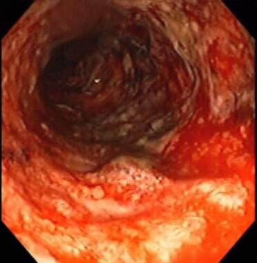 Inflammatory bowel disease. Severe colitis noted during colonoscopy. Mucosa is grossly denuded, with active bleeding noted. Patient had her colon resected very shortly after this view was obtained.
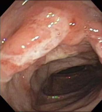 Colonoscopy. Colonoscopic image of large ulcer and inflammation of descending colon in 12-year-old boy with Crohn disease.
The cancer surveillance schedule varies in patients with inflammatory disease. Patients with pancolitis for more than 7-10 years and patients with left-side ulcerative colitis for more than 15 years are at an increased risk of developing colon cancer. The current recommendation for screening colonoscopy for these groups is every 1-2 years. For patients with Crohn disease of the colon, the same schedule of colonoscopic surveillance is warranted.
Ideally, because differentiating inflammatory changes from premalignant ones can be difficult, colonoscopy for surveillance purposes should not be performed during periods of active colitis, and biopsies from areas of less inflammation should be preferred. It has been suggested that as many as 64 biopsies are needed to achieve 95% sensitivity in surveying for dysplasia in patients with IBD.
Newer technologies, including chromoendoscopy, magnification endoscopy, and narrow-band imaging, may improve detection of dysplasia during surveillance colonoscopy and allow endoscopists to take fewer but higher-yield biopsies.
For additional information on these topics, see Ulcerative Colitis, Inflammatory Bowel Disease, and Crohn Disease.
Identification and treatment of acute bleeding sites
In the case of lower gastrointestinal (GI) bleeding, colonoscopy can be useful not only for localizing the site of bleeding but also, potentially, for enabling therapeutic intervention. Endoscopic therapy using injection of epinephrine, electrocauterization, argon plasma coagulation (APC), band therapy, and/or clips can be used to treat various causes of lower GI bleeding, including postpolypectomy coagulation syndrome, diverticula, arteriovenous malformations (AVMs), hemorrhoids, and radiation-induced mucosal injury.
In the acute setting, the endoscopist may be limited by poor visualization in an unprepared colon and by the risks of sedation in an acutely bleeding patient. A purge preparation may be considered, using 4 L of polyethylene glycol (eg, GoLYTELY, CoLyte) either orally over 2 hours or via a nasogastric tube, as tolerated by the patient.
If the bleeding source cannot be determined by means of colonoscopy, angiography or a nuclear medicine scan may be required. Radiographic studies should be performed before colonoscopy when perforation or obstruction is suspected.
Decompression of colon
A volvulus is a twist of a segment of intestine, most commonly in the sigmoid colon and cecum, which often causes a bowel obstruction and can lead to ischemia. Patients present with abdominal pain, nausea/vomiting, obstipation, and abdominal distention. Surgical intervention is generally recommended for a cecal volvulus. Colonoscopy/sigmoidoscopy can be used to decompress the colon in the case of sigmoid volvulus by advancing the endoscope through the torsed segment of bowel. A large expulsion of air indicates a successful reduction.
Acute colonic pseudo-obstruction (Ogilvie syndrome) is a clinical condition characterized by signs and symptoms of an acute large-bowel obstruction in the absence of a mechanical cause. When supportive treatment fails, endoscopic decompression may be considered to prevent bowel ischemia and perforation. This is a technically difficult procedure and should be performed by using minimal air insufflation and without preceding oral laxative preparation.
Whereas colonoscopy appears to be beneficial in the management of patients with Ogilvie syndrome, it is associated with a greater risk of complications, and randomized trials have not been done to establish its efficacy.
Contraindications
Pregnancy is considered to increase the risk of colonoscopy. Guidelines for colonoscopy during pregnancy are not available, because of insufficient data. The largest reported series included eight colonoscopies performed during pregnancy. In this study, six patients delivered healthy infants after colonoscopy. One patient suffered a miscarriage unrelated to colonoscopy, and another had an elective abortion.
In general, colonoscopy may be considered for severe life-threatening conditions during pregnancy when the only alternative is colonic surgery or when colon cancer is suspected. The procedure is best performed in a hospital setting rather than in a doctor’s office. Defer surveillance colonoscopy for prior history of cancer or polyps, abdominal pain, or change in bowel habits until the postpartum period.
Other relative contraindications for colonoscopy include known or suspected colonic perforation, toxic megacolon, and fulminant colitis or severe IBD with ulceration; these conditions increase the risk of perforation. [5]
Technical Considerations
Best practices
For a colonoscopy to be effective, the bowel preparation (cleansing) must be adequate, [6] or visualization suffers. Preprocedural patient instructions are important to ensure good colon preparation. Numerous regimens exist today, but polyethylene glycol (PEG) is still the most cost-effective preparation. PEG is an osmotic laxative and works by causing watery diarrhea so that the stool can be emptied from the colon. The medication also contains electrolytes to prevent dehydration and other serious side effects that may be caused by fluid loss as the colon is emptied.
Bowel preparation quality should be measured by endoscopy units, on a unit level, at least annually. Screening and surveillance colonoscopies should be associated with adequate bowel preparation (ie, a Boston Bowel Preparation Scale [BBPS] score ≥6, with each segment score ≥2) in at least 90% of procedures, with the aspirational target being 95% or above. [1]
In patients undergoing colonoscopy, split-dose bowel preparation should serve as the endoscopy unit's standard preparation strategy. [1]
High-definition colonoscopes should be used by endoscopy units for screening and surveillance colonoscopy. [1]
Many patients choose to have anesthesia for their colonoscopy. This can take the form either of conscious sedation with midazolam and fentanyl or moderate sedation handled by means of anesthesia.
Inflating the colon with air to facilitate visualization of the mucosa simulates abdominal cramps and can be uncomfortable.
The patient is most commonly positioned in the left lateral decubitus position. A digital rectal examination is mandatory. The scope is inserted gently and then advanced to the cecum. The cecum can be identified by three landmarks: the cecal strap, the appendiceal orifice, and the ileocecal valve. If it is difficult to identify one of the landmarks, transillumination (visualizing the light of the scope in the right lower quadrant) can be used as an aid for determining the location. The endoscopist should photographically document the cecum with a landmark.
The American Gastroenterological Association colonoscopy clinical practice guidelines include the following recommendations [1] :
-
To improve polyp detection, endoscopists should give the right colon a second look, either in retroflexed or forward view.
-
Endoscopy units should, at the endoscopist and unit level, routinely measure the adenoma detection rate and provide feedback on it, doing so at least annually or when 250 screening colonoscopies have been accrued by the endoscopists.
-
Individual endoscopists should have a goal adenoma detection rate of 30% or above, with the aspirational target being at least 35%. When these thresholds are not met, endoscopists "may consider extending withdrawal times, self-learning regarding mucosal inspection and polyp identification, peer feedback, and other educational interventions."
-
Serrated lesion detection rates should be measured by endoscopy units on an endoscopist and unit level, with the unit providing feedback on these values. For serrated lesion detection, an individual endoscopist should have a goal rate of 7% or higher, with the aspirational target being at least 10%. Low rates should be addressed with improvement efforts oriented toward colonoscopists and pathologists.
-
For nonpedunculated polyps 3-9 mm in size, cold snare polypectomy should be employed, with aim taken at "a small rim of normal tissue around the polyp." Polyps that are over 2 mm in size should generally not be addressed with forceps.
-
If overt malignant endoscopic features are not present and patient pathology is not consistent with invasive adenocarcinoma, individuals with complex polyps should be evaluated by an expert in polypectomy with regard to the use of endoscopic resection.
Withdrawal time should exceed 6 minutes so as to maximize the rate of polyp detection. Using minimal air during advancement keeps the colon length short and makes the procedure more comfortable for the patient.
Water immersion colonoscopy and water exchange colonoscopy are two newer techniques that use water instead of air. Use of these techniques results in less colon distention and more comfort and can enhance visualization.
Complication prevention
Several organizations and authors have made recommendations for safe colonoscopy during the COVID-19 pandemic. [7] The European Centre for Disease Prevention and Control (ECDC) has recommended using a filtering face piece of respiratory class 2 or 3, goggles or a face shield to protect the eyes, and long-sleeved water-resistant gowns and gloves. Use of a class 2 or 3 filtering face piece is recommended during interrogation and when the colonoscopy report is being written.
Outcomes
Colonoscopy is safe and effective and rarely leads to complications. There is a real risk of colon injury during the procedure, [8] but with only about one in 1750 cases resulting in perforation, colonoscopy is, on the whole, extremely safe. Currently, more aggressive techniques of polyp removal, such as endoscopic mucosal resection (EMR) and endoscopic submucosal dissection (ESD), are becoming more widely performed; these are associated with a higher rate of colonic perforation than routine screening is.
-
Inflammatory bowel disease. Severe colitis noted during colonoscopy. Mucosa is grossly denuded, with active bleeding noted. Patient had her colon resected very shortly after this view was obtained.
-
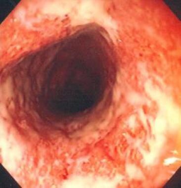
Colonoscopy. Stricture in terminal ileum noted during colonoscopy. Narrowed segment visible upon intubation of terminal ileum with colonoscope. Relatively little active inflammation is present, indicating that this is cicatrix stricture.
-
Colonoscopy. Inflammation in terminal ileum noted during colonoscopy. Areas of inflammation, friability, and ulceration in terminal ileum are consistent with mild-to-moderate Crohn disease.
-
Colonoscopy. Ulcerative colitis as visualized with colonoscope.
-
Colonoscopy. Colonoscopic image of large ulcer and inflammation of descending colon in 12-year-old boy with Crohn disease.
-
Colonoscopy. Colonoscopy video depicts arteriovenous malformation (AVM) in colon. Video courtesy of Dawn Sears, MD, and Dan C Cohen, MD, Division of Gastroenterology, Scott & White Healthcare.
-
Colonoscopy. Colonoscopy video shows removal of small polyp from cecum with snare polypectomy technique. Video courtesy of Dawn Sears, MD, and Dan C Cohen, MD, Division of Gastroenterology, Scott & White Healthcare.
-
Colonoscopy. Colonoscopy video shows mass in colon suspicious for colon cancer. Mass is too large to remove endoscopically and will have to be tattooed so that surgeons can find it easily. Video courtesy of Dawn Sears, MD, and Dan C Cohen, MD, Division of Gastroenterology, Scott & White Healthcare.
-
Colonoscopy. Colonoscopy video shows pseudopolyps in colon. This is usually seen in inflammatory bowel disease (eg, Crohn disease or ulcerative colitis). Video courtesy of Dawn Sears, MD, and Dan C Cohen, MD, Division of Gastroenterology, Scott & White Healthcare.
-
Colonoscopy. Colonoscopy video shows narrowed area in colon in setting of diverticulitis. Video courtesy of Dawn Sears, MD, and Dan C Cohen, MD, Division of Gastroenterology, Scott & White Healthcare.
-
Colonoscopy. Colonoscopy video shows narrowed area in colon in setting of diverticulitis. Video courtesy of Dawn Sears, MD, and Dan C Cohen, MD, Division of Gastroenterology, Scott & White Healthcare.
-
Colonoscopy. Colonoscopy video shows removal of large polyp by hot snare polypectomy technique. Video courtesy of Dawn Sears, MD, and Dan C Cohen, MD, Division of Gastroenterology, Scott & White Healthcare.
-
Colonoscopy. Colonoscopy video shows placement of tattoo adjacent to colon mass in order to make it visible and easy to locate for surgeons. Video courtesy of Dawn Sears, MD, and Dan C Cohen, MD, Division of Gastroenterology, Scott & White Healthcare.
-
Colonoscopy. Colonoscopy video shows diverticulosis (pockets within colon that can bleed or become infected). Video courtesy of Dawn Sears, MD, and Dan C Cohen, MD, Division of Gastroenterology, Scott & White Healthcare.


