Background
Lower eyelid blepharoplasty is often the treatment for patients who have bags or puffiness under the eyes. This deformity is frequently a result of pseudoherniation of the orbital fat. Skin laxity and rhytides may contribute to the overall perception of the deformity. Blepharoplasty rejuvenates and restores the lower lid area. Traditionally, the anterior transcutaneous approach is used for this procedure. This technique provides excellent exposure to the orbital fat, and the resultant scar is minimal. Unfortunately, the approach is associated with many complications. [1, 2, 3]
Lower eyelid malposition is a feared complication after lower lid blepharoplasty and may be associated with scleral show and ectropion of various degrees. These complications tend to limit the success of the transcutaneous blepharoplasty technique. To this end, the use of a posterior transconjunctival technique has been explored. By avoiding scarring at the anterior lamella and lower eyelid septum, retraction of the eyelid and ectropion can be avoided. Accordingly, the posterior transconjunctival approach has gained in popularity and acceptance. The transconjunctival incision through the fornix seems to offer less risk for postoperative lid malposition and provides for the best exposure to the orbital fat. [4]
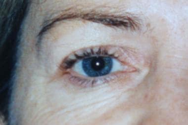
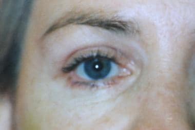
Images depicting preoperative and postoperative blepharoplasty can be seen below.
History of the Procedure
Tessier reported his use of the transconjunctival approach for craniofacial procedures in the 1970s. [5] He had been applying this approach for congenital abnormalities and trauma for almost 20 years when he presented his findings. In the 1920s, Bourget first described the transconjunctival approach in the French literature as a method for removing herniated orbital fat. [6] Tessier adapted the technique to approach the orbital floor without leaving a visible scar. He found the standard periorbital incisions to be limited to specific maneuvers and theorized that the transconjunctival technique was truly the most logical approach to the orbital walls.
Transconjunctival approaches to the lower eyelid have become very popular. In combination with other techniques, the application can be expanded to address skin laxity and rhytides as well as pseudoherniated fat.
Problem
The goal of blepharoplasty of the lower lid is to rejuvenate and thus restore the eyelid. This is achieved with special attention to lateral canthus definition and to eyelid position and function. Excess fat (pseudoherniated orbital fat) is removed, and excess skin is then addressed. Excess fat in this region may arise from a hereditary pseudoherniation of the fat or from the effects of aging. Additionally, systemic diseases, including allergies, cardiac or renal disease, liver cirrhosis, and hyperthyroidism, may contribute to puffiness around the lower eyelid.
A genetic condition known as blepharochalasis is another potential disorder leading to this deformity. Blepharochalasis is a familial disorder that results in chronic eyelid edema, which can progress to localized tissue breakdown and fat herniation. Clearly, these conditions are separate from the pseudoherniation observed in the typical patient evaluated for cosmetic blepharoplasty, and they must therefore be excluded.
The ideal patient is young and has excess pseudoherniated fat but minimal skin laxity. Older patients may benefit from the transconjunctival approach combined with a pinch excision of excess skin or a resurfacing technique such as laser resurfacing or deep chemical peel.
Pathophysiology
As humans age, the orbital septum loses its structural integrity and consequently fails to contain the orbital fat. The supporting structures of the orbit also become lax, allowing settling of the globe, which contributes to fat protrusion. Although the fat remains posterior to the orbital septum, the septum/fat complex bulges forward, giving the illusion of herniated fat (hence the term pseudoherniation). The transconjunctival approach delivers the fat without disrupting the orbital septum and removes the excess fat.
Presentation
Candidates for blepharoplasty typically present with fat herniation and are unhappy with the bags, puffiness, or dark circles under their eyes.
Indications
Assess candidates for blepharoplasty by evaluating pseudoherniated fat, excess eyelid skin, skin laxity, and rhytides. Because the transconjunctival approach is best for removal of pseudoherniated fat, patients who have fat pseudoherniation without additional skin laxity or orbicularis oculi hypertrophy are the best candidates. [2]
Because the technique has a lower risk of resultant scleral show and eyelid retraction (as with the transcutaneous approach), patients with pseudoproptosis and lower eyelid bulges are eligible for the transconjunctival approach. Additionally, patients with retracted eyelids from Graves disease are also good candidates for transconjunctival blepharoplasty.
The transconjunctival approach is a better option in patients who express concerns about scarring with the transcutaneous approach. Although the resultant transcutaneous scar is almost imperceptible, depigmentation at the scar line sometimes occurs in darker-skinned individuals. The transconjunctival approach obviates this possibility.
Relevant Anatomy
The lower eyelid is composed of 3 lamellae. At the level of the tarsus, the posterior lamella consists of the conjunctiva and the tarsus. Inferior to the tarsus, the posterior lamella is composed of conjunctiva, the retractor muscle, and the capsulopalpebral fascia. The middle lamella fuses with the posterior lamella at the tarsal plate and is composed of the orbital septum. The anterior lamella is the orbicularis oculi muscle and overlying skin. The orbital septum is a central component of the lower eyelid. The orbital septum inserts along the inferior aspect of the tarsus and extends inferiorly to its insertion at the arcus marginalis at the orbital rim. The orbital septum confines the fat pads. If the septum is weak, either congenitally or from aging, pseudoherniation of the orbital fat can begin.
Three fat compartments apparently exist: lateral, middle, and medial. Although these compartments are more practical than anatomical, the medial fat pad is separated from the middle pad by the inferior oblique muscle and the arcuate expanse dividing the middle from the lateral fat pad. The medial fat pad is whiter than the middle and lateral pads. The middle fat pad is more yellowish and is often removed in pieces, whereas the medial and lateral pads are often removed intact. In repose, the position of the lower eyelid conjunctiva is vertical at the lid margin and horizontal at the fornix. The average depth of the fornix from the lid margin is 12-14 mm, and the average height of the tarsal plate is approximately 4.5 mm.
Contraindications
Transconjunctival blepharoplasty is a technique that best addresses pseudoherniated orbital fat. In the patient with minimal fat pseudoherniation, this approach may not be best. In patients with significant skin laxity and redundancy, and in patients with hypertrophic orbicularis oculi muscle, the transconjunctival approach can offer little benefit unless combined with an adjunct procedure.
As with all blepharoplasty techniques, transconjunctival blepharoplasty is relatively contraindicated in the patient with severe lower eyelid laxity unless accompanied with lid-tightening procedures.
-
Preoperative photograph of the right eye.
-
Postoperative photograph of the right eye.


